
Acknowledgements Robert Stroud James Fraser Aina Cohen Ken
Acknowledgements Robert Stroud James Fraser Aina Cohen Ken Frankel John Tainer UCSF LBNL SLAC ALS 8. 3. 1 creator: Tom Alber UC Multicampus Research Programs and Initiatives (MRPI) UCSF Program for Breakthrough Biomedical Research (PBBR) one-time NIH-DOE Inter-agency agreement (IAA) Integrated Diffraction Analysis Technologies (IDAT) Plexxikon, Inc. M D Anderson CRC Synchrotron Radiation Structural Biology Resource (SLAC) The Advanced Light Source is supported by the Director, Office of Science, Office of Basic Energy Sciences, Materials Sciences Division, of the US Department of Energy under contract No. DE-AC 02 -05 CH 11231 at Lawrence Berkeley National Laboratory.

Acknowledgements Robert Stroud James Fraser Aina Cohen Ken Frankel John Tainer UCSF LBNL SLAC ALS 8. 3. 1 creator: Tom Alber UC Multicampus Research Programs and Initiatives (MRPI) UCSF Program for Breakthrough Biomedical Research (PBBR) one-time NIH-DOE Inter-agency agreement (IAA) Integrated Diffraction Analysis Technologies (IDAT) Plexxikon, Inc. M D Anderson CRC Synchrotron Radiation Structural Biology Resource (SLAC) The Advanced Light Source is supported by the Director, Office of Science, Office of Basic Energy Sciences, Materials Sciences Division, of the US Department of Energy under contract No. DE-AC 02 -05 CH 11231 at Lawrence Berkeley National Laboratory.
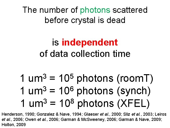
The number of photons scattered before crystal is dead is independent of data collection time 3 um 5 10 1 = photons (room. T) 1 um 3 = 106 photons (synch) 1 um 3 = 108 photons (XFEL) Henderson, 1990; Gonzalez & Nave, 1994; Glaeser et al. , 2000; Sliz et al. , 2003; Leiros et al. , 2006; Owen et al. , 2006; Garman & Mc. Sweeney, 2006; Garman & Nave, 2009; Holton, 2009
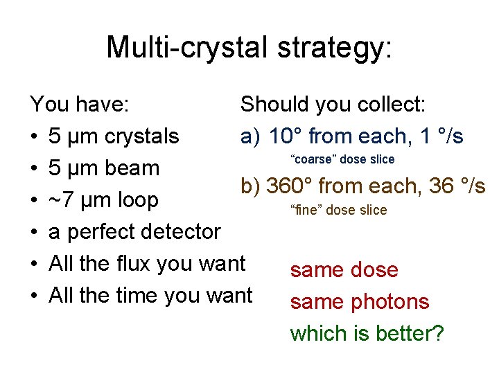
Multi-crystal strategy: Should you collect: You have: a) 10° from each, 1 °/s • 5 μm crystals “coarse” dose slice • 5 μm beam b) 360° from each, 36 °/s • ~7 μm loop “fine” dose slice • a perfect detector • All the flux you want same dose • All the time you want same photons which is better?
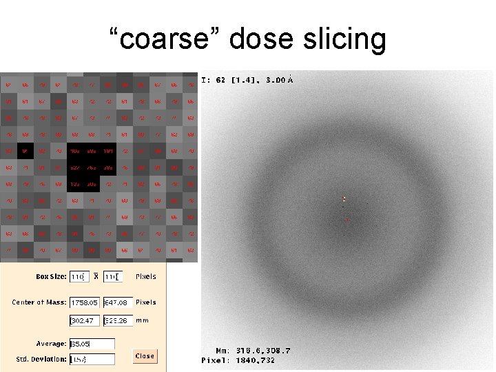
“coarse” dose slicing
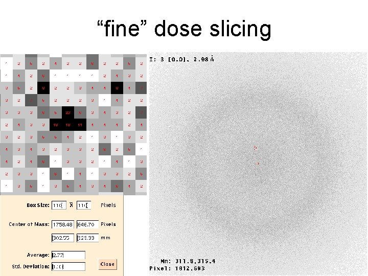
“fine” dose slicing
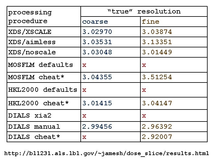
XDS/XSCALE XDS/aimless XDS/noscale “true” resolution coarse fine 3. 02970 3. 03874 3. 03531 3. 13351 3. 03048 3. 01449 MOSFLM defaults x x MOSFLM cheat* 3. 04355 3. 51254 processing procedure HKL 2000 defaults x x HKL 2000 cheat* 3. 01415 3. 04147 DIALS xia 2 x x DIALS manual DIALS cheat* 2. 99456 x 2. 96392 2. 92007 http: //bl 1231. als. lbl. gov/~jamesh/dose_slice/results. html
Overcoming non-isomorphism not same structure with fancy math thing
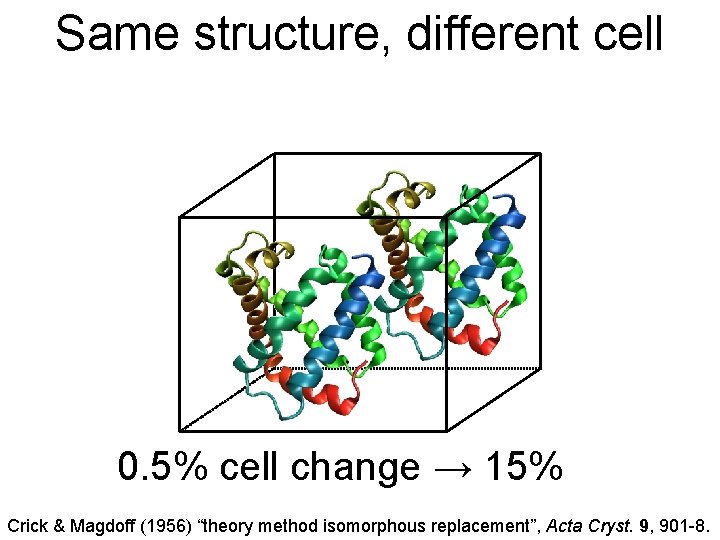
Same structure, different cell 0. 5% cell change → 15% Crick & Magdoff (1956) “theory method isomorphous replacement”, Acta Cryst. 9, 901 -8.
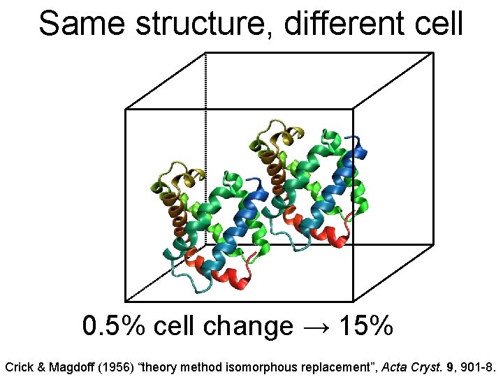
Same structure, different cell 0. 5% cell change → 15% Crick & Magdoff (1956) “theory method isomorphous replacement”, Acta Cryst. 9, 901 -8.
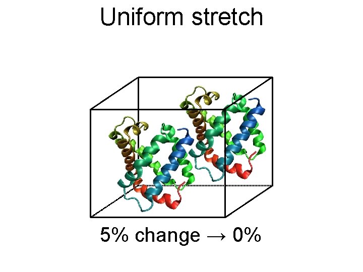
Uniform stretch 5% change → 0%
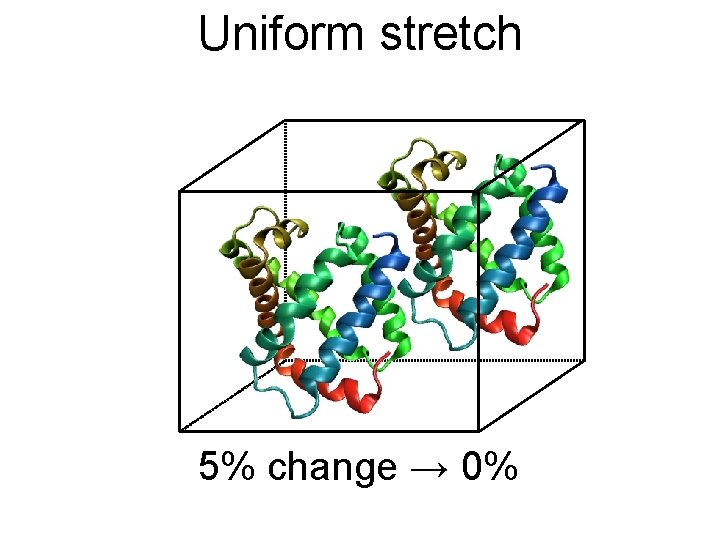
Uniform stretch 5% change → 0%
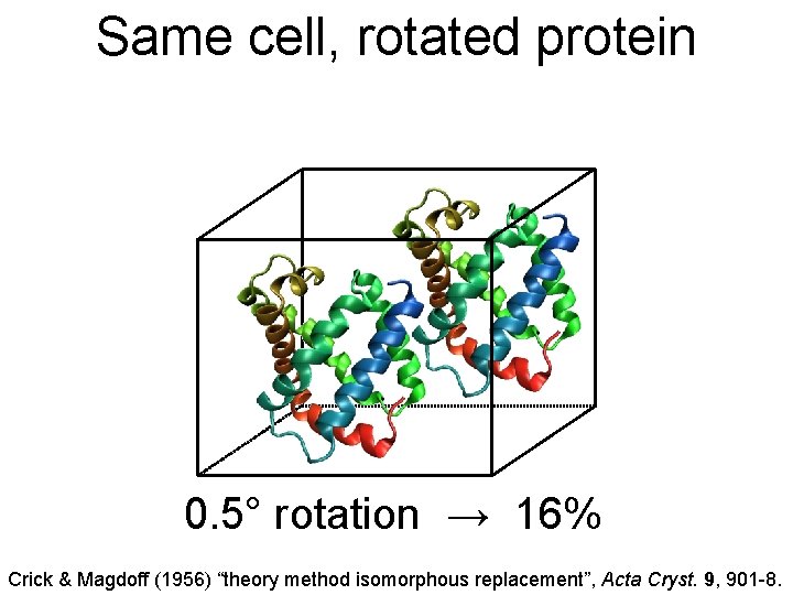
Same cell, rotated protein 0. 5° rotation → 16% Crick & Magdoff (1956) “theory method isomorphous replacement”, Acta Cryst. 9, 901 -8.
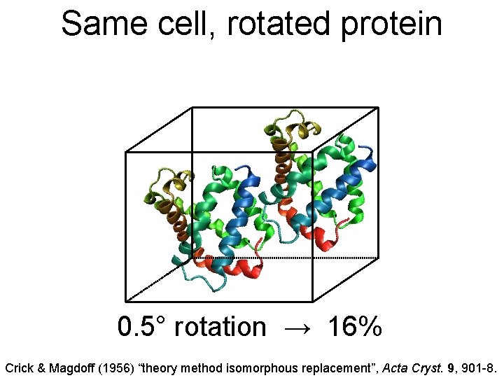
Same cell, rotated protein 0. 5° rotation → 16% Crick & Magdoff (1956) “theory method isomorphous replacement”, Acta Cryst. 9, 901 -8.
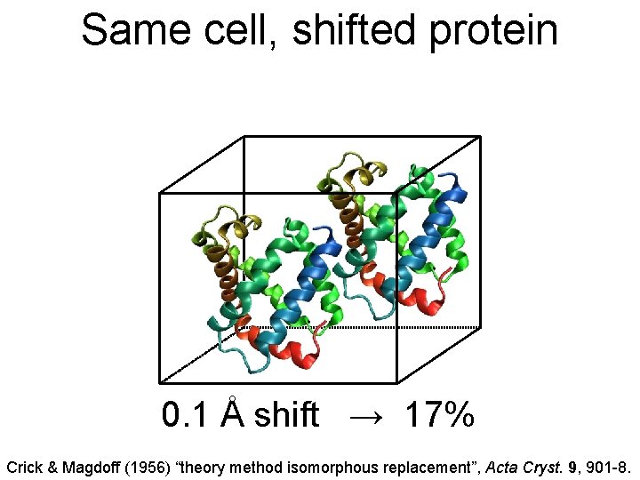
Same cell, shifted protein 0. 1 Å shift → 17% Crick & Magdoff (1956) “theory method isomorphous replacement”, Acta Cryst. 9, 901 -8.
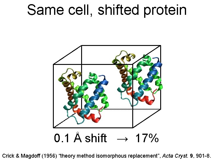
Same cell, shifted protein 0. 1 Å shift → 17% Crick & Magdoff (1956) “theory method isomorphous replacement”, Acta Cryst. 9, 901 -8.
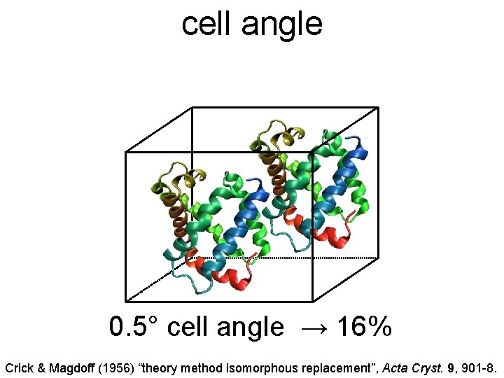
cell angle 0. 5° cell angle → 16% Crick & Magdoff (1956) “theory method isomorphous replacement”, Acta Cryst. 9, 901 -8.
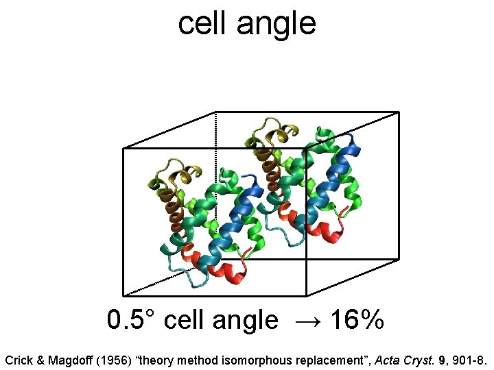
cell angle 0. 5° cell angle → 16% Crick & Magdoff (1956) “theory method isomorphous replacement”, Acta Cryst. 9, 901 -8.
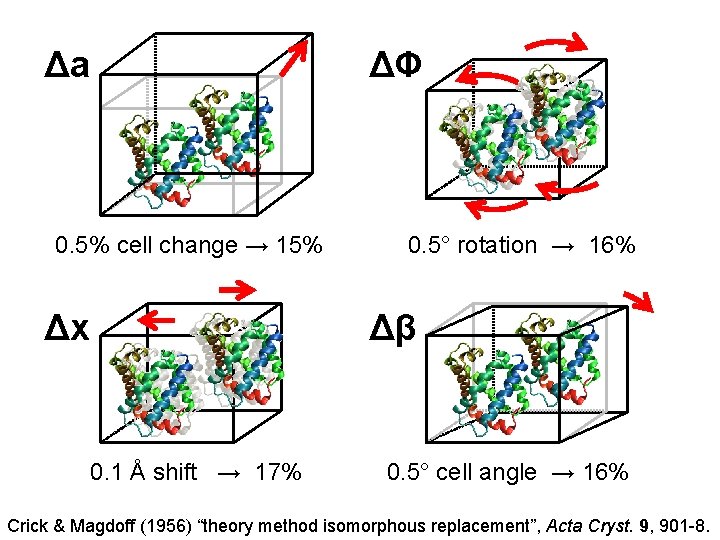
Δa ΔΦ 0. 5% cell change → 15% Δx 0. 5° rotation → 16% Δβ 0. 1 Å shift → 17% 0. 5° cell angle → 16% Crick & Magdoff (1956) “theory method isomorphous replacement”, Acta Cryst. 9, 901 -8.
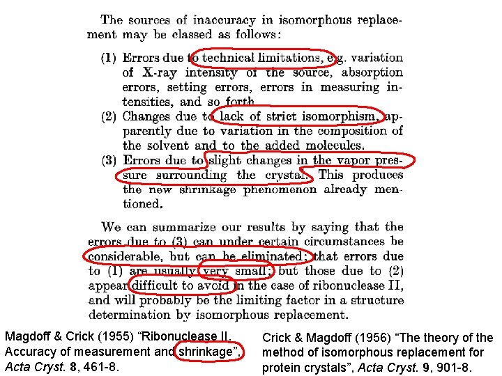
Magdoff & Crick (1955) “Ribonuclease II. Accuracy of measurement and shrinkage”, Acta Cryst. 8, 461 -8. Crick & Magdoff (1956) “The theory of the method of isomorphous replacement for protein crystals”, Acta Cryst. 9, 901 -8.
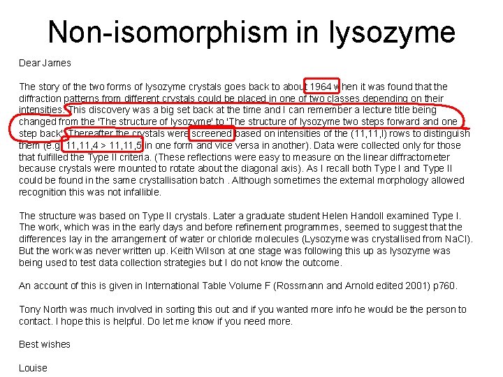
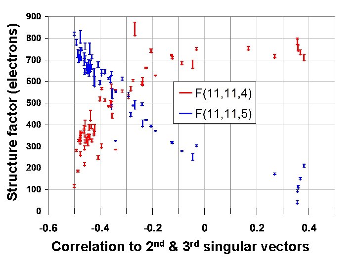
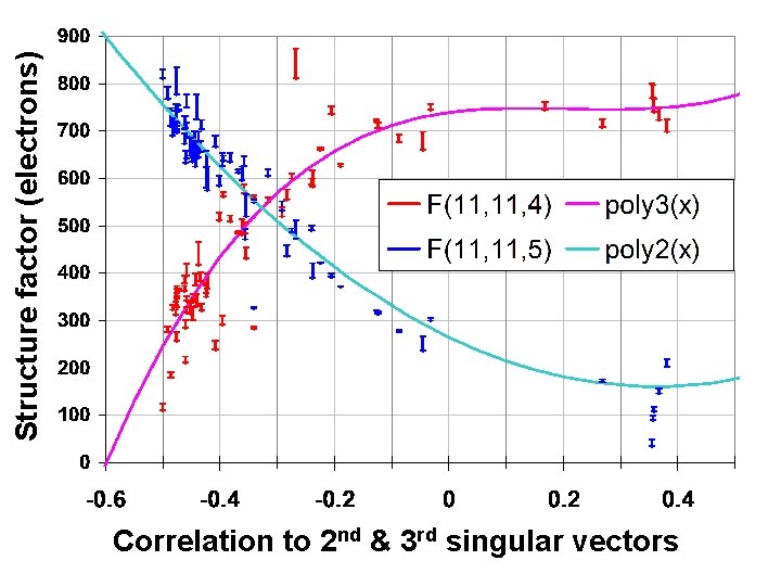
Non-isomorphism in lysozyme Dear James The story of the two forms of lysozyme crystals goes back to about 1964 when it was found that the diffraction patterns from different crystals could be placed in one of two classes depending on their intensities. This discovery was a big set back at the time and I can remember a lecture title being changed from the 'The structure of lysozyme' to 'The structure of lysozyme two steps forward and one step back'. Thereafter the crystals were screened based on intensities of the (11, l) rows to distinguish them (e. g. 11, 4 > 11, 5 in one form and vice versa in another). Data were collected only for those that fulfilled the Type II criteria. (These reflections were easy to measure on the linear diffractometer because crystals were mounted to rotate about the diagonal axis). As I recall both Type I and Type II could be found in the same crystallisation batch. Although sometimes the external morphology allowed recognition this was not infallible. The structure was based on Type II crystals. Later a graduate student Helen Handoll examined Type I. The work, which was in the early days and before refinement programmes, seemed to suggest that the differences lay in the arrangement of water or chloride molecules (Lysozyme was crystallised from Na. Cl). But the work was never written up. Keith Wilson at one stage was following this up as lysozyme was being used to test data collection strategies but I do not know the outcome. An account of this is given in International Table Volume F (Rossmann and Arnold edited 2001) p 760. Tony North was much involved in sorting this out and if you wanted more info he would be the person to contact. I hope this is helpful. Do let me know if you need more. Best wishes Louise
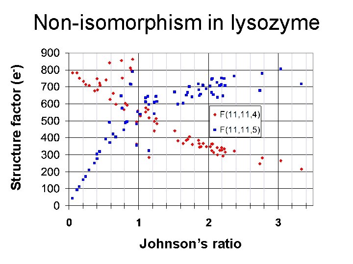
Structure factor (e-) Non-isomorphism in lysozyme Johnson’s ratio
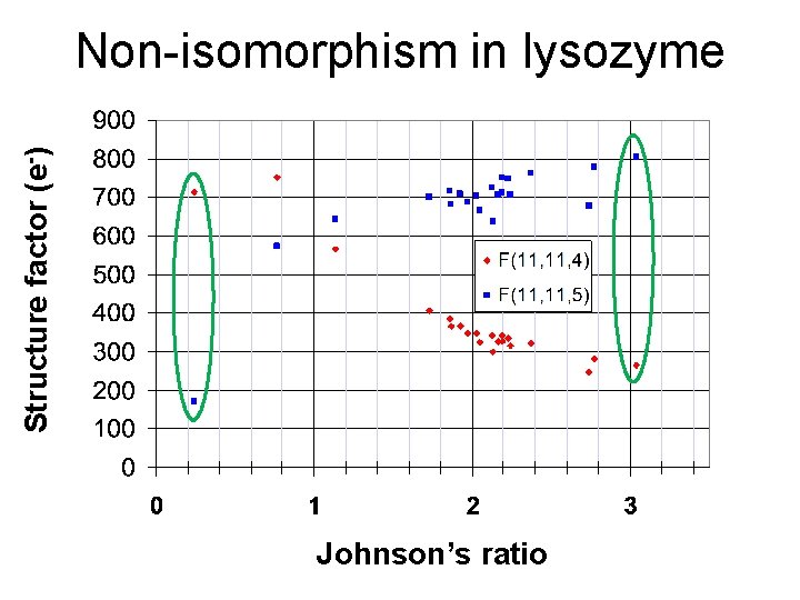
Structure factor (e-) Non-isomorphism in lysozyme Johnson’s ratio
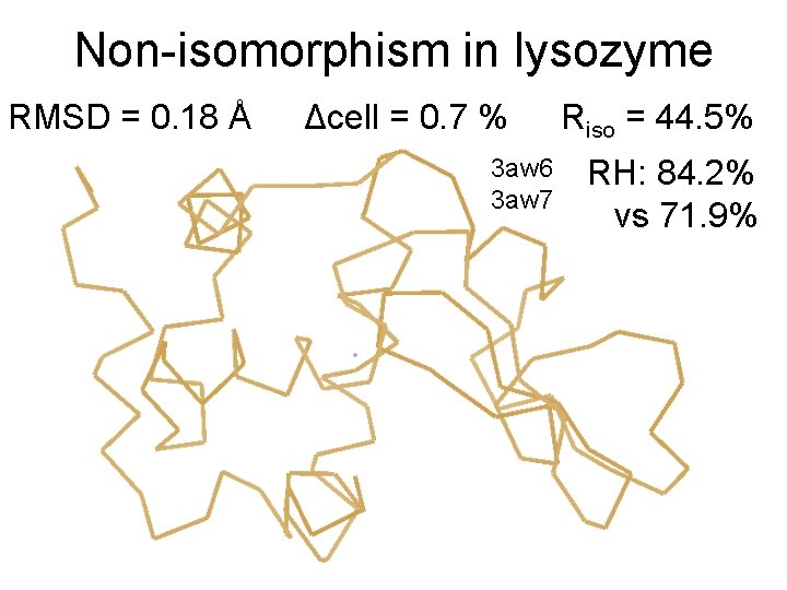
Non-isomorphism in lysozyme RMSD = 0. 18 Å Δcell = 0. 7 % 3 aw 6 3 aw 7 Riso = 44. 5% RH: 84. 2% vs 71. 9%
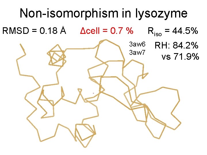
Non-isomorphism in lysozyme RMSD = 0. 18 Å Δcell = 0. 7 % 3 aw 6 3 aw 7 Riso = 44. 5% RH: 84. 2% vs 71. 9%
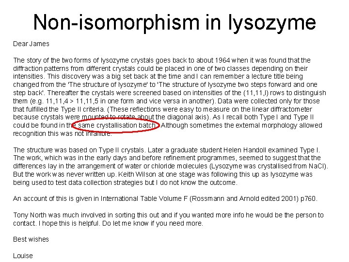
Non-isomorphism in lysozyme Dear James The story of the two forms of lysozyme crystals goes back to about 1964 when it was found that the diffraction patterns from different crystals could be placed in one of two classes depending on their intensities. This discovery was a big set back at the time and I can remember a lecture title being changed from the 'The structure of lysozyme' to 'The structure of lysozyme two steps forward and one step back'. Thereafter the crystals were screened based on intensities of the (11, l) rows to distinguish them (e. g. 11, 4 > 11, 5 in one form and vice versa in another). Data were collected only for those that fulfilled the Type II criteria. (These reflections were easy to measure on the linear diffractometer because crystals were mounted to rotate about the diagonal axis). As I recall both Type I and Type II could be found in the same crystallisation batch. Although sometimes the external morphology allowed recognition this was not infallible. The structure was based on Type II crystals. Later a graduate student Helen Handoll examined Type I. The work, which was in the early days and before refinement programmes, seemed to suggest that the differences lay in the arrangement of water or chloride molecules (Lysozyme was crystallised from Na. Cl). But the work was never written up. Keith Wilson at one stage was following this up as lysozyme was being used to test data collection strategies but I do not know the outcome. An account of this is given in International Table Volume F (Rossmann and Arnold edited 2001) p 760. Tony North was much involved in sorting this out and if you wanted more info he would be the person to contact. I hope this is helpful. Do let me know if you need more. Best wishes Louise
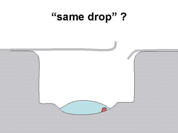
“same drop” ?
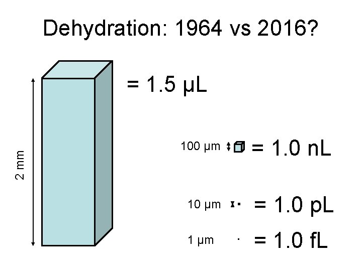
Dehydration: 1964 vs 2016? 2 mm = 1. 5 μL 100 μm = 1. 0 n. L 10 μm = 1. 0 p. L 1 μm = 1. 0 f. L
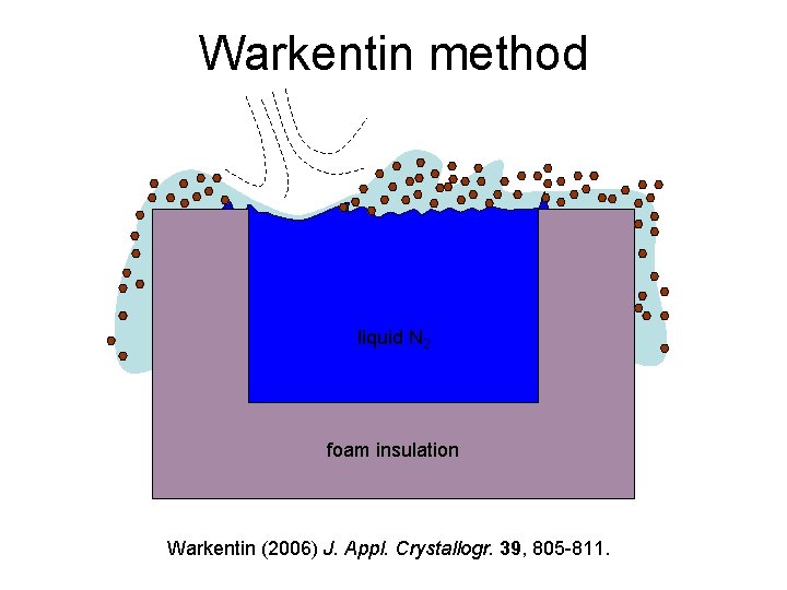
Warkentin method liquid N 2 foam insulation Warkentin (2006) J. Appl. Crystallogr. 39, 805 -811.
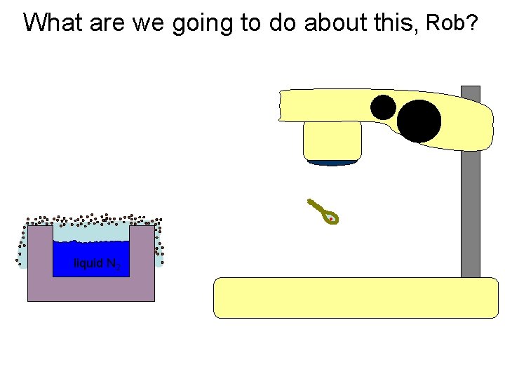
What are we going to do about this, Rob? liquid N 2
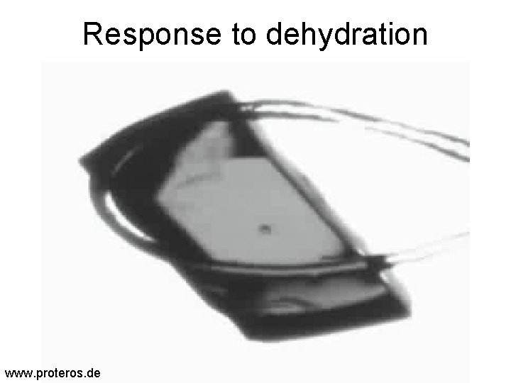
Response to dehydration www. proteros. de

Response to dehydration www. proteros. de
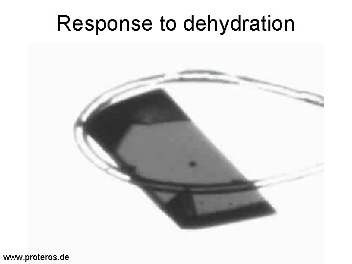
Response to dehydration www. proteros. de
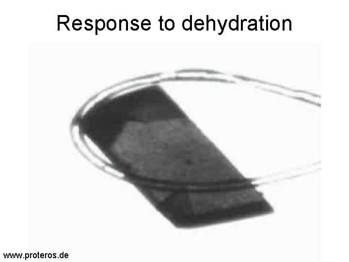
Response to dehydration www. proteros. de
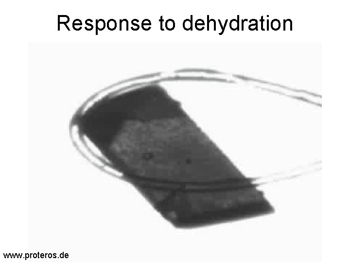
Response to dehydration www. proteros. de
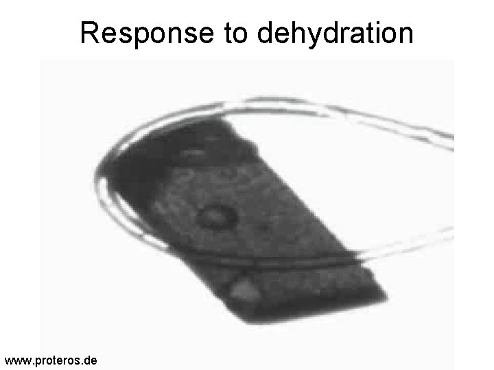
Response to dehydration www. proteros. de
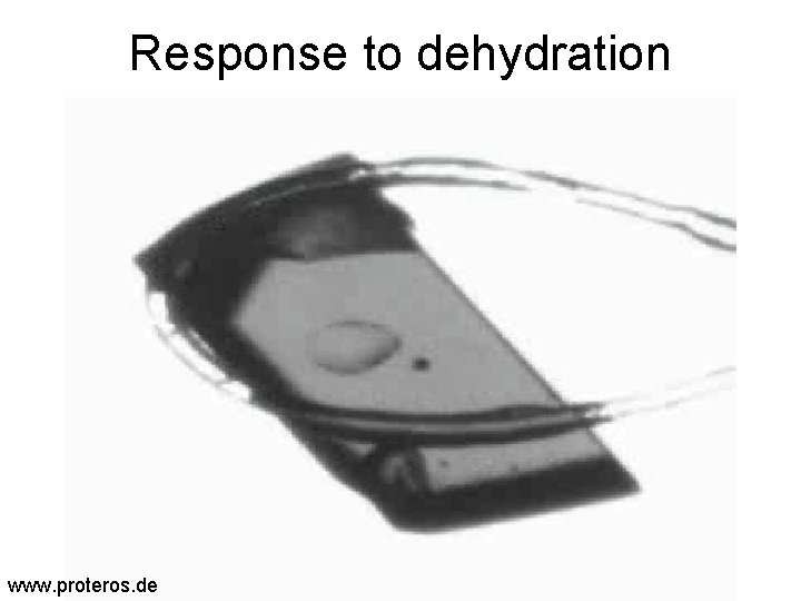
Response to dehydration www. proteros. de
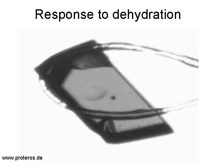
Response to dehydration www. proteros. de
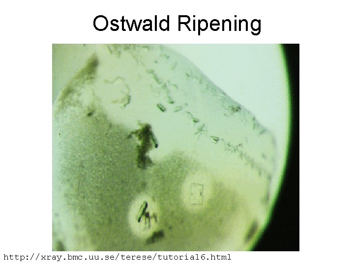
Ostwald Ripening http: //xray. bmc. uu. se/terese/tutorial 6. html
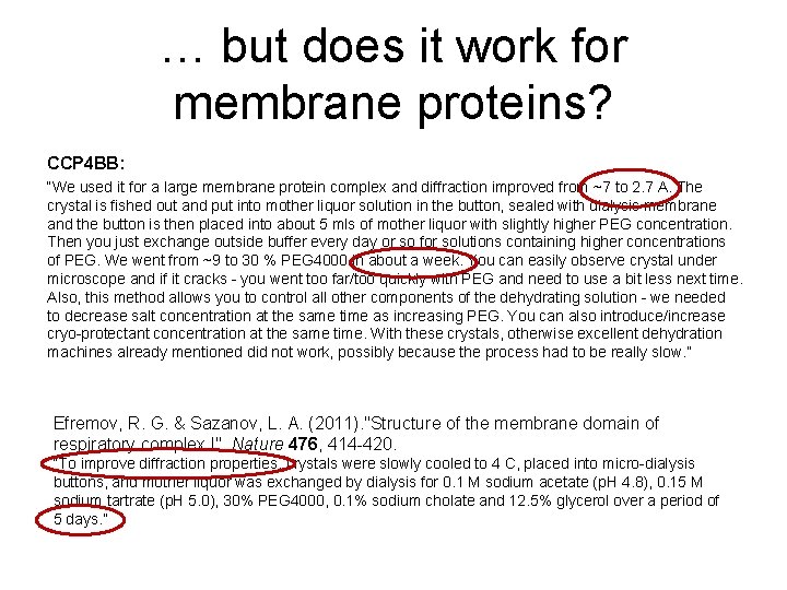
… but does it work for membrane proteins? CCP 4 BB: “We used it for a large membrane protein complex and diffraction improved from ~7 to 2. 7 A. The crystal is fished out and put into mother liquor solution in the button, sealed with dialysis membrane and the button is then placed into about 5 mls of mother liquor with slightly higher PEG concentration. Then you just exchange outside buffer every day or so for solutions containing higher concentrations of PEG. We went from ~9 to 30 % PEG 4000 in about a week. You can easily observe crystal under microscope and if it cracks - you went too far/too quickly with PEG and need to use a bit less next time. Also, this method allows you to control all other components of the dehydrating solution - we needed to decrease salt concentration at the same time as increasing PEG. You can also introduce/increase cryo-protectant concentration at the same time. With these crystals, otherwise excellent dehydration machines already mentioned did not work, possibly because the process had to be really slow. ” Efremov, R. G. & Sazanov, L. A. (2011). "Structure of the membrane domain of respiratory complex I", Nature 476, 414 -420. “To improve diffraction properties, crystals were slowly cooled to 4 C, placed into micro-dialysis buttons, and mother liquor was exchanged by dialysis for 0. 1 M sodium acetate (p. H 4. 8), 0. 15 M sodium tartrate (p. H 5. 0), 30% PEG 4000, 0. 1% sodium cholate and 12. 5% glycerol over a period of 5 days. ”
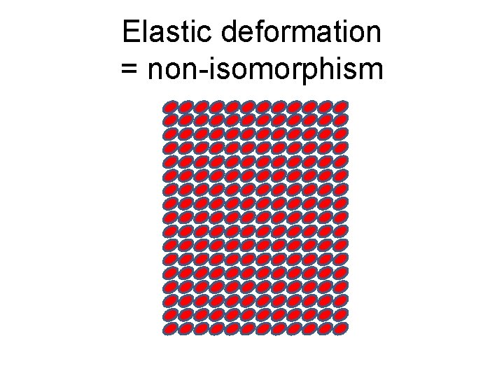
Elastic deformation = non-isomorphism
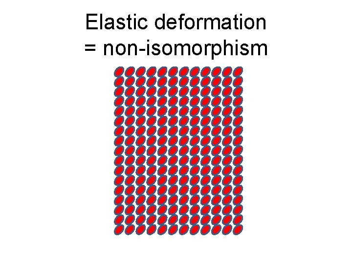
Elastic deformation = non-isomorphism
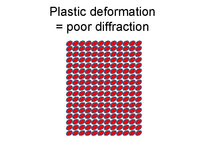
Plastic deformation = poor diffraction
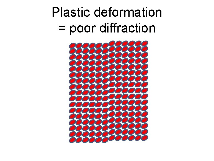
Plastic deformation = poor diffraction

What if we can’t control it?
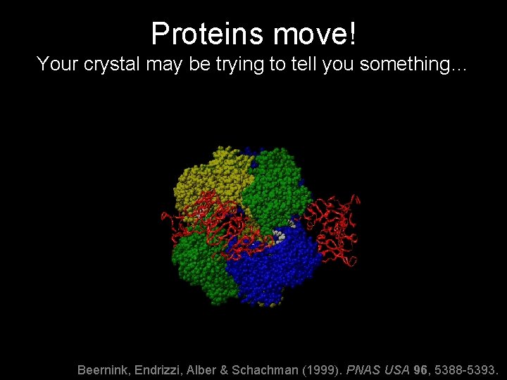
Proteins move! Your crystal may be trying to tell you something… Beernink, Endrizzi, Alber & Schachman (1999). PNAS USA 96, 5388 -5393.
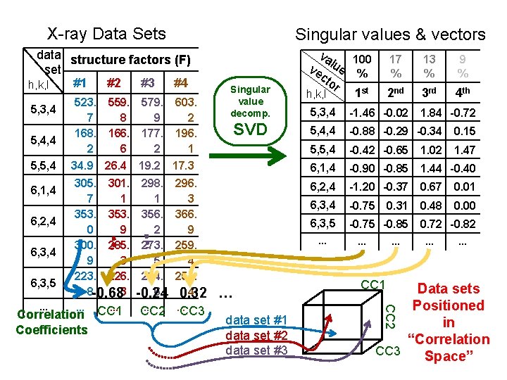
X-ray Data Sets Singular values & vectors data structure factors (F) set #1 #2 #3 #4 h, k, l 5, 3, 4 5, 4, 4 5, 5, 4 6, 1, 4 6, 2, 4 6, 3, 5 523. 7 168. 2 559. 8 166. 6 579. 9 177. 2 603. 2 196. 1 34. 9 26. 4 19. 2 305. 301. 7 1 353. 0 9 300. 285. 9 3 223. 226. 8 -0. 683 17 % 13 % 9 % 2 nd 3 rd 4 th 5, 3, 4 -1. 46 -0. 02 5, 4, 4 -0. 88 -0. 29 -0. 34 0. 15 5, 5, 4 -0. 42 -0. 65 1. 02 1. 47 17. 3 6, 1, 4 -0. 90 -0. 85 1. 44 -0. 40 298. 1 356. 2 273. 5 234. 6 -0. 24 296. 3 366. 9 259. 4 251. 4 0. 32 6, 2, 4 -1. 20 -0. 37 0. 67 0. 01 6, 3, 4 -0. 75 0. 48 0. 00 6, 3, 5 -0. 75 -0. 85 … CC 2 … CC 3 SVD … … data set #1 data set #2 data set #3 0. 31 … … CC 1 1. 84 -0. 72 -0. 82 … … Data sets Positioned in “Correlation CC 3 Space” CC 2 … … … Correlation CC 1 Coefficients Singular value decomp. va 100 ve lue % cto r 1 st h, k, l
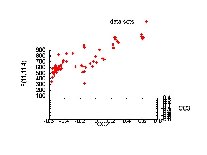
Structure factor (electrons) Correlation to 2 nd & 3 rd singular vectors
Structure factor (electrons) Correlation to 2 nd & 3 rd singular vectors
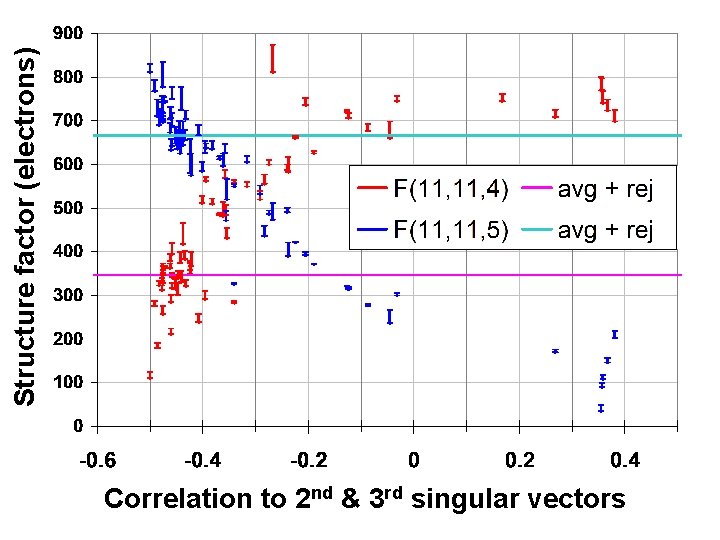
Structure factor (electrons) Correlation to 2 nd & 3 rd singular vectors
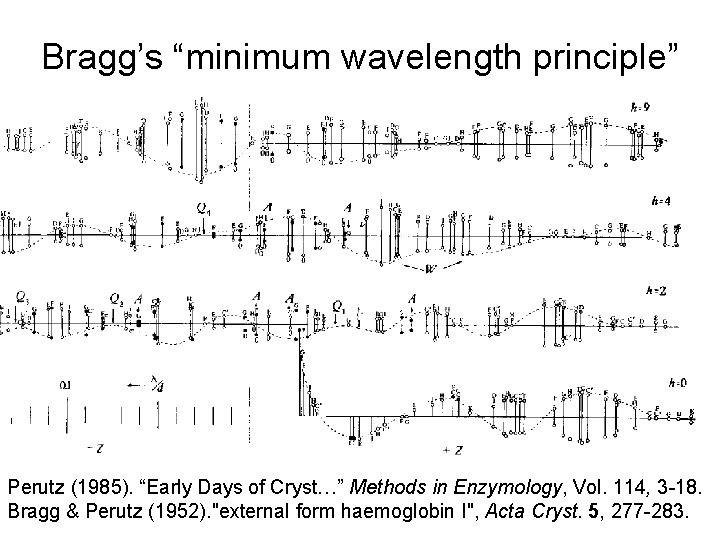
Bragg’s “minimum wavelength principle” Perutz (1985). “Early Days of Cryst…” Methods in Enzymology, Vol. 114, 3 -18. Bragg & Perutz (1952). "external form haemoglobin I", Acta Cryst. 5, 277 -283.
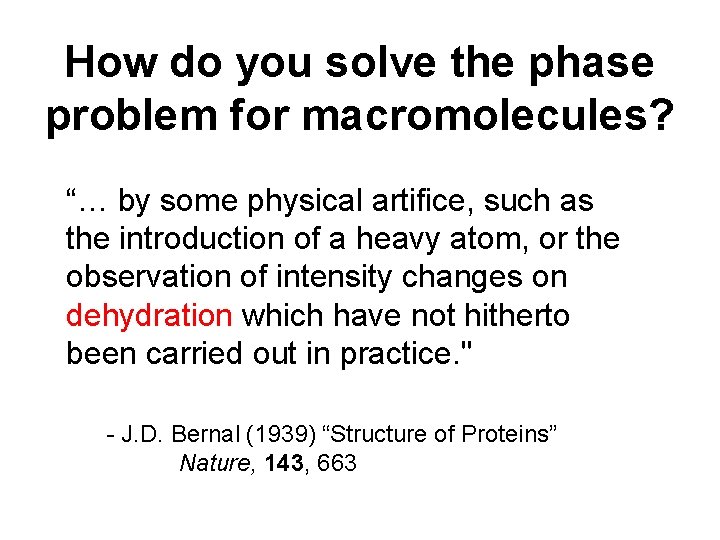
How do you solve the phase problem for macromolecules? “… by some physical artifice, such as the introduction of a heavy atom, or the observation of intensity changes on dehydration which have not hitherto been carried out in practice. " - J. D. Bernal (1939) “Structure of Proteins” Nature, 143, 663
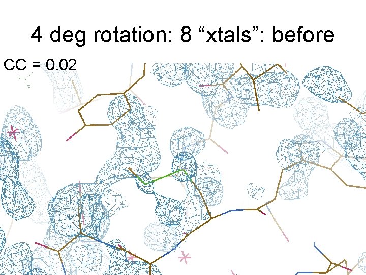
4 deg rotation: 8 “xtals”: before CC = 0. 02
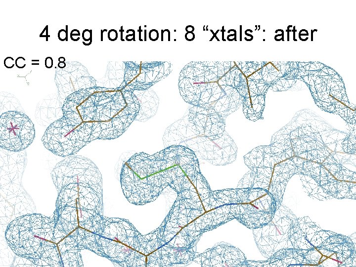
4 deg rotation: 8 “xtals”: after CC = 0. 8
Summary • Careful with dehydration! • Weak images: wave of future? • SVD/PCA instead of averaging • Non-isomorphism phasing (NIP)? http: //bl 831. als. lbl. gov/~jamesh/ powerpoint/ACA_multixtal_2016. pptx
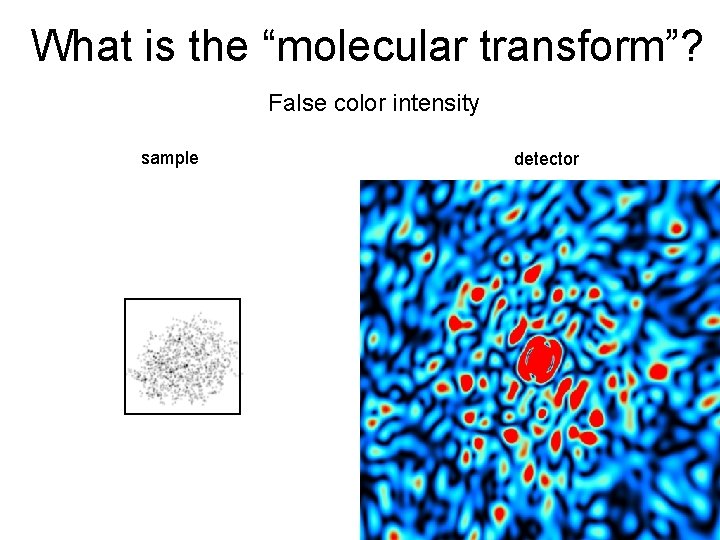
What is the “molecular transform”? False color intensity sample detector
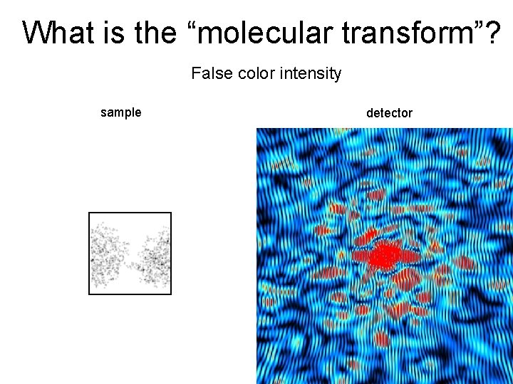
What is the “molecular transform”? False color intensity sample detector
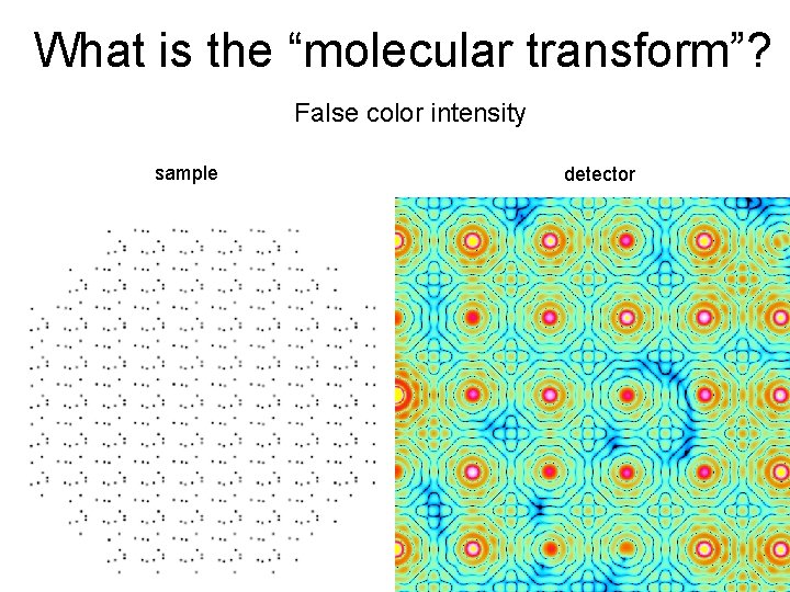
What is the “molecular transform”? False color intensity sample detector
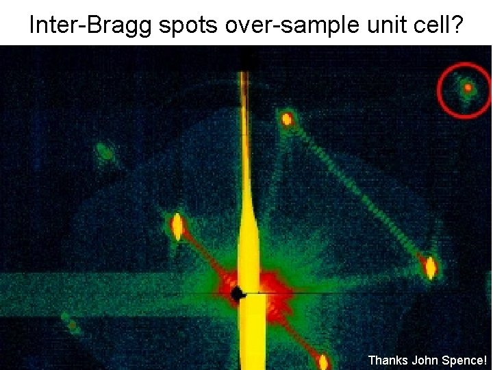
Inter-Bragg spots over-sample unit cell? Thanks John Spence!
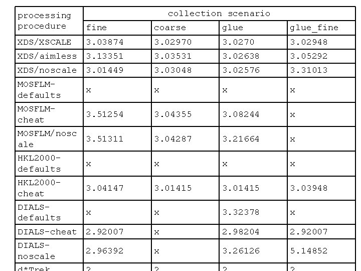
collection scenario processing procedure fine coarse glue_fine XDS/XSCALE 3. 03874 3. 02970 3. 02948 XDS/aimless 3. 13351 3. 03531 3. 02638 3. 05292 XDS/noscale 3. 01449 3. 03048 3. 02576 3. 31013 MOSFLMdefaults x x MOSFLMcheat 3. 51254 3. 04355 3. 08244 x MOSFLM/nosc 3. 51311 ale 3. 04287 3. 21664 x HKL 2000 defaults x x 3. 04147 3. 01415 3. 03948 x x 3. 32378 x DIALS-cheat 2. 92007 x 2. 98204 2. 92007 DIALSnoscale x 3. 26126 5. 14852 HKL 2000 cheat DIALSdefaults 2. 96392
- Slides: 62