Introduction Uveitis inflammation of uveal tract iris ciliary
 and of")


, ischaemia (BRAO, B/CRVO, retinal oedema),")


 2+ Moderate")


 Chronic")


 may be associated with uveitis, particularly in")


- Slides: 43
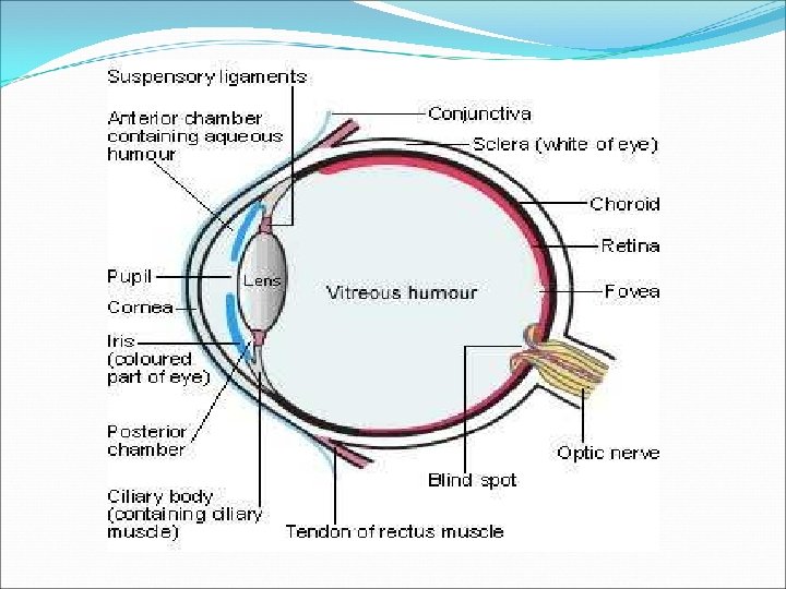
Introduction Uveitis : inflammation of uveal tract (iris, ciliary body & choroid) and of neighbouring structures(retina, vitreous, optic nerve)
IUSG classification Group Sub. Group Infectious Bacterial Fungal Viral Parasitic Others Non Infectious Known Systemic Association No Known Systemic Association Masquerade Neoplastic Non Neoplastic
Anatomical classification Type Primary Site Includes Anterior Uveitis Anterior Chamber Iritis, Iridocyclitis Anterior cyclitis Intermediate Uveitis Vitreous Posterior Uveitis Retina or Choroid Pan Uveitis Anterior Chamber, Vitreous and Retina or choroid Pars Planitis Posterior cyclitis Hyalitis Focal, Multi Focal or Diffuse Choroiditis Chorioretinitis Retinochoroiditis Neuroretinitis
Pathological Classification Granulomatous Non granulomatous Large greasy mutton fat KPs Iris nodules-Koeppe & Busacca Confluent, fine white coloured small(lymphocyte, plasma cells pigments)
Descriptors of Clinical Behaviour Type Descriptor Onset Sudden Insidious Duration Limited Persistent ≤ 3 Months ≥ 3 Months Course Acute Recurrent Sudden Onset+Limited Duration Repeated Episodes, inactive periods ≥ 3 months off treatment Persistent, Relapse in <3 months off treatment Chronic Definition
Approach to patient of Uveitis Symptoms Anterior : photophobia, redness, watering, pain, ↓VA; may be asymptomatic Intermediate: floaters, photopsia, ↓VA Posterior: ↓VA, Photopsia, floaters, scotomata POH Previous episodes and investigation; surgery/trauma PMH Arthropathies (e. g. Ankylosing sponylitis), Chronic infections (e. g. HSV, tuberculosis), systemic inflammation(e. g. sarcoid, Behcet, s disease) SR Detailed review of all systems
Approach to patient of Uveitis FH Family members with Uveitis or related diseases SH Travel/residence abroad, pets IV drugs, sexual Hx DX Including any systemic immunosuppresion AX Allergies or relevant drug contraindictions Visual acuity Best-corrected/pin-hole; near Visual Function Check for RAPD, colour vision Conjunctiva Circumcorneal injection Cornea Band keratopathy, keratic precipitates (distribution, size, pigment)
Approach to patient of Uveitis AC Flare/ cells, fibrin, hypopyon Gonioscopy PAS (consider if ↑IOP) Iris Loss of pattern, Transillumination defects/sectoral atrophy, miosis, posterior synechiae, heterochromia, Koeppe or Busacca nodules , NVI Lens Pigment on ALS, cataract, aphakia/ pseudophakia Tonometry Dilated fundoscopy (non-contact handheld lens ±indirect/indenting) Vitreous Haze, cells, snowballs, opacities, subhyaloid precipitates (KP-like but on posterior vitreous face) Optic disc Disc swelling, glaucomatous changes, atrophy
Approach to patient of Uveitis Vessels Inflammation (sheathing, leakage), ischaemia (BRAO, B/CRVO, retinal oedema), occlusion Retina CMO, uni/multifocal retinitis (blurred white lesions may progress oedema, )occlusion Choroid Uni/multifocal choroiditis (deeper yellow-white lesions), associated exudative retinal detachment
Ocular Signs Anatomical location Condition Forehead & Adnexa Vesicles Herpes Zoster Ophthalmicus Poliosis VKH Nodules Sarcoid, Leprosy Madarosis Leprosy Conjunctiva Granulomas Foreign body granulomas Sarcoid Cornea Dendritic keratitis, SPK Viral uveitis Sclero Kerato uveitis Syphilis, tuberculosis, Hansen’s & viral Exposure and neurotrophic keratitis Leprosy Band Keratopathy Juvenile rheumatoid arthritis, Sarcokdosis
Ocular signs Anatomical location Condition Iris/Pupil Miotic and irregular pupils posterior synechiae (but the response of the pupil to light and near is symmetric), Festooned pupil, seclusio pupillae, occlusio pupillae, ectropion pupillae Relative Afferent Pupillary Defect Asymmetric disc involvement as a result of disc edema due to uveitis or optic atrophy as a result of chronic uveitis Sectoral iris atrophy Herpetic uveitis (irregular constriction of pupil) Argyl Robinson pupil Neurosyphilis
Ocular signs Anatomical Location Condition Gonioscopic evaluation Peripheral Anterior Synechiae Sarcoid, Tuberculosis Iris nodules Sarcoid, Tuberculosis Hyphema Herpetic Foreign body Trumatic uveitis
Grading of AC cells Grade Description 0 None 1+ Faint (just detactable) 2+ Moderate (iris+lens clear) 3+ Marked (iris+lens hazy) 4+ Intense (fibrin or plastic aqueous)
Grading of AC Flare Activity Cells 0 <1 0. 5+ 1 -5 1+ 6 -15 2+ 16 -25 3+ 26 -50 4+ >50
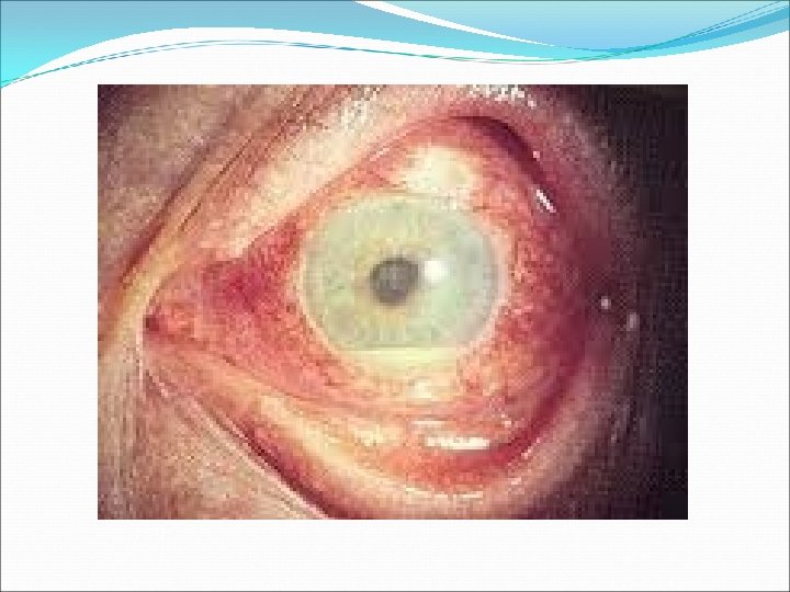
Signs
Signs KP: fibrous deposits on the posterior surface of the cornea, usually associated with uveitis. Both the size and distribution of keratic precipitates are helpful in the differential diagnosis. White, yellowish greasy precipitates of inflammatory cells Typically distributed in a wedge-shaped region on the inferior corneal endothelium, known as Arlt's triangle
AS Fibrin clot and posterior synechiae in a patient with acute, anterior uveitis and ankylosing spondylitis.
Signs
Iris atrophy in a patient with herpes simplex virus–associated anterior uveitis.
Etiology of anterior uveitis Ø Acute § Idipathic § HLA-B 27 associated v v Ankylosing Spondylitis Reiter’s syndrome IBD Psoariasis § Herpetic anterior uveitis(HSV and VZV) § Posner Schlossman Syndrome § Systemic diseases associated v Diabetes v Sarcoidosis v TINU, Ig. A Nephropathy
Etiology of anterior uveitis Ø Chronic § Fuch’s Heterochromic uveitis § JIA
Systemic signs Systemic sign Differentials Poliosis VKH, Sympathetic ophthalmia Loss of Hair SLE, VKH and Syphillis Hypo-pigmentation of the skin Leprosy, Sympathetic ophthalmia, VKH harada’s Rash Vascuitic disease, SLE, Behcet’s Disease, Syphilis Erythema nodosum-tender violaceous subcutaneous nodules in lower extremities I BD, sarcoidosis, TB, Behcet’s Scaling of the skin SLE, Psoriatic arthritis, Syphilis and Reiter’s syndrome Discoid lesions SLE, Psoriatic arthritis, Syphilis, Reiter’s Nail abnormalities Psoriatic arthritis, reiter’s, Vasculitis Oral ulcers alone SLE& IBD Urethral discharge Reiter’s s, Syphilis, HSV, Gonococcal Urethritis
Systemic sign Differentials Epididymitis Behcet’s , TB Prostatitis Reiter’s , AS and Gonococcal Nephritis Vasculitis (Wegener’s, SLE, Behcet’s), Sarcoidosis, TB Arthritis Sero negative spondyloarthropathies, JRA, Behcet’s, Sarcoidosis, SLE, Leprosy, Relapsing polychondritis Cartilage loss Relapsing polychondritis, Syphilis, Gonococcal, Wegener’s Nasopharyngeal manifestation Wegener’s , Sarcoidosis, Whipple’s, Mucormycosis Cystitis Whipple’s, Reiter’s Lymph nodes TB, Sarcoidosis, Lymphoma Neuropathy Leprosy, HZV, Sarcoidosis, MS, Syphilis
Systemic sign Differentials Hearing loss VKH, Sarcoidosis Respiratory Symptoms TB, Sarcoidosis, Wegener’s Bowel disease Whipple’s, Crohn’s, Ulcerative colitis Fever Collagen vascular disease, TB, Leptospirosis
Nongranulomatous Vs granulomatous Uveitis Non-granulomatous Granulomatous Onset Acute Insidious Evolution Spontaneous regression (mostly) Chronic Keratic precipitates Confluent, fine white coloured small(lymphocyte, plasma cells pigments) Non confluent, large mutton fat keratic precipitates (epithelioid cells, hystiocytes) Iris Occasionally Koeppe’s nodules Frequent Koeppe’s and Busacca’s nodules Flare Intense Mild Synecthiae Easy to break with Dens broad based, mydriatics in early stages. difficult to break Vitreous exudates Fine punctuate opacities in vitreous Heavy vitreous exudates
Differential Dignosis Feature Acute conjunctivitis Acute iridocyclitis Acute congestive glaucoma Onset Gradual Usually gradual Sudden Pain Mild discomfort Moderate in eye and along the first division of trigeminal nerve Severe in eye and the entire trigeminal area Discharge Mucopurulent Watery Coloured halos May be present Absent Present Vision Good Slightly impaired Markedly impaired Congestion Superficial Conjunctival Deep ciliary Tenderness Absent Marked Pupil Normal Small and irregular Large and vertically oval
Differential dignosis Anterior chamber Normal May be deep Very shallow Iris Normal Muddy Oedematous Intraocular pressure Normal Usually normal Raised Media Clear Hazy due to KPs, aqueous flare and pupillary exudates Hazy due to oedematous Constitutional symptoms Absent Little Prostration and vomiting
When to Investigate In general, Investigations may be performed for: Diagnosis : by identifying causative or associated systemic disease; by identifying a definite aetiology Management: monitoring disease activity/complication(e. g. OCT for macular oedema); monitoring potential side-effects of treatment (e. g. Blood tests for some immunosuppressants. )
Role of investigation Monitoring disease • OCT • FFA • EDTs • Visual fields
Role of Investigation Monitoring • Regular BP • Weight • BM • Urinalysis • Blood test
Investigations in diagnosis of Uveitis types Investigation Base-line Consider FBC, ESR Syphilis serology Syphilis ANA (in children) JIA Urinalysis TINU (Protein), diabetes (glucose) CXR TB, sarcoidosis ACE Sarcoidosis ANCA Wegener’s(PR 3) Toxoplasma serology Toxoplasmosis Toxocara ELISA Toxoplasmosis Borrelia serology Lyme disease HLA-B 27 -associated disease HLA-A 29 Birdshot retinochoroidopathy Mantoux test TB, sarcoidosis Fundus Fluorescein angiography
Investigations in diagnosis of uveitis type Electrophysiology Ultrasound B-scan High- resolution CT thorax CT orbits MRI head scan Sarcoidosis Gallium scan Lumbar puncture Conjunctival biopsy PCR of intraocular fluid Vitreous biopsy Choroidal biopsy Sarcoidosis Demyelination, Lymphoma Sarcoidosis Infection, Lymphomia Lymphoma Demyelination, sarcodosis, lymphoma
Complications • • • Band keratopathy Cataract CMO Glaucomaous optic neuropathy Vitreous debris Retinal detachment Non-glaucomatous optic Neuropathy Choroidal neovascularization Subretinal fibrosis
Treatment Medical or surgical Medical therapy Ø Corticosteroid i. Topically ii. Perioculary iii. Intravitreally iv. Systemically Ø Cycloplegics Ø Systermic immunosuppressants -Resistant, sight-threatening cases Ø The use of anti-VEGF agents -Macular oedema
Secondary ↑IOP i. Topical therapies -Avoid those causing uveitis and CMO Surgery Ø Cataract, glaucoma Ø Vitreoretinal procedures
Fuchs heterochromic uveitis Rare , chronic , young adults Unknown cause , no systemic association Mild ant uveitis, no signs of conjunctival inflamation, no ant synechia KPs diffusely distributed over the cornea Heterochromic iris due to loss of pigment epithelial cells. Inflamed vitreous 70% catarct Steroids are not effective and not prescribed, cataract surgery is done when indicated, and patients usually respond well
Immune-mediated systemic disorders Spondyloarthritides such as ankylosing spondylitis and reactive arthritis , are the most common systemic immune disorders associated with uveitis v 20 -40 % v Male >female v typically unilateral, v tends to resolve within three months of its onset. v Recurrences are common, and can occur in the contralateral eye. v The prognosis for this form of uveitis is generally excellent provided that acute attacks are treated early and vigorously
Immune-mediated systemic disorders 7% psoriatic arthritis and 2 to 9 % of patients with IBD may develop uveitis, vis frequently bilateral, vposterior to the lens, vinsidious in onset, vchronic in duration vmore common in females than males
Immune-mediated systemic disorders Juvenile idiopathic arthritis (JIA) may be associated with uveitis, particularly in the subset of patients with pauciarticular disease and a positive antinuclear antibody. v majority of patients asymptomatic. (white eye but with signs of uveitis present ) v usually bilateral v insidious in onset, v chronic in duration, v anterior. v commonly associated with complications such as band keratopathy, posterior synechiae , cataract formation, and glaucoma.
Immune-mediated systemic disorders 80 percent of patients with Behcet's disease develop uveitis v typically bilateral. v frequently episodic, v generally does not resolve completely between episodes v Retinal vasculitis is a frequent manifestation
Behcet Male, young, bilateral , hypopyon