Central retinal artery occlusion Central retinal vein occlusion
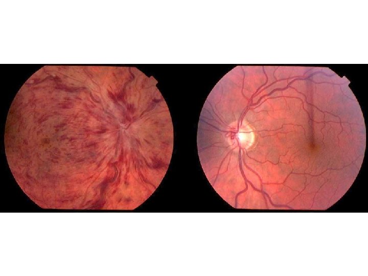

- Slides: 82
Central retinal artery occlusion Central retinal vein occlusion Hypertensive retinopathy Dr. Soujanya K Assistant Professor, YMCH
CENTRAL RETINAL ARTERY OCCLUSION
Pathogenesis
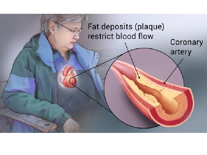
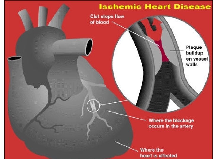
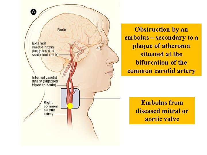
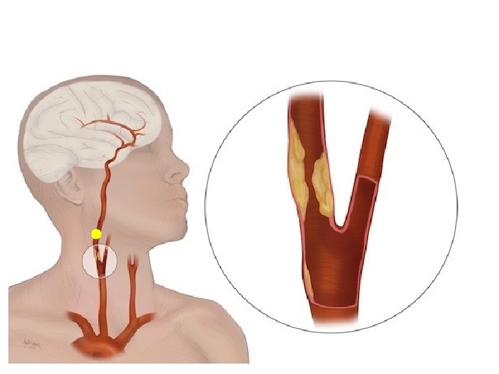
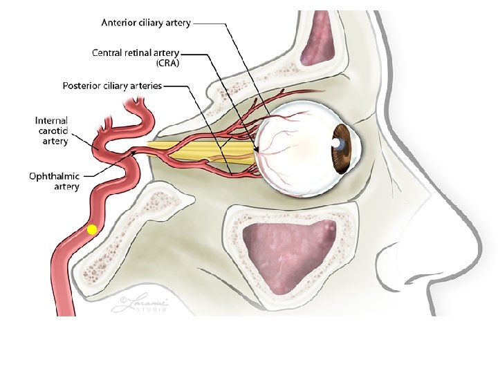
Obstruction by an embolus – secondary to a plaque of atheroma situated at the bifurcation of the common carotid artery Embolus from diseased mitral or aortic valve
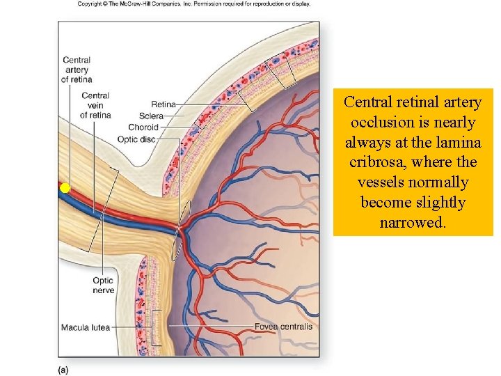
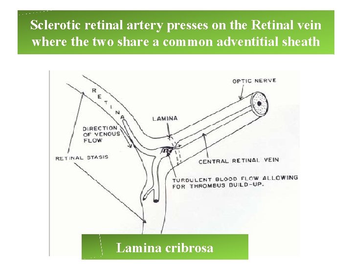
Central retinal artery occlusion is nearly always at the lamina cribrosa, where the vessels normally become slightly narrowed.
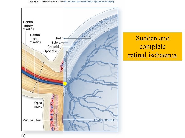
Sudden and complete retinal ischaemia
Symptoms • Sudden painless loss of vision
Signs • Direct pupillary light reflex is absent
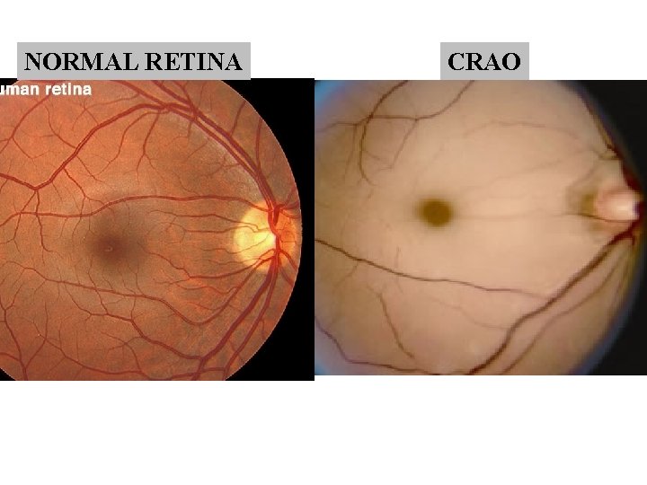
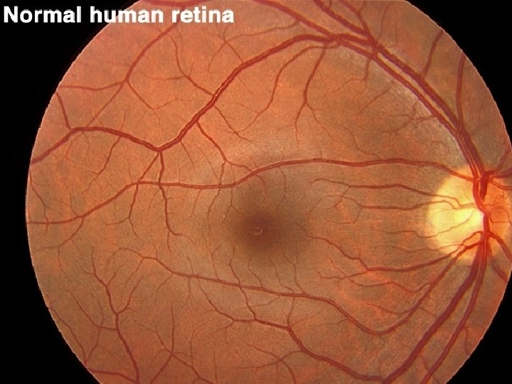
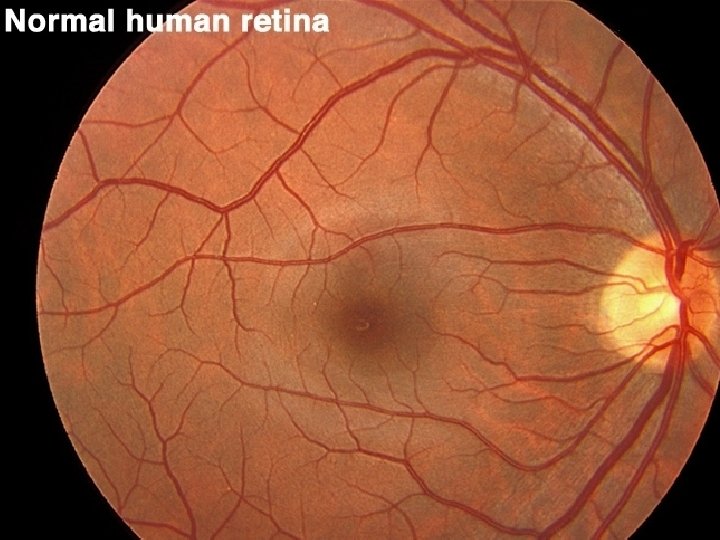
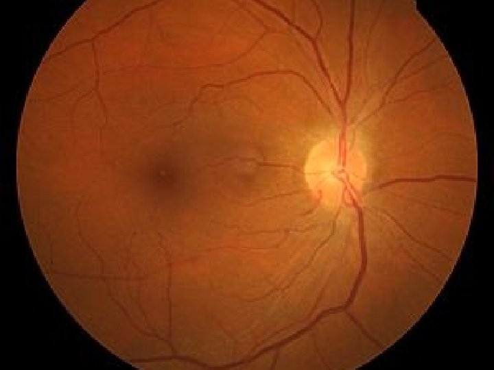
NORMAL RETINA CRAO
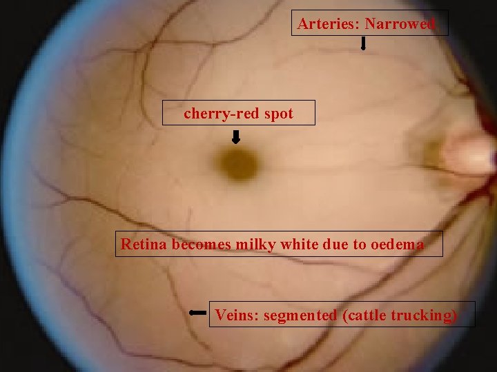
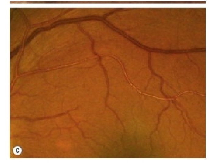
Arteries: Narrowed cherry-red spot Retina becomes milky white due to oedema Veins: segmented (cattle trucking)
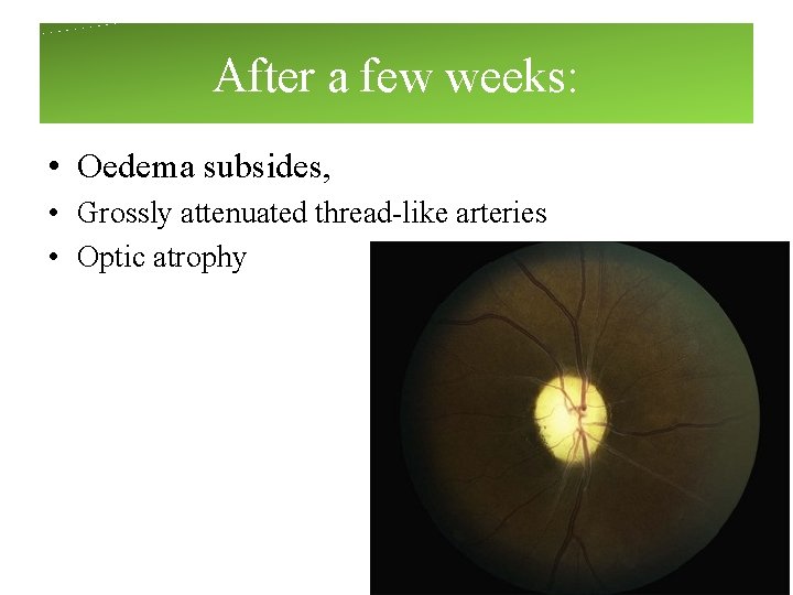
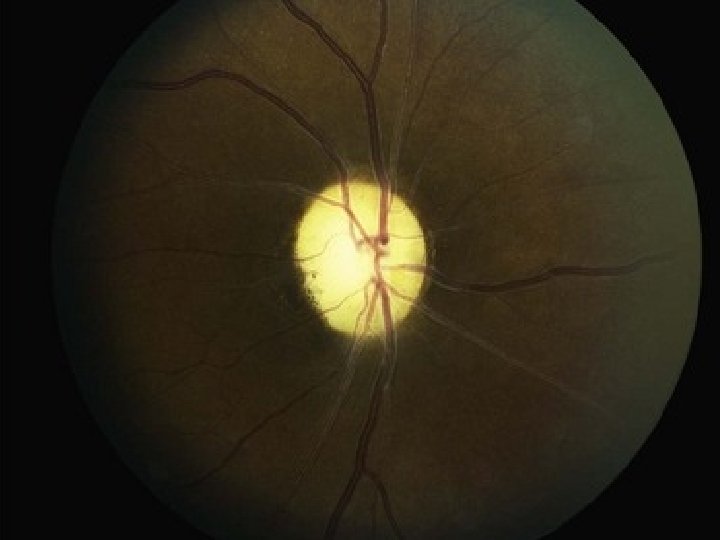
After a few weeks: • Oedema subsides, • Grossly attenuated thread-like arteries • Optic atrophy
Management • Retinal tissue – cannot survive ischemia more than few hours • EMERGENCY TREATMENT
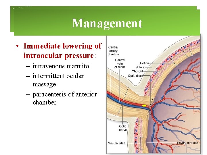
Management • Immediate lowering of intraocular pressure: – intravenous mannitol – intermittent ocular massage – paracentesis of anterior chamber
Management • Vasodilators (Amyl nitrite) and inhalation of a mixture of 5% CO 2 and 95% O 2.
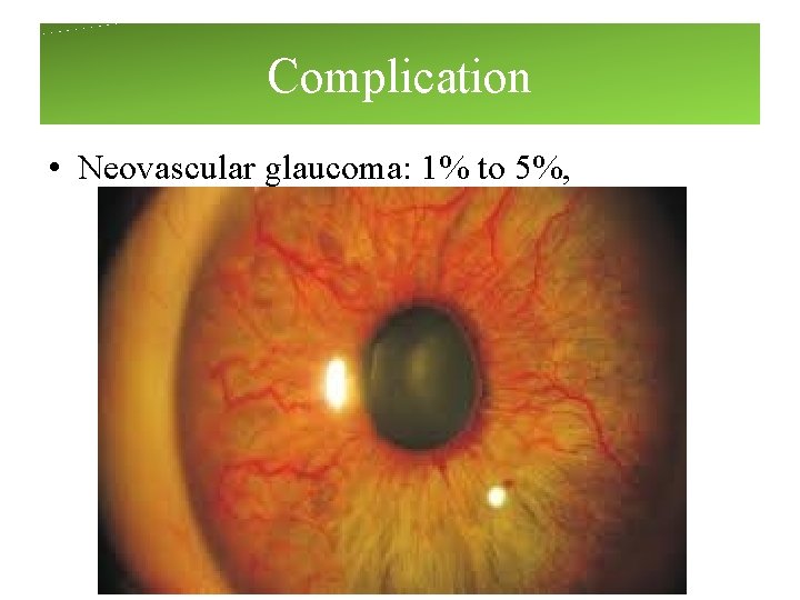
Complication • Neovascular glaucoma: 1% to 5%,
CENTRAL RETINAL VEIN OCCLUSION
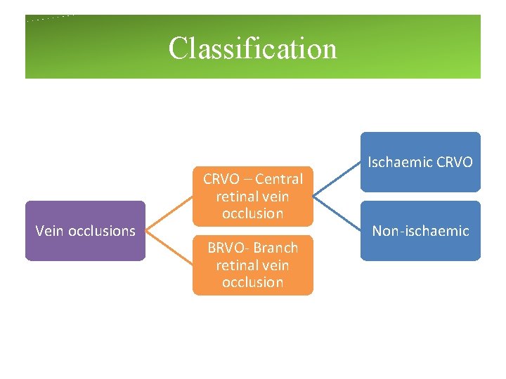
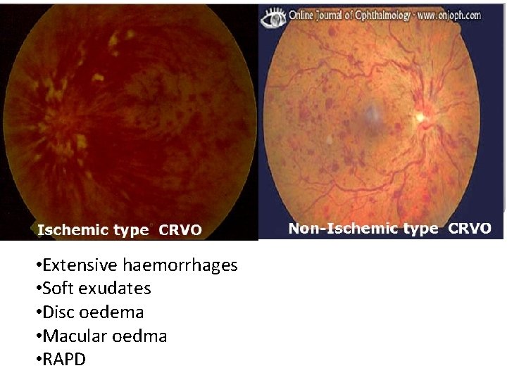
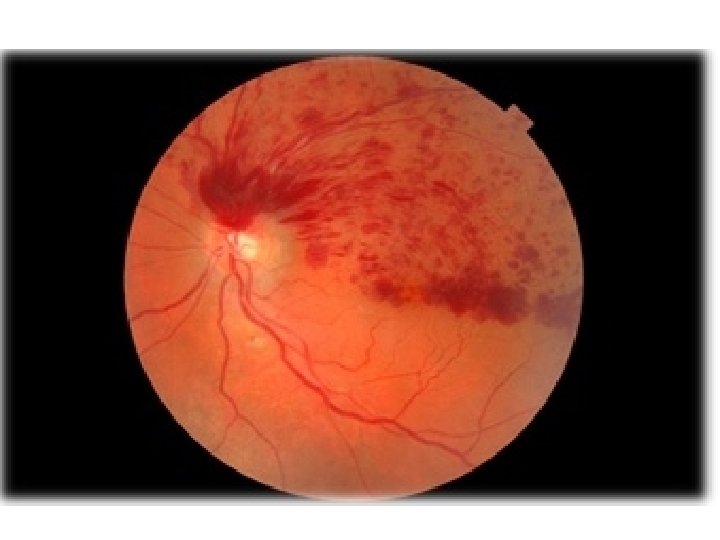
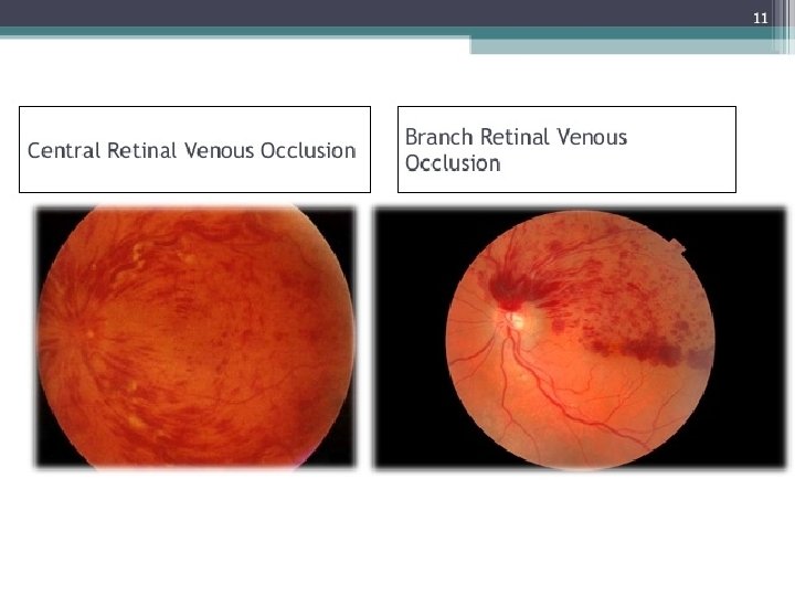
Classification Vein occlusions CRVO – Central retinal vein occlusion BRVO- Branch retinal vein occlusion Ischaemic CRVO Non-ischaemic
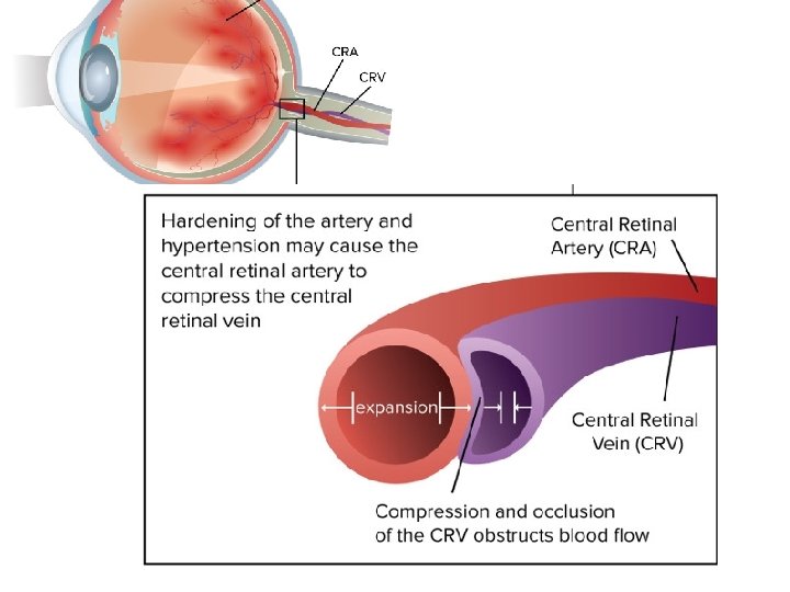
Pathogenesis • Arteriosclerosis
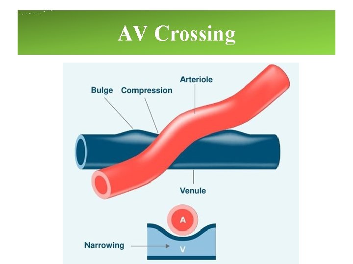
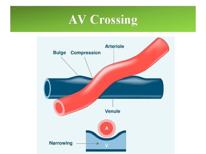
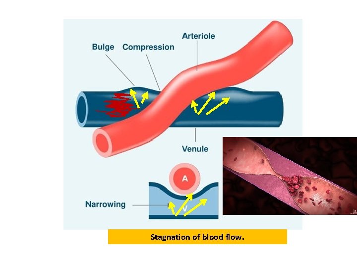
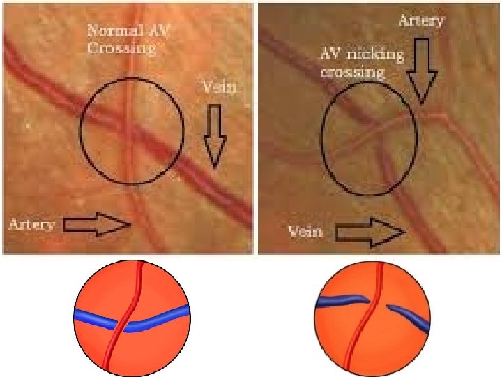
Sclerotic retinal artery presses on the Retinal vein where the two share a common adventitial sheath Lamina cribrosa
AV Crossing
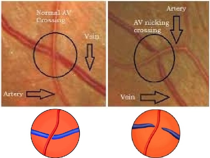
Normal Vs Arteriosclerosis
AV Crossing
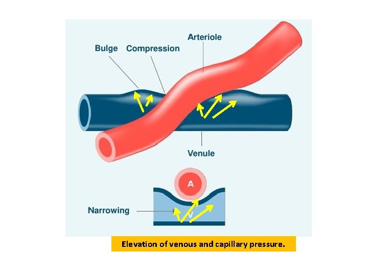
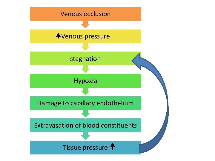
Elevation of venous and capillary pressure.
Stagnation of blood flow.
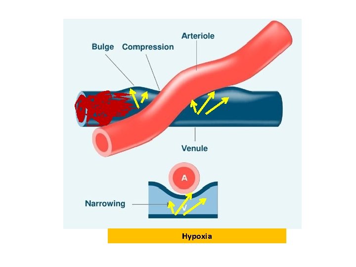
Hypoxia
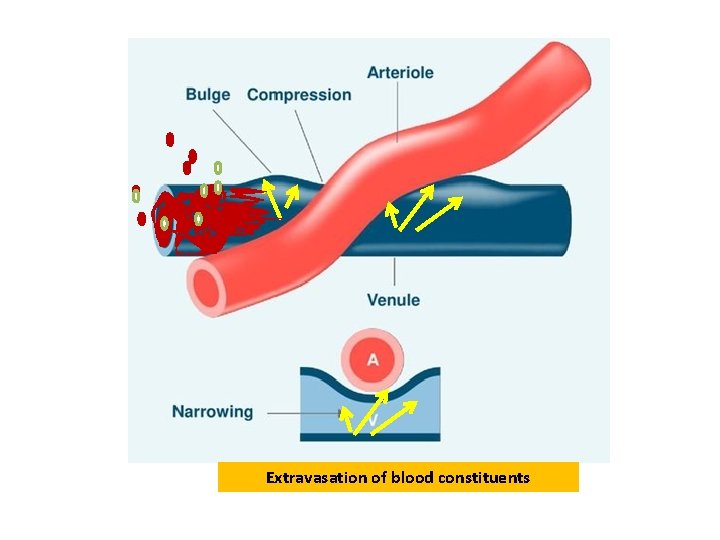
Extravasation of blood constituents
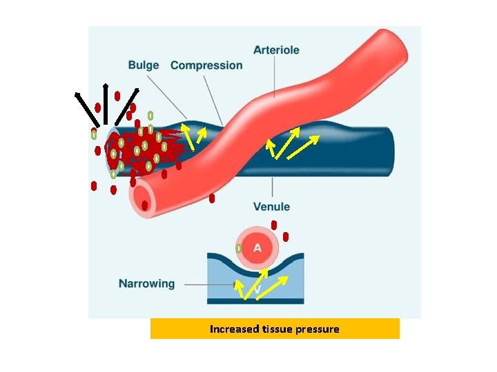
Increased tissue pressure
Venous occlusion Venous pressure stagnation Hypoxia Damage to capillary endothelium Extravasation of blood constituents Tissue pressure
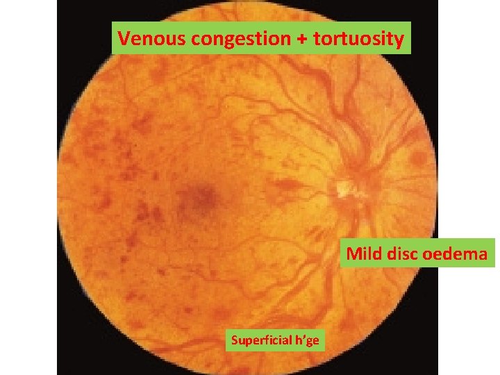
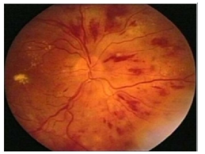
Venous congestion + tortuosity Mild disc oedema Superficial h’ge
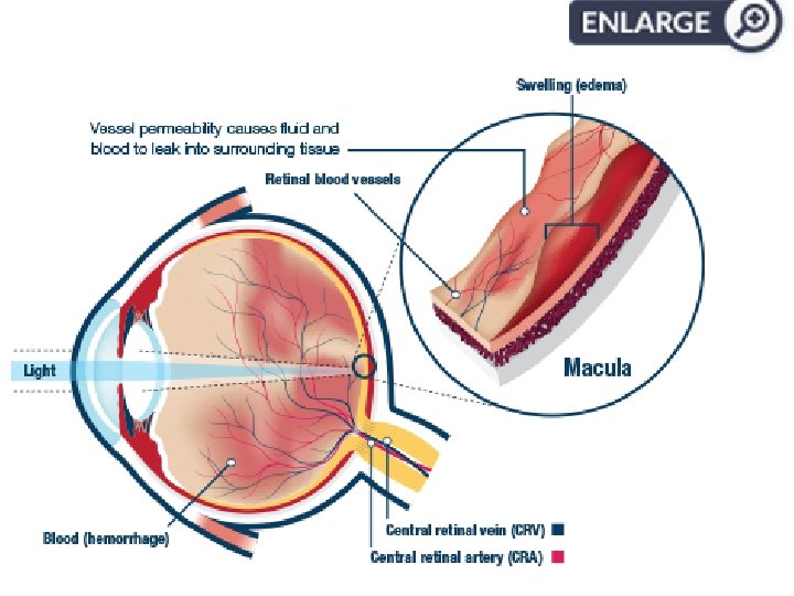
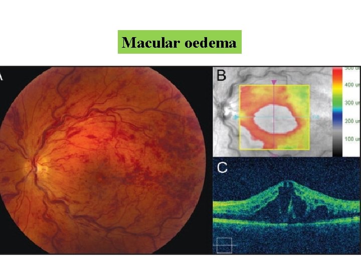
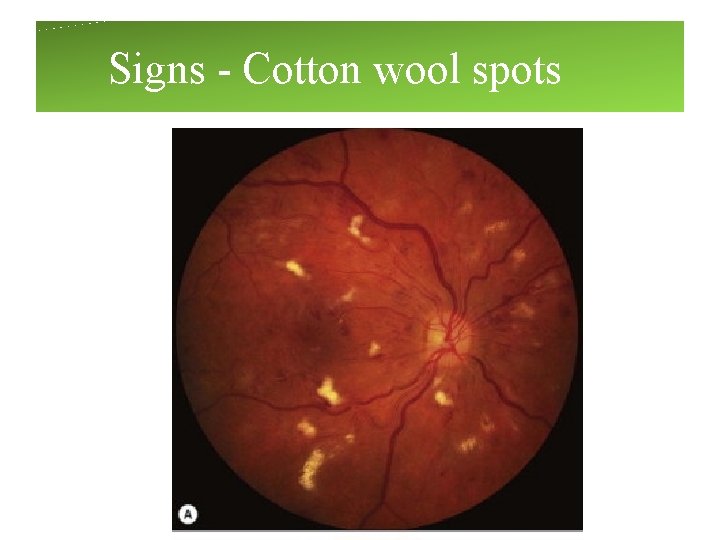
• Extensive haemorrhages • Soft exudates • Disc oedema • Macular oedma • RAPD
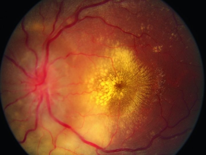
Macular oedema
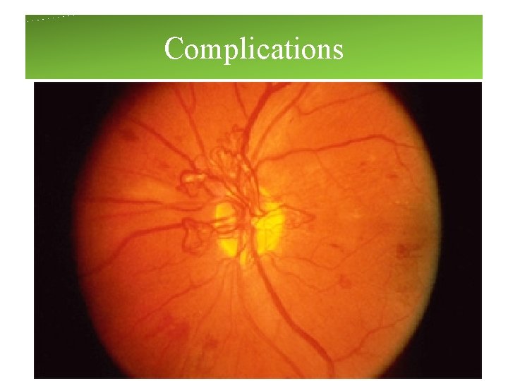
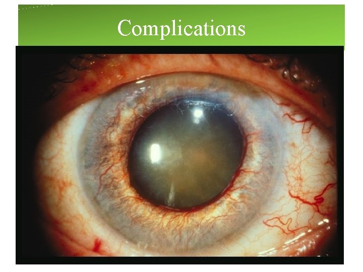
Complications
Complications
Management • Screening monthly- for 6 months : to detect anterior segment neovascularisation
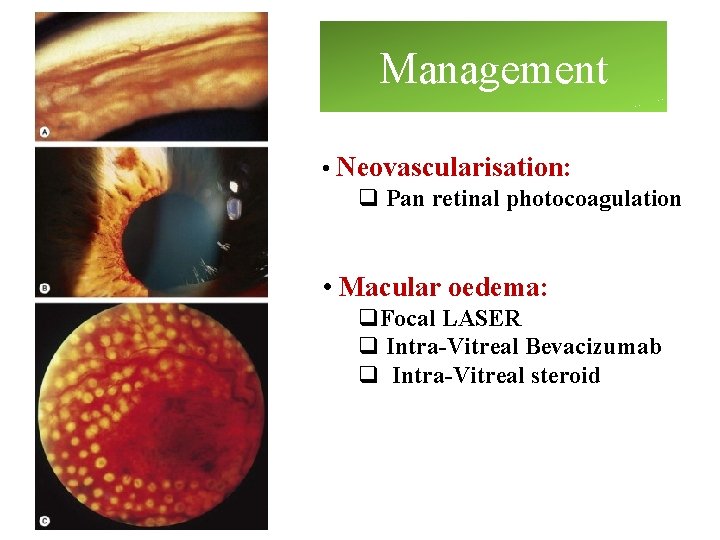
Management • Neovascularisation: q Pan retinal photocoagulation • Macular oedema: q. Focal LASER q Intra-Vitreal Bevacizumab q Intra-Vitreal steroid
HYPERTENSIVE RETINOPATHY
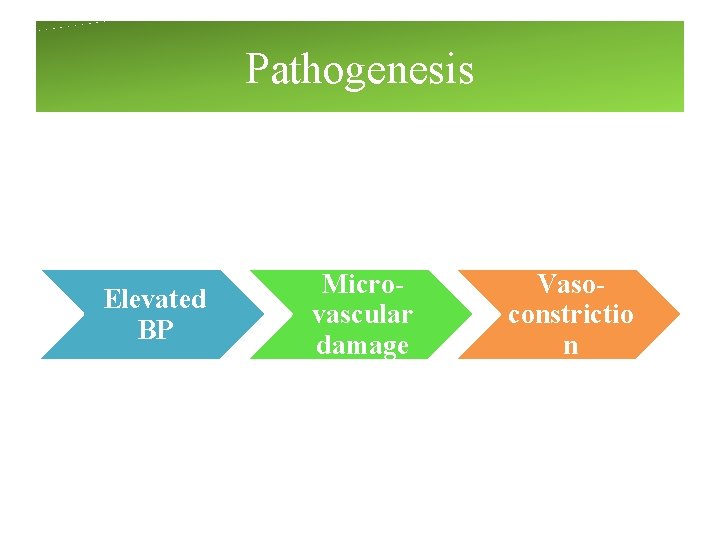
Pathogenesis Elevated BP Microvascular damage Vasoconstrictio n
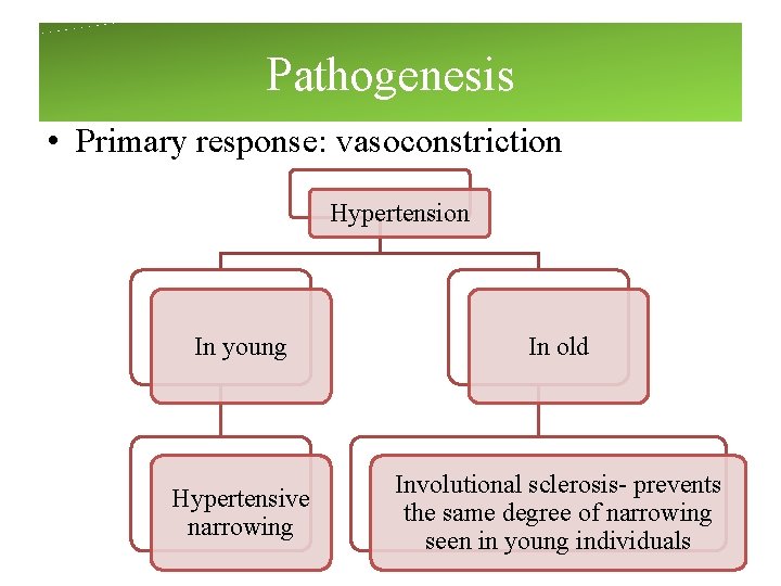
Pathogenesis • Primary response: vasoconstriction Hypertension In young In old Hypertensive narrowing Involutional sclerosis- prevents the same degree of narrowing seen in young individuals
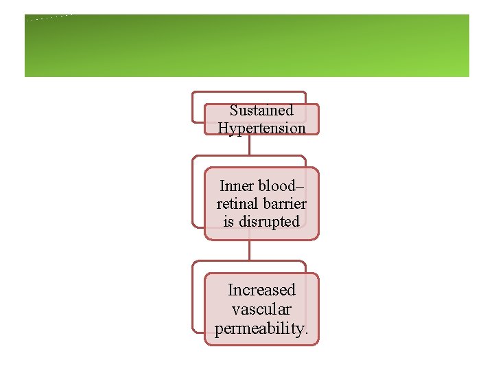
Sustained Hypertension Inner blood– retinal barrier is disrupted Increased vascular permeability.
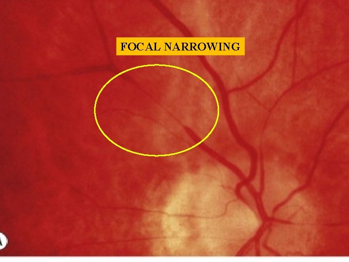
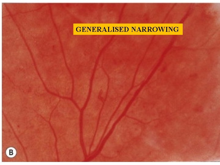
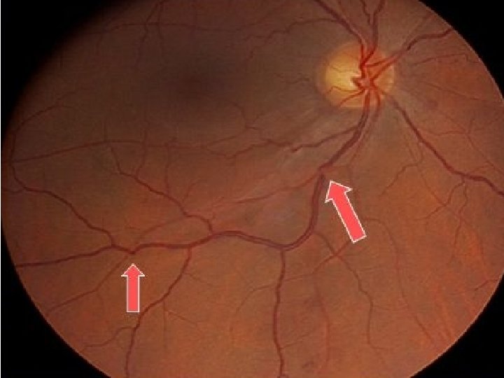
Signs 1. Arteriolar narrowing: Focal or generalised
FOCAL NARROWING
GENERALISED NARROWING
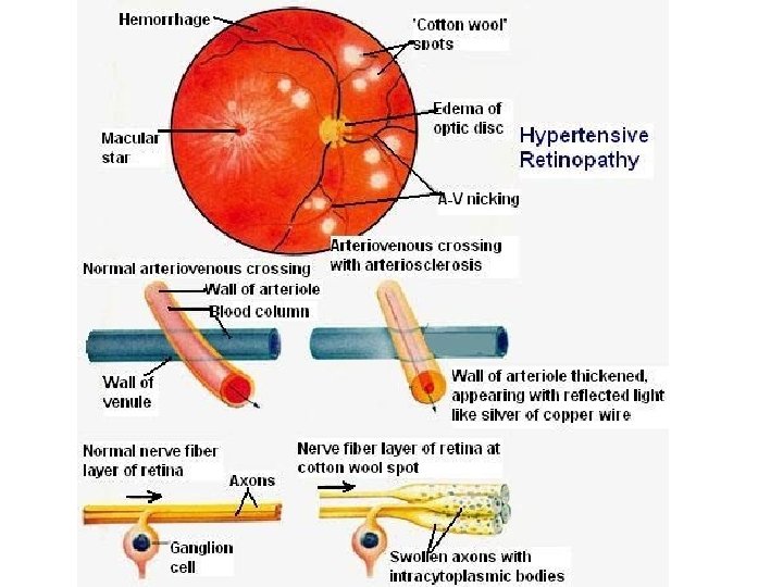
Signs - Cotton wool spots
Signs- Vascular leakage
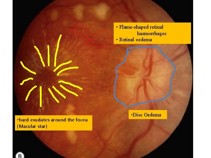
• Flame-shaped retinal haemorrhages • Retinal oedema • hard exudates around the fovea (Macular star) • Disc Oedema
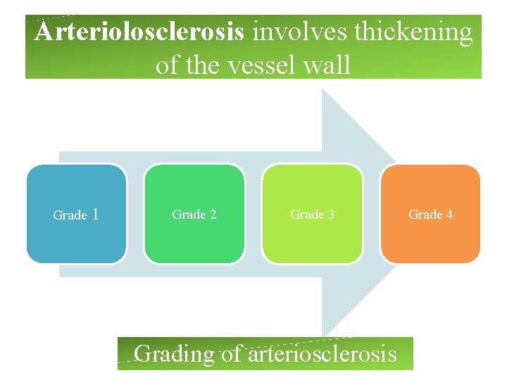
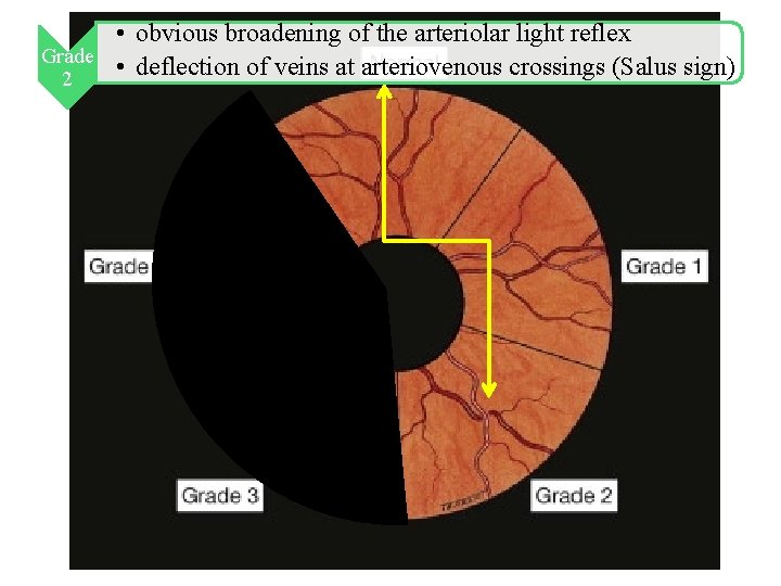
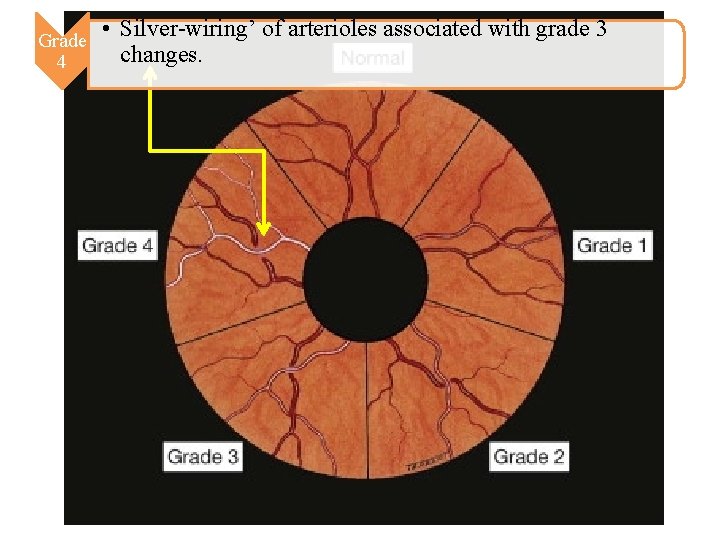
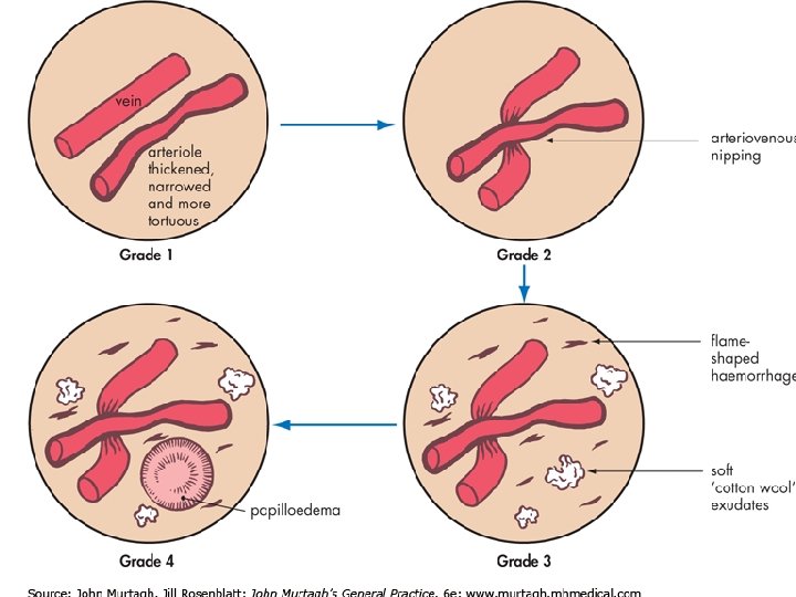
Arteriolosclerosis involves thickening of the vessel wall Grade 1 Grade 2 Grade 3 Grading of arteriosclerosis Grade 4
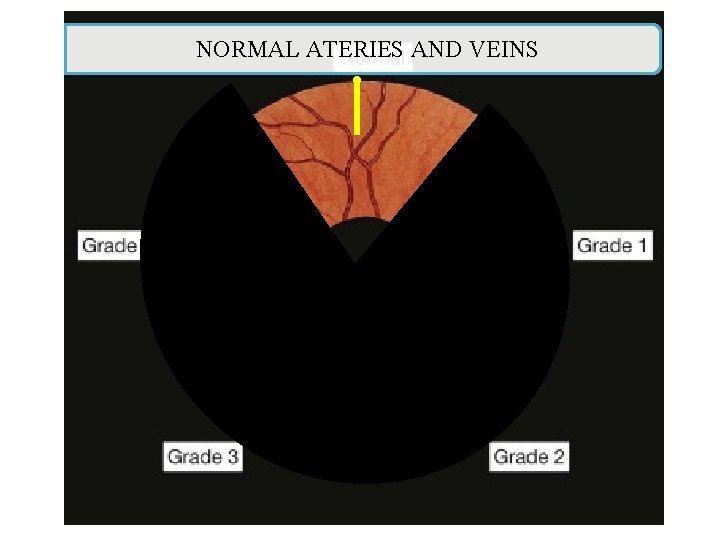
NORMAL ATERIES AND VEINS
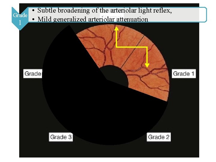
• Subtle broadening of the arteriolar light reflex, Grade 1 • Mild generalized arteriolar attenuation
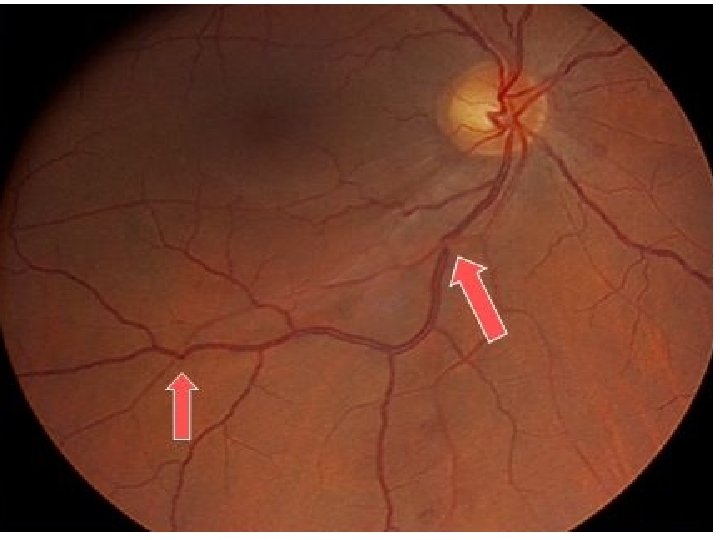
• obvious broadening of the arteriolar light reflex Grade • deflection of veins at arteriovenous crossings (Salus sign) 2
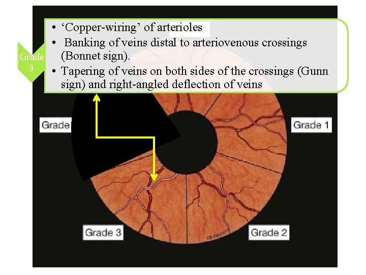
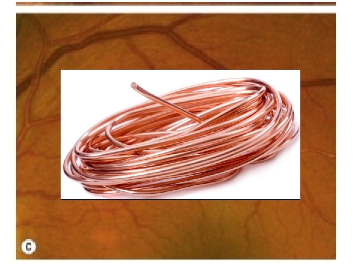
• ‘Copper-wiring’ of arterioles • Banking of veins distal to arteriovenous crossings (Bonnet sign). Grade 3 • Tapering of veins on both sides of the crossings (Gunn sign) and right-angled deflection of veins
Normal Vs Arteriosclerosis
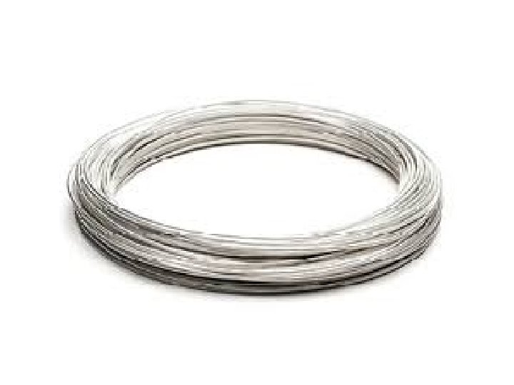
Grade 4 • Silver-wiring’ of arterioles associated with grade 3 changes.
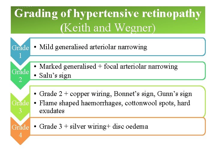
Grading of hypertensive retinopathy (Keith and Wegner) Grade • Mild generalised arteriolar narrowing 1 • Marked generalised + focal arteriolar narrowing Grade • Salu’s sign 2 • Grade 2 + copper wiring, Bonnet’s sign, Gunn’s sign Grade • Flame shaped haemorrhages, cottonwool spots, hard exudates 3 Grade • Grade 3 + silver wiring+ disc oedema 4
Summary
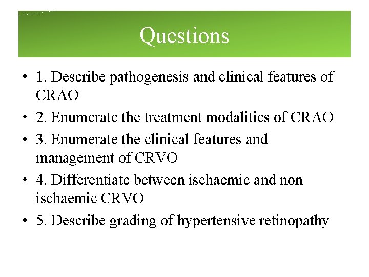
Questions • 1. Describe pathogenesis and clinical features of CRAO • 2. Enumerate the treatment modalities of CRAO • 3. Enumerate the clinical features and management of CRVO • 4. Differentiate between ischaemic and non ischaemic CRVO • 5. Describe grading of hypertensive retinopathy