Peripheral Vein Catheterization Indication Drug administration Hydration Transfusion


 Sclerosed veins Previous intravenous infiltration Burns")


 • Less resistance to blood flow")


- Slides: 23
Peripheral Vein Catheterization
Indication • • • Drug administration Hydration Transfusion of blood components Surgery Emergency care Other situations (blood samples etc)
Contraindications • • Infection Phlebitis (inflammation of vein) Sclerosed veins Previous intravenous infiltration Burns or traumatic injuries Arteriovenous fistula Surgical procedures
Difficult access • • Dehydration Shock Chemotherapy Intravenous substances abuse
Anatomy • Veins of upper extremity • Cephalic vein – radial side • Basilic vein – ulnar side • Communicates in cubital fossa and wrist
Site selection • Accessibility of vein • Patient age • Comfort • Urgency of the situation In general upper extremity veins are preferred • More durable • Fewer complication
Preferred veins
Lower extremity veins used
Lower extremity veins • Saphenous vein • Dorsal veins of the foot • Higher risk of thrombosis and embolism
Veins used for cannulation in children • Leg and feet veins • Scalp veins • External jugular vein
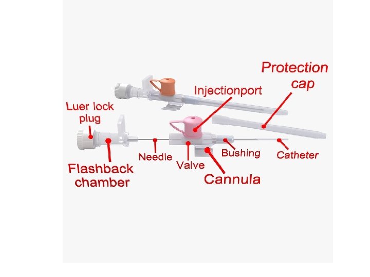
Equipment • • • Gloves Eye protector Tourniqet Antiseptic solution and sterile gauze Saline flush Occlusive dressing and tape Catheter Intravenous fluid bag with tubing Sharps container Local or topical anesthetic in some cases
Size of catheteters Small size (20 -24 g) • Less resistance to blood flow • Fewer complications Big sizes (14 -16 g) • Acute situations
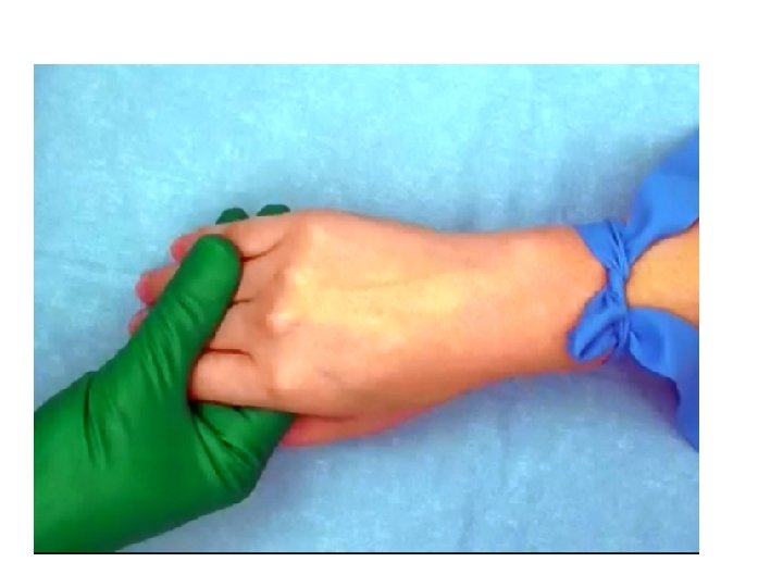
Preparation • Explain the procedure • Potential complications: Bleeding Bruising Infection • Washing hands • Wearing gloves • Using Eye protection • Patient in supine position • Inspect and Palpate vein, use the tourniqet • Use the antiseptic
Ideal vein for catheterizaion • • Round Firm Flexible Full Clean the site with antiseptic solution Allow area to dry completely Local anesthetic can be used Inspect the catheteter for damage Superficial veins must be stabilized
Insert the needle
Do not insert catheter too deeply
• When catheter in the lumen watch for initial flashback of blood • Then lower the catheter – it must be almost parallel to the skin • Stabilize the needle and move only plastic part of the catheter forward • Remove the tourniqet • Slowly remove the needle, • to prevent blood loss from the open plastic canula use direct pressure on the vein proximal to catheter • Never attempt to reinsert the neddle into the plastic canula • Remove blood from the chamber of catheter with flash of saline
Signs of extravasation • • Swelling Redness Leakage Discomfort
Transparent occlusive dressing
Complications • • Pain Bruising Infection Extravasation Phlebitis Thrombosis Embolism Nerve damage
Avoiding complications • Proper sterile technique • Selection of the appropriately sized catheter