Medication Administration in Cats and Dogs CTVT pages

 Sublingual Via feeding tube ›")
 o Subcutaneous (SC")


")


")
")

 � Usually blood enters the hub of the needle at")


- Slides: 39
Medication Administration in Cats and Dogs CTVT pages 588 -590
Routes of Medication Administration � � � Oral (PO) Sublingual Via feeding tube › Esophagostomy › Gastrostomy › Jejunostomy � � � Topical ophthalmic Transdermal Intranasal Inhalation › Nebulized or volatilized � � � Orogastric intubation � Intratracheal Nasogastric intubation Rectal (PR) � Aural
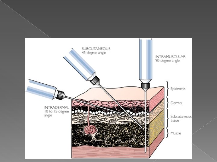
Parenteral Routes: administered with a needle and syringe o Intradermal (ID) o Subcutaneous (SC or SQ) o Intramuscular (IM) o Intravenous (IV) o Intraosseous (IO) o Intraperitoneal (IP) o Intra-arterial (IA) o Epidural/subdural o Intracardiac (IC) o Intramammary
Route Depends on Many Factors Patient Factors � Health conditions › Symptoms › Absorption rate of drug required Temperament � Ease of administration for client � Drug Factors � Type of medication/fluid � Formulation of drug � Cost � Systemic vs. local effect desired
Other Factors to Consider � Does this medication require that special precautions be followed during administration (i. e. gloves, mask)? › Examples? � Potential side effects? › Pain from injection › Vomiting/GI upset/constipation
Comparison of Common Parenteral Routes of Drug Administration Intramuscular 90° Subcutaneous 45° Intravenous 25° Intradermal Epidermis Dermis Subcutaneous tissue Muscle 10°– 15°
Mini A&P Review of Skin Layers � The skin is made up of three layers: the epidermis, and subcutaneous layers › The epidermis is several cell layers thick and does not contain blood vessels. Its thickness varies greatly from region to region in any animal and varies from species to species. › The dermis is composed of blood vessels, lymph, nerve fibers, and accessory organs of the skin (glands and hair follicles). › The subcutaneous layer (hypodermis) is composed of connective tissue and contains a large amount of fat. � Muscle lies underneath all of these layers
Before the Injection Gather supplies � Needles � Syringe � Medication to be injected � Proficient person to restrain the animal For venipuncture only: � Cotton ball with alcohol � Hydrogen Peroxide (optional)
Syringes are available in various sizes (even more than this)
Needles � Needle gauge is determined › Consistency of drug by: › Route of administration › Patient size � At least two needles are required › One to draw up the medication › One to administer the medication Why is this?
Process for Changing Needle � Draw up exact amount of drug › Can use smallest needle available � Aspirate all drug into the syringe (out of the needle) › Hub loss� Remove needle and replace with appropriate needle � Carefully ensure all air is out of the syringe by slowly depressing the plunger.
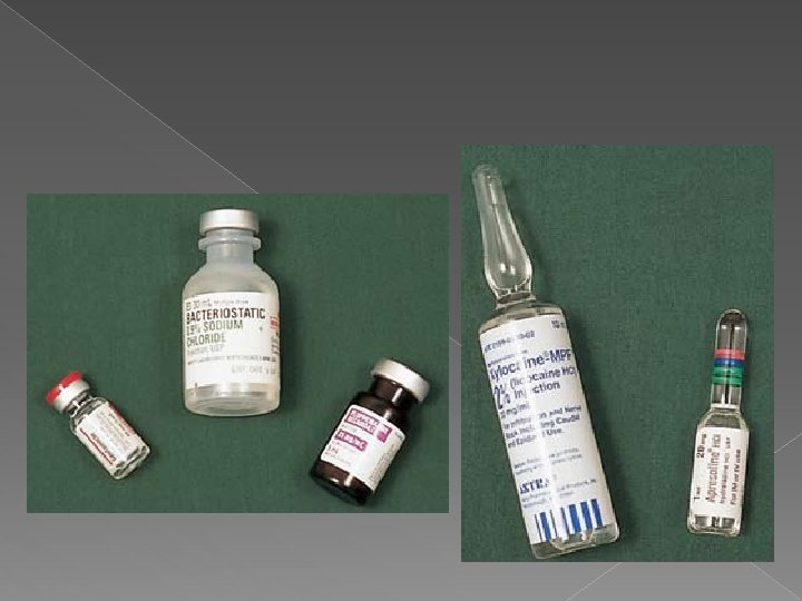
Injectable Drugs � Supplied as: › Sterilized solutions › Powders that must be reconstituted with sterile solution � May be stored in: › Vials (single or multi-dose) › Ampules › Fluid bags
Intradermal Injections � Common use of ID route: › Desensitize skin › Allergy skin testing � Skin is usually shaved before performing an ID injection � Drug is not dispersed throughout the body
Intradermal Administration
ID Injection Procedure �A fold of skin is lifted or skin is pulled taught and a 25 - to 27 - gauge needle attached to a syringe is inserted with the bevel up into the dermis. › If allergy testing: A 0. 1 ml volume of allergen is injected. › If locally anesthetizing skin: dose of drug � The injection site will look like a translucent lump if the injection is performed correctly.
Subcutaneous Injections Easiest route to perform technically � Common use of SQ route: � › Vaccine administration › Fluid administration › Pain medications, antibiotic injections, Insulin � Absorption rate is slow compared to other routes › May be slower in obese animals � Some substances are harmful if injected SC › Example:
Subcutaneous Administration
SC Injection Sites � Preferred site for most SC injections is the dorsolateral region from the neck to the hips. › The dorsal region of the neck and back should be avoided for drug administration. � Cat vaccines: the ____________ should be avoided because of the incidence of vaccineinduced tumors. › Feline vaccinations should be administered in as distal a portion of an extremity as possible.
SC Injection Sites
SC Injection Procedure � Fold of skin is tented and the needle is inserted at the base of the tent. › Insert needle as if walking INTO the tent � Aspirate › Why? � After injection, briefly massage skin to facilitate drug distribution.
Skin Tent
Intramuscular Injections � Appropriate route for injection of small volumes of medication. � Common uses of IM route: › Anesthetics/sedatives �Convenient route of administration for fractious animals › Pain medications › Heartworm treatment � Generally, or IV. › Why? more painful for animals than SC
Intramuscular Administration
IM Injection Sites � Drugs are most often administered in the: › Lumbosacral musculature lateral to the dorsal spinous processes › Semimembranosus/ semitendinosus muscles of the rear leg � In the hind limb: needle should enter the lateral aspect of the muscle and be directed caudally › Why?
Avoid the sciatic nerve!
IM Injection Sites � Deep IM injections in the third to fifth lumbar region of the __________ are used to administer adulticide heartworm treatment (Immiticide®).
IM Injection Procedure Isolate the muscle between the fingers and thumb. � A 22 to 25 gauge needle attached to a syringe is embedded in the muscle at a ______ angle. � As with a SQ injection, the needle hub is checked for blood before administration of medication to make certain a vessel is not inadvertently penetrated. � › How did we do this? Once in the muscle, inject the medication slowly. � Massage the site for a few seconds after the injection to help distribute the substance if possible. � › Exception:
Complications from IM Injection � Tissue trauma � Pain at injection site � Nerve damage
Intravenous Administration Drugs and/or fluids may be injected directly into a vein or through an IV catheter. � IV route produces an immediate response � › Usually given slowly � Common use of IV route: › › › � Inducing anesthesia Chemotherapeutic agents Anti-convulsant drugs Irritating drugs Emergency/resuscitation drugs Large amounts of _____ needed Requires new needle for administration
IV Injection Sites CAT DOG Cephalic � Lateral saphenous � Cephalic � Medial saphenous � Femoral � Note: The jugular vein is used to administer injections in small animals IF an intravenous jugular catheter is in place.
Cephalic Vein
Femoral/Medial Saphenous Vein (cat)
Lateral Saphenous Vein (dog)
IV Injection Instructions Expel all air bubbles from the syringe prior to inserting into the vein. � Restrainer should occlude the vessel with digital pressure or use a tourniquet. � Grasp the extremity and pull the skin taut in a distal direction. � Swab the skin and hair with an alcohol-soaked cotton ball (go with the fur). � Insert a 20 - to 25 - gauge needle, bevel up into the vein. �
IV Injection Instructions (cont’d) � Usually blood enters the hub of the needle at penetration of the vein (flash), BUT, placement is confirmed by aspirating. › What should we see? › Venipuncturist: communicate to restrainer “I’m in” › Restrainer: release pressure from the vein � Inject the medication into the vein. › If large volume of drug or movement of needle: Communicate with restrainer and remove needle � Apply firm pressure to the injection site until hemostasis/coagulation occurs �
If Using a Tourniquet � Most common: Nye Tourniquet or Penrose drain/rubber material � Can be very dangerous if used improperly � Goal is to visualize and palapte vein. � Must be able to remove before injecting!
Possible Complications with IV Injections � Injecting drug outside of vein � Hematoma formation � Intra-arterial injection of drug � Hitting a nerve (pain, lameness, paralysis) � Air-embolus � Phlebitis � Septicemia