Peritoneum and the peritoneal cavity A thin membrane
 lines the walls of")
Peritoneum and the peritoneal cavity A thin membrane (the peritoneum) lines the walls of the abdominal cavity and covers much of the viscera. The parietal peritoneum lines the walls of the cavity and the visceral peritoneum covers the viscera.

Between the parietal and visceral layers of peritoneum is a potential space (the peritoneal cavity) the cavity contains a thin film of serous fluid allows free movement of one peritoneal surface over another.

• Abdominal viscera are either suspended in the peritoneal cavity by folds of peritoneum or are outside the peritoneal cavity. • Organs suspended in the cavity are referred to as intraperitoneal (Such organs are mobile); organs outside the peritoneal cavity, with only one surface or part of one surface covered by peritoneum, are retroperitoneal (Such organs are fixed).

The peritoneal cavity is subdivided further into the greater sac and the omental bursa: greater sac accounts for most of the space in the peritoneal cavity, beginning superiorly at the diaphragm and continuing inferiorly into the pelvic cavity-it is entered once the parietal peritoneum has been penetrated; omental bursa is a smaller subdivision of the peritoneal cavity posterior to the stomach and liver and is continuous with the greater sac through an opening, the omental foramen

Omenta, mesenteries, and ligaments • Throughout the peritoneal cavity numerous peritoneal folds (Omenta, mesenteries, and ligaments) connect organs to each other or to the abdominal wall. • peritoneal folds provide pathways for passage of vessels, nerves and lymphatics
 two layers of peritoneum, which pass from the stomach to")
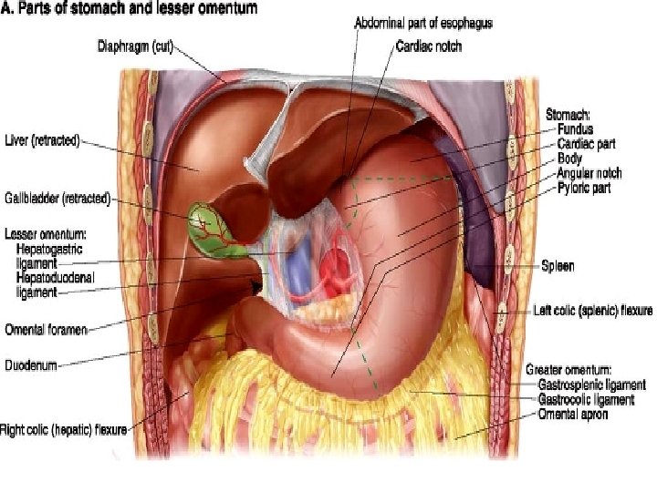
The omenta (singular omentum) two layers of peritoneum, which pass from the stomach to other viscera. There are two: greater omentum attaches to the greater curvature of the stomach; lesser omentum attaches to the lesser curvature of the stomach

Mesenteries are peritoneal folds that attach viscera to the posterior abdominal wall and include: • the mesentery-associated with parts of the small intestine; • the transverse mesocolon-associated with the transverse colon; • the sigmoid mesocolon-associated with the sigmoid colon

Peritoneal ligaments consist of two layers of peritoneum that connect two organs to each other or attach an organ to the body wall, and may connects organ to the diaphragm. They are usually named after the structures being connected. For example, the splenorenal ligament connects the left kidney to the spleen and the gastrophrenic ligament connects the stomach to the diaphragm.

Peritoneal membrane distributionas seen in sagittal section Passing upwards from the umbilicus: Peritoneum lining the anterior abdominal wall, and under surface of diaphragm, including the falciform ligament.

• Peritoneum lining the anterior and inferior surfaces of the liver and then the two layers leave the liver to form the lesser omentum

The double layers of lesser omentum split to cover the anterior and posterior surfaces of the stomach, the peritoneal membrane of the two surfaces meet at the greater curvature of the stomach to form the greater omentum.
then reflected backward(called")
The greater omentum descends downward (called anterior 2 layer of greater omentum)then reflected backward(called posterior 2 layer of the greater omentum) which pass to the posterior abdominal wall and become attached to the anterior border of

As the posterior layer of the greater omentum extend backward it become adherent to the transverse mesocolon so the greater omentum appear to attach the greater curvature of the stomach to the transverse colon

As the Peritoneal membrane reach the pancreas it extend and cover the posterior abdominal wall above the pancreas (forming the posterior wall of the lesser sac) and below the pancreas.

Below the pancreas the Peritoneal membrane extend to passing round the transverse colon and small intestine to form the transverse mesocolon and mesentery of small intestine respectively

Peritoneum lining the posterior abdominal wall then descending into the pelvis forming mesentery of pelvic colon (pelvic mesocolon). The Peritoneum then extend downward and has different distribution in the male and female pelvis
 ØThis is a large recess of the peritoneal cavity behind")
Lesser sac (Omental Bursa) ØThis is a large recess of the peritoneal cavity behind the stomach, the lesser omentum and the caudate lobe of the liver Øit communicates with the greater sac through the epiploic foramen
 peritoneum covering the caudate")
Lesser sac Boundaries The anterior wall is formed by: 1) peritoneum covering the caudate lobe of the liver; 2) lesser omentum; 3) peritoneum on the postero-inferior surface of the stomach; and 4)anterior 2 layers of the greater omentum.
 the posterior two layers")
Lesser sac Boundaries The posterior wall is formed by 1) the posterior two layers of the greater omentum; and 2) Peritoneum covering structures forming the stomach bed
")
EPIPLOIC FORAMEN (aditus to the lesser sac, or the foramen of Winslow)
 Boundaries: Anteriorly:")
EPIPLOIC FORAMEN (aditus to the lesser sac, or the foramen of Winslow) Boundaries: Anteriorly: Right free margin of the lesser omentum containing the portal vein, the hepatic artery, and the bile duct.

Posteriorly: peritoneum covering the inferior vena cava, the right suprarenal gland vertebra T 12 Superiorly: Caudate process of the liver. Inferiorly: First part of the duodenum and the horizontal part of the hepatic artery.

Abdominal Viscera

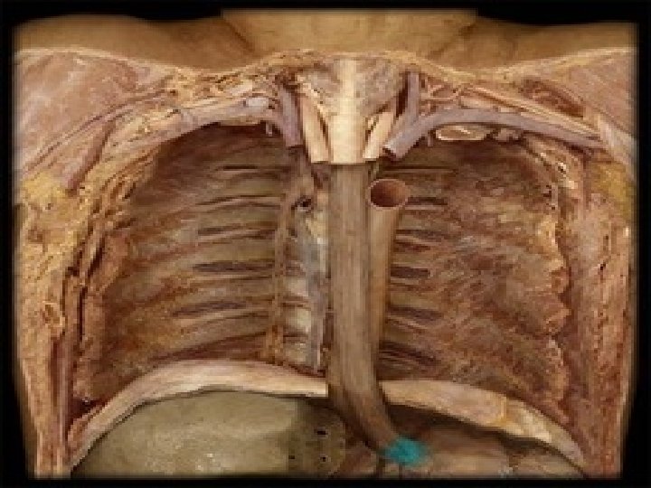
Abdominal part of esophagus • about half an inch long. • It enters the abdomen through the esophageal opening of the diaphragm T 10, slightly to the left of the median plane. • and ends by opening into the cardiac end of the stomach at the level of vertebra T 11, about an inch to the left of the median plane
 plexus • left")
Abdominal part of esophagus • Is encircled by the esophageal (nerve) plexus • left border is separated from the fundus of the stomach by the cardiac notch. • Peritoneum covers the oesophagus only anteriorly and on the left side

Abdominal part of esophagus Arterial supply derived from left gastric branch of coeliac trunk and from left inferior phrenic branch of the abdominal aorta. The veins drain into the left gastric vein The lymphatic drainage into the left gastric lymph nodes.

Abdominal part of esophagus innervated by the esophageal plexus, formed by the vagal trunks and the greater splanchnic nerves and periarterial plexuses around the left gastric arteries

Stomach • The stomach is the most dilated part of the gastrointestinal tract and has a J-like shape. • Positioned between the abdominal esophagus and the small intestine

• the stomach is in the epigastric, umbilical, and left hypochondrium regions of the abdomen

The stomach is divided into four regions: • Cardia: the part surrounding the cardial orifice (esophageal opening), • the fundus of stomach, which is the area above the level of the cardial orifice; • the body of stomach, which is the largest region of the stomach; Cardiac notch angular notch pyloric canal • the pyloric part, which is divided into the pyloric antrum and pyloric canal and is the distal end of the stomach
 is marked on the surface of the")
The outlet of the stomach (pyloric orifice) is marked on the surface of the organ by the pyloric constriction and surrounded by a thickened ring of gastric circular muscle (the pyloric sphincter). The pyloric orifice is just to the right of midline in a plane that passes through the lower border of vertebra LI (the transpyloric plane).

The lesser curvature is concave and forms the right border of the stomach. It provides attachment to the lesser omentum The most dependent part of the curvature is marked by the angular notch Cardiac notch angular notch pyloric canal The greater curvature is convex and forms the left border of the stomach. It is 4 - 5 times as long as the lesser curvature.
 surfaces forwards and upwards. The posterior (posteroinferior) surfaces backwards and")
Surfaces: The anterior' (anterosuperior) surfaces forwards and upwards. The posterior (posteroinferior) surfaces backwards and downwards.

Peritoneal Relations of the stomach • The stomach is covered by peritoneum on both of its surfaces. At the lesser curvature the layers of peritoneum covering the anterior and posterior surfaces meet and become continuous with the lesser omentum.
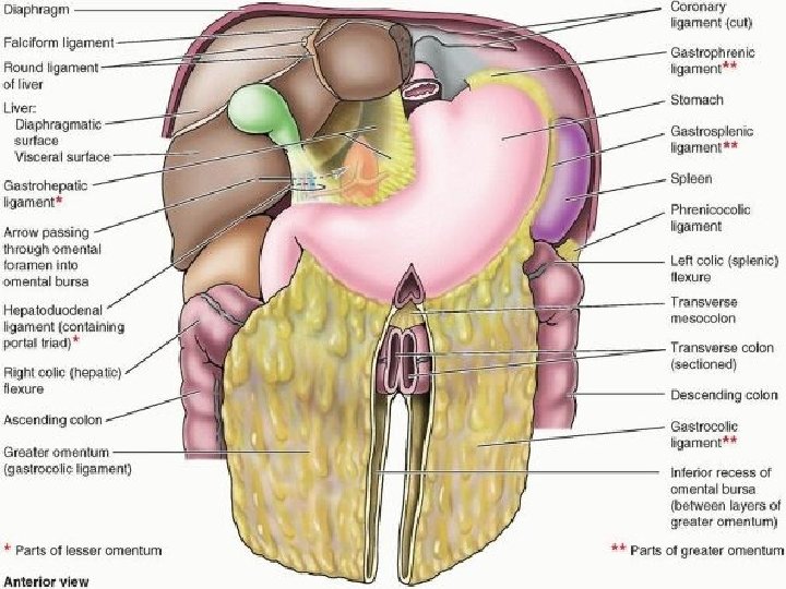

• Along the greater part of the greater curvature, the two layers meet to form the greater omentum. Near the cardiac end of the greater curvature, the two layers meet to form the gastrosplenic ligament. • Near the cardiac end, the peritoneum on the posterior surface is reflected on to the diaphragm as the gastrophrenic ligament.

Cranial to this ligament a small part of the posterior surface of the stomach is in direct contact with the diaphragm (left crus). This is the bare area of the stomach.
 of the stomach arranged into three layers • Outer")
The muscular layer (muscularis externa) of the stomach arranged into three layers • Outer longitudinal • Middle circular Inner oblique

The mucosa of an empty stomach is thrown into folds termed gastric rugae.
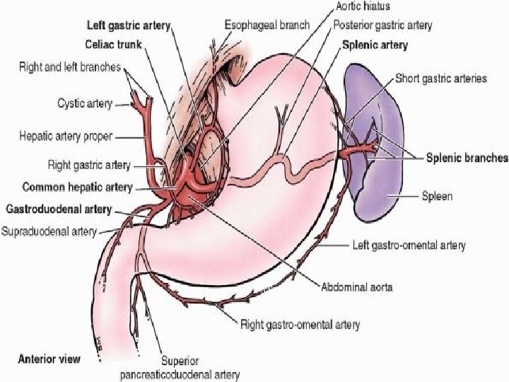

Arterial Supply: The stomach is supplied by: Øthe left gastric artery, a branch of the coeliac trunk; Øthe right gastric artery, a branch of the common hepatic;

Arterial Supply: The stomach is supplied by: Øthe right gastroepiploic artery, a branch of the gastroduodenal; Øthe left gastroepiploic artery, a branch of the splenic; and Ø 5 to 7 short gastric arteries, which are branches of the splenic

The veins. The right and left gastric veins drain into the hepatic portal vein; the short gastric veins and left gastroepiploic veins drain into the splenic vein, The right gastro-omental vein empties in the superior mesenteric vein.

Lymph from the superior two thirds of the stomach drains along the right and left gastric vessels to the gastric lymph nodes; . Lymph from the right two thirds of the inferior third of the stomach drains along the right gastroepiploic vessels to the pyloric lymph nodes. Lymph from the left one third of the inferior third drains to the pancreaticoduodenal lymph nodes, which are located along the short gastric and splenic vessels.

Nerve Supply The sympathetic nerve supply of the stomach from the T 6 through T 9 segments of the spinal cord passes to the celiac plexus through the greater splanchnic nerve and is distributed through the plexuses around the gastric and gastroepiploic arteries The parasympathetic nerve supply of the stomach is from the anterior and posterior vagal trunks and their branches, which enter the abdomen through the esophageal hiatus.

Anterior Relations: Related to the liver, the diaphragm, and the anterior abdominal wall. The diaphragm separates the stomach from the left pleura, the pericardium, and the 6 th to 9 th ribs- Upper and left part of the anterior surface is related to spleen

Posterior relations: The posterior surface of the stomach is related to structures forming the stomach bed, all of which are separated from the stomach by the cavity of the lesser sac
 the diaphragm; 2) the left kidney;")
Structures forming the stomach bed include: are 1) the diaphragm; 2) the left kidney; 3) the left suprarenal gland; 4) the pancreas; 5) the transverse mesocolon; 6) the splenic flexure of the colon; and 7) the splenic artery.

Sometimes the spleen is also included in the stomach bed, but it is separated from the stomach by the cavity of the greater sac (and not of the lesser sac).

Lesser omentum: This is a fold of peritoneum, which extends from stomach and the first 2 cm of the duodenum to the liver.

The portion of the lesser omentum between the stomach and the liver is called the hepatogastric ligament and the portion between the duodenum and the liver is called the hepatoduodenal ligament
 The hepatic artery proper")
The right free margin of the lesser omentum contains: 1) The hepatic artery proper (anterior to the left); 2) the bile duct (anterior to the right); the portal vein (posteriorly); 4) lymph nodes and lymphatics; and 5) the hepatic plexus of nerves along the arteries.

Along the lesser curvature of the stomach and along the upper border of the adjoining part of the duodenum it contains: 1) the right gastric vessels; 2) the left gastric vessels; 3) the gastric group of lymph nodes and lymphatics; and 4) branches from the gastric nerves.

Contents: The greater omentum §The right and left gastroepiploic vessels §Right gastroepiploic lymph nodes. §Sympathetic plexus of nerves accompanying gastro epiploic arteries.

Contents: Fat. The greater omentum

Functions of the greater omentum: • It is a storehouse of fat. • It protects the peritoneal cavity against infection because of the presence of macrophages in it. Collections of macrophages form small, dense patches, known as 'milky spots', which are visible to the naked eye • It also limits the spread of infection by moving to the site of infection and sealing it off from the surrounding areas. It is also known as the policeman of the abdomen.
- Slides: 58