THE CIRCULATORY SYSTEM Introduction The circulatory system is


 separate the atrium and ventricle on each side of")

 control the beat of")
 – Parasympathetic")


: 2. ARTERI SEDANG (MUSCULAR ARTERY): 3. ARTERI")


 Direction Blood Away from of flow Heart Pressure Higher")


")


- Slides: 73
THE CIRCULATORY SYSTEM
Introduction The circulatory system is comprised of: -the heart, - veins, -capillaries, -arteries, -lymph vessels, -lymph glands, which work together to supply the body tissues with nourishment and collect waste materials.
What is the circulatory system? Ø The cardiovascular system carries blood and dissolved substances to and from different places Ø in the body. The Heart has the job of pumping these things around the body. Ø The Heart pumps blood and substances around the body in tubes called blood vessels. Ø The Heart and blood vessels together make up the cardiovascular System.
Functions of the circulatory system: Distribute nutrients, Transport and exchange oxygen and carbon dioxide, Remove waste materials, Distribute secretions of endocrine glands,
Prevent excessive bleeding, Prevent infection, and Regulate body temperature.
How does this system work? pulmonary vein pulmonary artery lungs head & arms aorta main vein Right Left liver digestive system kidneys legs Circulatory System
Our cardiovascular system is a double circulatory system. This means it has two parts. Lungs the right side of the left side of the system deals with oxygenated blood. deoxygenated blood. Body cells
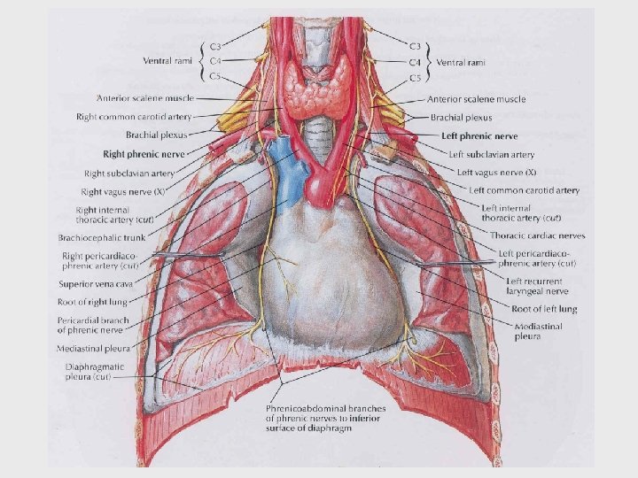
Anatomy of the Heart The heart is a funnel-shaped, hollow, muscular organ that is responsible for pumping blood to all parts of the body. The heart is located near the center of the thoracic cavity between the lungs and is contained in the pericardial sac. The pericardial sac supports the heart and contains some fluid for lubrication.
Location of Heart in Thorax
Location of Heart in Chest • • Oblique Position Apex = Left of Midline (5 th ICS), Anterior to rest of heart Base (posterior surface) sits on vertebral column Superior Right = 3 rd Costal Cartilage, 1” right midsternum Superior Left = 2 nd Costal Cartilage, 1” left midsternum Inferior Right = 6 th Costal Cartilage, 1” right midsternum Inferior Left = 5 th Intercostal Space at Midclavicular line
The broad end, or base, of the heart is also supported by large arteries and veins. The pointed end, or apex, of the heart is directed toward the abdomen.
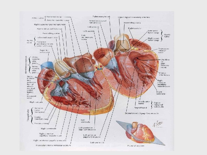
The heart wall is made up of three layers. • Epicardium – outer layer of heart wall, which is also the inner layer • of epicardial sac; Endocardium – inner layer that endothelial cells, which heart . consists of line the heart, covers the valves, and lines the blood vessels. Myocardium – middle layer composed of cardiac muscle. The cardiac muscle is an involuntary, striated muscle with fibers that intertwine.
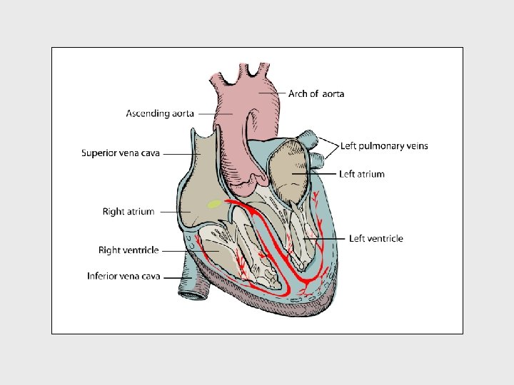
In mammals and birds, the heart is divided into a right and left side and each side is divided into an atrium and ventricle. Therefore, the heart is said to have four chambers (right atrium, right ventricle, left atrium, and leftventricle).
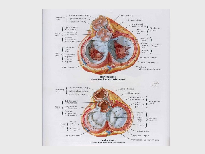
The atrioventricular valves (AV valve) separate the atrium and ventricle on each side of the heart. The AV valves have flaps of tissues, called leaflets or cusps, which open and close to ensure that the blood flows only in one direction and does not backflow into the atriums. The AV valve on the right side of the heart is called the tricuspid valve because it has three leaflets (cusps). The AV valve on the left side of the heart is called the bicuspid valve (or mitral valve) because it has two leaflets. The pulmonary valve and the aortic valve prevent blood from back-flowing into their respective ventricles.
The pulmonary valve is located between the right ventricle and the pulmonary artery. The aortic valve is located between the left ventricle and the aortic artery.
A group of cells called the sinoatrial node (SA node) control the beat of the heart by sending out electrical signals to make the heart pump.
Heart Innervation • Heart receives visceral motor innervation – Sympathetic (speeds up) – Parasympathetic (slows down) p. 534 Larry M. Frolich, Ph. D. , Human Anatomy
The Heart This is a vein. It brings blood from the body, except the lungs. These arteries. They carry blood away from the heart. 2 atria 2 ventricles Coronary arteries, the hearts own blood supply The heart has four chambers now lets look inside the heart
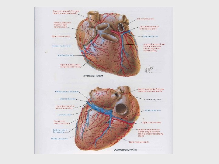
Blood supply to heart wall • Rt and Lft Coronary Arteries – – Branch from Ascending Aorta Have multiple branches along heart Sit in Coronary Sulcus Coronary Heart Disease • Cardiac Veins – Coronary Sinus (largest) – Many branches feed into sinus – Sit in Coronary Sulcus
A heart attack often involves a clot in the coronary arteries or their branches. In this illustration, a clot is shown in the location of #1. Area #2 shows the portion of the damaged heart that is affected by the .
The Heart Artery to Lungs Vein from Head and Body Right Atrium valve Right Ventricle Artery to Head and Body Vein from Lungs Left Atrium valve Left Ventricle
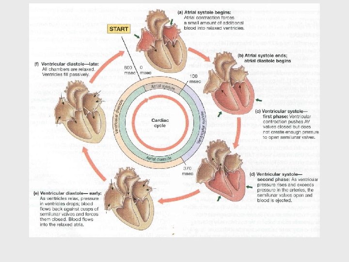
How does the Heart work? STEP ONE blood from the body blood from the lungs The heart beat begins when the heart muscles relax and blood flows into the atria.
How does the Heart work? STEP TWO The atria then contract and the valves open to allow blood into the ventricles.
How does the Heart work? STEP THREE The valves close to stop blood flowing backwards. The ventricles contract forcing the blood to leave the heart. At the same time, the atria are relaxing and once again filling with blood. The cycle then repeats itself.
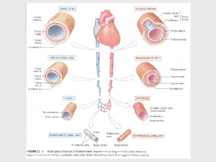
blood from the heart gets around the body through blood vessels There are 3 types of blood vessels a. ARTERY b. VEIN c. CAPILLARY
Walls of Arteries and Veins • Tunica externa – Outermost layer – CT w/elastin and collagen – Strengthens, Anchors • Tunica media – Middle layer – Circular Smooth Muscle – Vaso-constriction/dilation • Tunica intima – Innermost layer – Endothelium – Minimize friction • Lumen
ARTERI mempunyai 3 lapisan : 1. TUNICA ADVENTITIA/EXTERNA , MERUPAKAN LAPISAN TERLUAR , TERDIRI DARI JARINGAN IKAT FIBROUS DAN BERFUNGSI SEBAGAI LAPISAN PELINDUNG. 2. TUNICA MEDIA, TERDIRI DARI JARINGAN OTOT DAN ELASTIK, MERUPAKAN LAPISAN YANG KUAT; MEMBUAT PEMBULUH DARAH TETAP TERBUKA DAN KONTRAKSI JARINGAN OTOTNYA MEMBERIKAN TEKANAN YANG TETAP TERHADAP ALIRAN DARAH. 3. TUNICA INTIMA, MERUPAKAN LAPISAN TERDALAM DAN TERDIRI DARI LAPISAN ENDOTHELIUM.
ARTERI : 1. ARTERI BESAR (ELASTIC ARTERY): 2. ARTERI SEDANG (MUSCULAR ARTERY): 3. ARTERI KECIL (ARTERIOLE):
VENA • JUGA MEMPUNYAI 3 LAPISAN SEPERTI ARTERI • TUNICA MEDIA LEBIH TIPIS (JAR OTOT DAN ELASTIK), SEHINGGA LEBIH MUDAH KEMPIS DAN KURANG ELASTIS DIBANDING DENGAN ARTERI • MEMPUNYAI KATUB YANG DISUSUN SEDEMIKIAN RUPA SEHINGGA DARAH NORMAL HANYA MENGALIR KE COR DAN TIDAK SEBALIKNYA • TERDIRI VENA BESAR, VENA SEDANG DAN VENULE
The VEIN Veins carry blood towards from the heart. veins have valves which act to stop the blood from going in the wrong direction. thin muscle and elastic fibres body muscles surround the veins so that when they contract to move the body, they also squeeze the veins and push the blood along the vessel.
Artery/Vein differences Arteries (aa. ) Direction Blood Away from of flow Heart Pressure Higher Veins (vv. ) Blood to Heart Walls Lumen THICKER: Tunica media thicker than tunica externa Smaller THINNER: Tunica externa thicker than tunica media Larger Valves No valves Valves (see next) y M. Frolich, Ph. D. , Human Anatomy Lower
KAPILER • Ialah pembuluh darah yg sangat kecil tempat arteriole berakhir dan venule mulai • Makin kecil arteri ketiga lapisan diatas makin tak jelas, sehingga ketika sampai di kapiler yang sehalus rambut dindingnya tinggal 1 lapis saja yaitu endothelium. • Lapisan yang tipis ini memungkinkan pertukaran bahan dengan sel, dimana bahan makanan dan O 2 masuk ke dalam sel sedangkan CO 2 dan sisa metabolisme masuk ke dalam kapiler.
Capillaries • Microscopic--one cell layer thick • Network • Bathed in extracellular matrix of areolar tissue • Entire goal of C-V system is to get blood into capillaries where diffusion takes place Larry M. Frolich, Ph. D. , Human Anatomy
The CAPILLARY Capillaries link Arteries with Veins they exchange materials between the blood and other body cells. the wall of a capillary is only one cell thick The exchange of materials between the blood and the body can only occur through capillaries.
The CAPILLARY A collection of capillaries is known as a capillary bed artery body cell vein capillaries
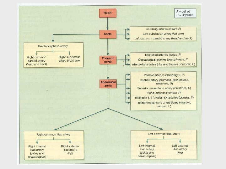
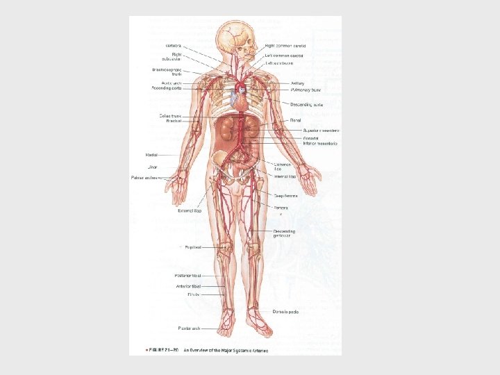
AORTA I. III. A. B. AORTA ASCENDENS : A. CORONARIA DEXTRA ET SINISTRA ARCUS AORTA DESCENDENS : AORTA THORACALIS AORTA ABDOMINALIS
II. ARCUS AORTA : 1. A. BRACHIOCEPHALICA: a. A. SUBCLAVIA DEXTRA b. A. CAROTIS COMMUNIS DEXTRA: - A. CAROTIS INT. DEXTR. - A. CAROTIS EXT. DEXTR. 2. A. CAROTIS COMMUNIS SINISTRA: - A. CAROTIS INT. SIN. - A. CAROTIS EXT. SIN. 3. A. SUBCLAVIA SINISTRA.
III. A. AORTA THORACALIS: 1. Aa. INTERCOSTALIS 2. Aa. BRONCHIALIS 3. Aa. ESOPHAGEALIS
III. B. AORTA ABDOMINALIS 1. 2. 3. 4. 5. 6. 7. 8. 9. - Aa. PHRENICA ABD. DEXTRA ET SINISTRA A. COELIACA Aa. SUPRARENALIS MEDIA DEXTRA ET SINISTRA Aa. RENALIS DEXTRA ET SINISTRA Aa. TESTICULARIS / OVARICA DEXT. ET SIN. A. MESENTERICA SUP. A. MESENTERICA INF. Aa. LUMBALIS Aa. ILIACA COMMUNIS: Aa. ILIACA EXTERNA Aa. ILIACA INTERNA
III. B. AORTA ABDOMINALIS 1. 2. 3. 4. 5. 6. 7. 8. 9. - Aa. PHRENICA ABD. DEXTRA ET SINISTRA A. COELIACA Aa. SUPRARENALIS MEDIA DEXTRA ET SINISTRA Aa. RENALIS DEXTRA ET SINISTRA Aa. TESTICULARIS / OVARICA DEXT. ET SIN. A. MESENTERICA SUP. A. MESENTERICA INF. Aa. LUMBALIS Aa. ILIACA COMMUNIS: Aa. ILIACA EXTERNA Aa. ILIACA INTERNA
ARTERI UNTUK LEHER DAN KEPALA 1. 2. 3. 4. A. CAROTIS EXTERNA A. CAROTIS INTERNA A. VERTEBRALIS A. MENINGICA MEDIA
ARTERI UNTUK EXTREMITAS SUPERIOR • • A. AXILLARIS A. BRACHIALIS A. RADIALIS A. ULNARIS
ARTERI UNTUK EXTREMITAS INFERIOR • • • A. FEMORALIS A. POPLITEA A. TIBIALIS ANTERIOR A. TIBIALIS POSTERIOR A. DORSALIS PEDIS Aa. PLANTARIS MEDIALIS ET LATERALIS
TEMPAT-TEMPAT UNTUK PALPASI ARTERI (NADI)
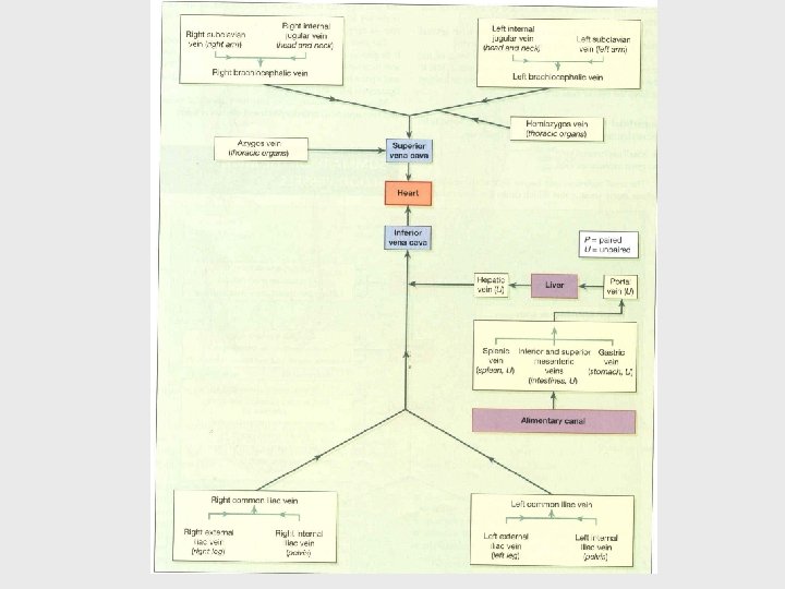
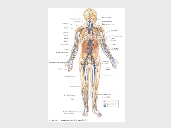
VENNAE BESAR ATRIUM DEXTRM I. V. CAVA SUPERIOR V. AZYGOS Vv. BRACHIOCEPHALICA DEXTRA ET SINISTRA Vv. JUG INT. Vv. SUBCLAVIA DEXT/SIN
II. V. CAVA INFERIOR: 1. Vv. PHRENICA INFERIOR 2. Vv. HEPATICA 3. Vv. SUPRARENALIS 4. Vv. RENALIS 5. V. TESTICULARIS/ OVARICA DEXTRA 6. Vv. LUMBALIS 7. Vv. ILIACA COMMUNIS DEXTRA ET SINISTRA
VENNAE LEHER DAN KEPALA • V. JUGULARIS EXTERNA • V. JUGULARIS INTERNA
VENNAE EXTREMITAS SUPERIOR V. SUBCLAVIA V. AXILLARIS V. CEPHALICA----- V. BASILICA VENNAE MANUS ------ V. MEDIANA CUBITI
VENNAE EXTREMITAS INFERIOR V. ILIACA EXTERNA V. FEMORALIS V. POPLITEA SAPHENA V. SAPHENA PARVA V. MAGNA VENNAE PEDIS
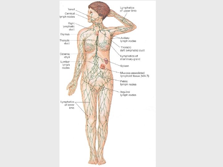
Lymphatic System
Functions of the lymphatic system: • remove excess fluids from body tissues, • absorb fatty acid and transport circulatory system, and fat to • produce immune cells (lymphocytes, monocytes, and plasma cells).
Blood fluid escapes through the thin-walled capillaries into spaces between body tissue cells. Lymph vessels, which have very thin walls, pick up these fluids called lymph.
The lymph vessels join to form larger ducts that pass through lymph nodes (or glands). Each lymph node has a fibrous outer covering (capsule), a cortex, and a medulla.
Lymph nodes filter foreign substances, such as bacteria and cancer cells, from the lymph before it is re-entered into the blood system through the larger veins. Lymph nodes, which are scattered among the lymph vessels, act as the body’s first defense against infection.
AREA LYMPHATIC LAIN • STRUKTUR LAIN PADA SISTEM LYMPHATIC SEBAGIAN BESAR TERDIRI DARI JARINGAN LYMPHE YANG SAMA DENGAN YANG TERDAPAT PADA LYMPHONODI. • ANTARALAIN : - TONSILLA PALATINA - TONSILLA LINGUALIS - TONSILLA PHARYNGICA - PEYER’S PATCHES
Lymph nodes produce the following cells: • Lymphocytes – a type of white blood cell, • Monocytes – a leukocyte that protects against bloodborne pathogens, and • Plasma cells – produce antibodies.
Each lymph node has its own blood supply and venous drainage. The lymph nodes usually have names that are related to their location in the body.
ALIRAN CAIRAN LYMPHE • CAIRAN LYMPHE YANG BERASAL DARI LENGAN KANAN, SISI KANAN KEPALA DAN LEHER, DIALIRKAN KE DUCTUS LYMPHATICUS DEXTER YANG BERMUARA SUDUT PADA PERTEMUAN ANTARA V. JUG INT DEXTRA DENGAN V. SUBCLAV DEXTRA (ANGULUS VENOSUS JUGULUM DEXTRA) • CAIRAN LYMPHE DARIBAGIAN TUBUH YANG LAI DIALIRKAN KE DUCTUS THORACICUS YANG BERJALAN MULAI DARI CYSTERNA CHYLLI (PADA CAVUM ABDOMEN DAN BERMUARA PADA ANGULUS VENOSUS JUGULUM SINISTRA).
When a specific location gets infected, the lymph nodes in that area will enlarge to fight the infection. If the lymph node closest to an infected area is unable to eliminate the infection, other lymph nodes in the system will attempt to fight the infection.
This is particularly critical in the case of cancer, which can be spread from its point of origin to all parts of the body through the lymphatic system.
THANK YOU