CIRCULATORY SYSTEM The circulatory system is divided into


: • It is divided into atrial & ventricular portions, separated")


 Left")


 • It drains venous blood")


- Slides: 47
CIRCULATORY SYSTEM
The circulatory system is divided into two main parts which communicate with one another CARDIOVASCULAR SYSTEM • Heart: acts as a pump • Blood Vessels: through which blood flows LYMPHATIC SYSTEM • Lymphatic Vessels: through which lymph flows • Lymphatic Tissue: collections of lymphocytes
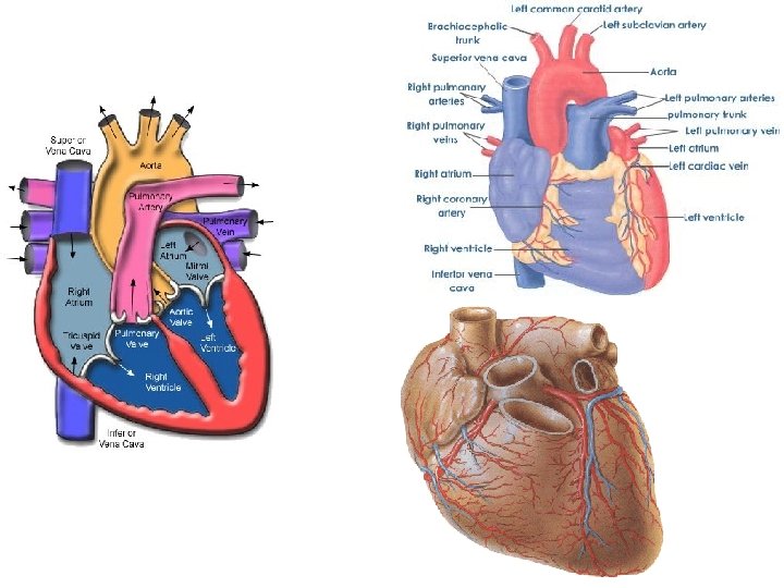
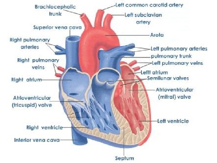
CARDIOVASCULAR SYSTEM • This system is concerned with the continuous flow of blood to all body cells. • It consists of the heart & blood vessels HEART • It is a muscular organ that pumps the blood to various parts of the body • It is located in the mediastinum (the space between the lungs) • It is surrounded by the Pericardium • It is formed of 4 chambers: 2 atria and 2 ventricles • The atria & ventricles are separated by the atrioventricular groove (coronary groove) • The right and left atria are separated by the interatrial septum • The right and left ventricles are separated by the interventricular septum
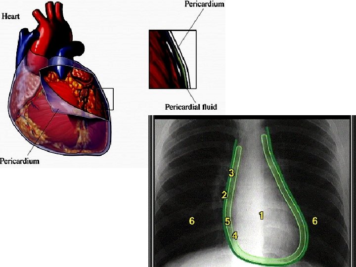
Pericardium The covering of the heart. There are 2 types of pericardium: Fibrous pericardium: This is the outer tough fibrous conical sac which surrounds the serous pericardium. • Its apex surrounds the beginning and termination of big vessels • Its base rests on the central tendon of the diaphragm. • The fibrous pericardium protects the heart and prevents its overdistension. Serous pericardium: this is a closed serous sac which is formed of: Visceral layer: adherent to the heart Parietal layer: lines the fibrous pericardium • The cavity between the 2 layers is called the pericardial cavity • Accumulation of excessive fluid in the pericardial cavity is called “Pericardial Effusion”. It leads to compression of the heart.
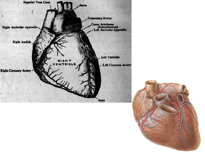
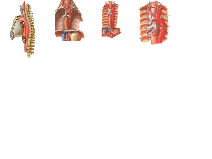
External Features of the Heart The heart is cone shaped. It has: Apex • It is directed downward, forward and to the left • It is formed by the left ventricle only • It lies opposite the 5 th intercostal space, 9 cm from the midline Base • It is directed upward, backward and to the right • It formed by the 2 atria, mainly the left atrium • It lies in front of the middle 4 thoracic vertebrae Base
Sterno-costal surface (anterior surface): • It is divided into atrial & ventricular portions, separated by the atrioventricular groove • The atrial portion is formed mainly by the right atrium • The ventricular portion is formed by the 2 ventricles separated by the anterior interventricular groove.
Diaphragmatic surface: • It rests on the diaphragm • It is formed of the 2 ventricles separated by the posterior interventricular groove. The heart has 4 borders • Upper border: formed by the 2 atria • Lower border: formed by the right ventricle & apical part of left ventricle • Right border: formed by the right atrium • Left border: formed by the left ventricle and left auricle Bas eo f th eh ear t Coronary groove e ac f r u cs ra ti a gm D h iap
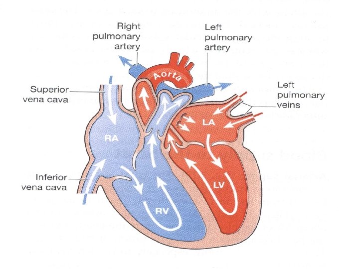
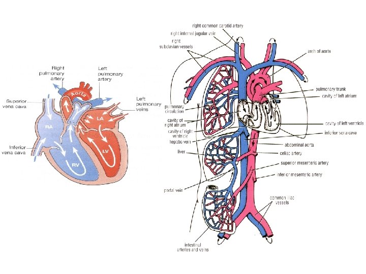
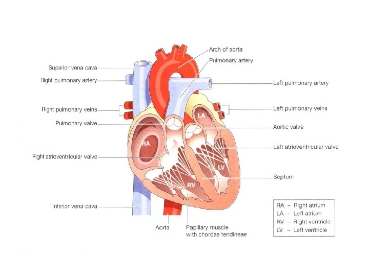
Flow of Blood in the Heart Right Atrium • • • It receives venous blood (non-oxygenated) from the whole body through: 1. The superior vena cava (SVC) 2. The inferior vena cava (IVC) 3. The coronary sinus (which drains the heart) Blood flows from the right atrium into the right ventricle through the right atrioventricular orifice The right atrioventricular orifice is guarded by the right atrioventricular (tricuspid) valve Right Ventricle • • • It receives venous blood from the right atrium It contracts to pump the venous blood to the lungs through the pulmonary trunk The pulmonary trunk divides into right and left branches which lead to the right and left lungs, respectively. N. B. When the right ventricle contracts the tricuspid valve closes to prevent blood from moving back into the right atrium.
Left atrium • It receives oxygenated blood from the lungs through 4 pulmonary veins (2 from each lung) • Blood flows from the left atrium to the left ventricle through the left atrio-ventricular orifice • The left atrio-ventricular orifice is guarded by the left atrio-ventricular (mitral) valve. Left ventricle • It receives oxygenated blood from the left atrium through the left atrio-ventricular orifice • It contracts to pump the blood through the Aorta to be distributed to the whole body • When the left ventricle contracts the mitral valve closes to prevent blood from moving back into the left atrium
Valves of the Heart 1. 2. 3. 4. Right atrio-ventricular valve (tricuspid valve) Left atrio-ventricular valve (mitral valve) Pulmonary valve (semilunar valve): between right ventricle and pulmonary trunk Aortic valve (semilunar valve): between the left ventricle and aorta N. B. • • The atrio-ventricular valves are open during flow of blood from atria to ventricles The atrio-ventricular valves close during ventricular contraction to prevent back flow of blood into the atria • • The semilunar valves (pulmonary & aortic) open during contraction of ventricles The semilunar valves close at the end of ventricular contraction to prevent back flow of blood into the ventricles
INTERIOR OF THE HEART Right Atrium • It has a smooth posterior wall which receives the openings of SVC, IVC, Coronary sinus & anterior cardiac veins • Its anterior wall is rough due to the presence of muscular projections • The 2 walls are separated by a raised band called “crista terminalis” • Its septal wall shows an oval depression called “fossa ovalis” Left Atrium • Its walls are smooth, except at the auricle • It receives the 4 pulmonary veins Crista terminalis
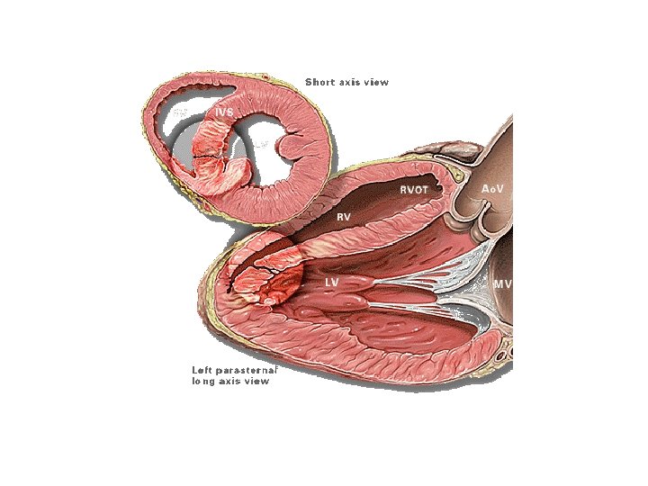
Right Ventricle • It is thin-walled • Its lumen is large and curved in cross section • Its formed of: • Rough inflowing part due to the presence of muscular projections “trabeculae carnae” & 3 papillary muscles • Smooth outflowing part “infundibulum” which leads to the pulmonary trunk Trabeculae Left Ventricle Papillary muscle • It is thick-walled • Its lumen is small & circular in cross section • It is formed of • Rough inflowing part due to the presence of trabeculae carnae and 2 papillary muscles • Smooth outflowing part “vestibule” which leads to the aorta
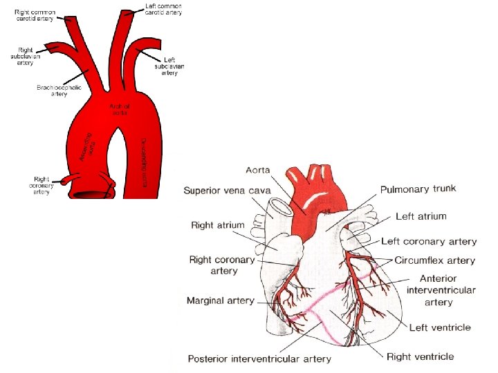
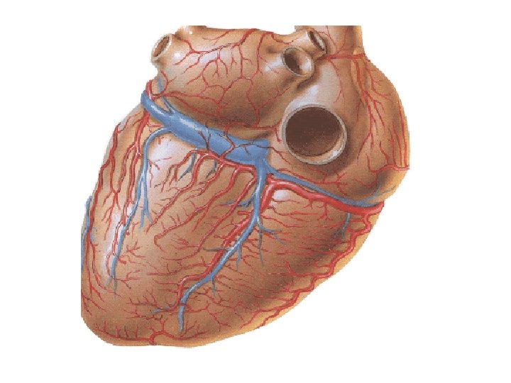
Blood Supply of the Heart Arterial supply Right coronary artery • Arises from the beginning of the ascending aorta • Runs in the coronary groove then curves around the right side of the heart to reach the posterior interventricular groove • Ends by anastomosing with the circumflex branch of left coronary artery Left coronary artery • Arises from the beginning of the ascending aorta • Divides into “anterior interventricular” and “circumflex” arteries • The anterior interventricular artery descends in the anterior interventricular goove • The circumflex artery continues around the left side of the heart in the coronary groove and ends by anastomosing with the right coronary artery
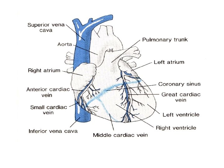
Venous Drainage 1. Coronary sinus • A wide & short venous channel which collects the majority of venous drainage of the heart • It is lodged in the posterior part of coronary groove and opens in the right atrium • It receives: • Great cardiac vein • Middle cardiac vein • Small cardiac vein • Oblique vein of left atrium • Posterior vein of left ventricle 2. Anterior cardiac veins 3. Venae cordis minimi
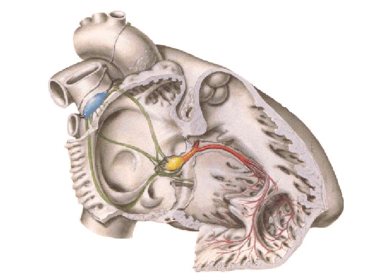
Conductive System of the Heart • This system is concerned with the initiation and propagation of the cardiac impulse. • It consists of specialized cardiac muscle fibers. • The conductive system includes: Sino-Atrial Node (SAN) • Located at the junction of the right atrium and the SVC • It is the site where the cardiac impulse is initiated (pace maker of the heart) Atrio-Ventricular Node (AVN) • Located in the lower part of the inter-atrial septum • Receives the impulse from the SAN Atrio-Ventricular Bundle (AVB) • Begins from the AVN and descends in the inter-ventricular septum • It divides into two bundle branches, right and left • It transmits the impulse from the AVN to the wall of the ventricles
Blood Circulations Three types of circulations can be distinguished: Pulmonary circulation Right ventricle veins pulmonary trunk left atrium lungs pulmonary Systemic circulation Left ventricle venules aorta arteries veins arterioles right atrium capillaries Portal circulation A circulation which starts by capillaries and ends in capillaries (or sinusoids) without entering the pulmonary or systemic circulation Veins from GIT IVC Hepatic portal circulation portal vein liver sinusoids 2 hepatic veins
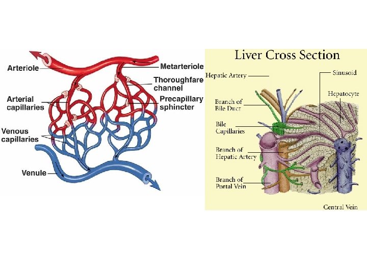
Blood Vessels Arteries • These are vessels which carry blood away from the heart • They are elastic & branching giving medium sized arteries which give small sized arteries, arterioles, and finally arterial capillaries • Arteries may communicate forming “arterial anastomosis” Types of arteries: *Elastic arteries • These are the largest arteries (e. g. aorta) • They contain elastic tissue which enables them to expand recoil *Muscular arteries • These are medium sized arteries (e. g. arteries in the limbs) • They contain smooth muscle fibers which allow vasoconstriction or vasodilatation *Arterioles • These are small arteries • They contain smooth muscle fibers which allow vasoconstriction or vasodilatation
Veins • They are wide and thin walled vessels • They carry blood toward the heart • Veins begin as venous capillaries which collect into venules then small veins and finally large veins • They maybe superficial or deep • The deep veins accompany arteries and are called “venae comitantes” • Many veins have valves which prevent backflow of blood
Connections between arteries and veins Capillaries • Microscopic blood vessels with very thin walls • They are lined by a single layer of endothelium • They form a network in body tissues called “capillary bed” • The thin walls of the capillaries allow diffusion of oxygen and nutrients out of the capillaries and carbon dioxide and waste products into the capillaries Sinusoids • These are similar to capillaries, except they form wider spaces • They are seen in the liver, spleen, bone marrow Arterio-venous anatomosis (shunts) • These are direct connections between arterioles and venules without the intervention of capillaries • They are present in the skin of lips, nose, tips of fingers and toes & intestinal mucosa • The shunts contain sphincters which open and close to control the blood supply to the involved organs
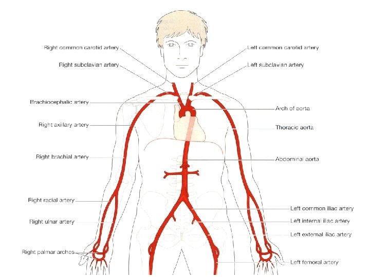
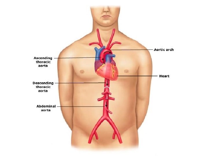
Major Arteries of the Body Aorta • • It arises from the left ventricle of the heart It has 3 parts: 1. Ascending Aorta: it gives right & left coronary arteries 2. Arch of Aorta: it gives: • Brachiocephalic artery (which gives right common carotid & right subclavian arteries) • Left common carotid artery • Left subclavian artery • Each common carotid artery divides into internal & external carotid arteries to the head & neck The subclavian artery enters the upper limb. • 3. Descending Aorta It descends first in the thorax (descending thoracic aorta), then it pierces the diaphragm and enters the abdomen (descending abdominal aorta)
Descending Thoracic Aorta: gives bronchial arteries to the lungs, branches to trachea, osophagus and intercostal spaces. Descending Abdominal Aorta: gives: *Single branches • Celiac trunk • Superior mesenteric artery • Inferior mesenteric artery *Paired branches: • Renal arteries to the kidneys • Gonadal (testicular or ovarian) arteries Termination of the Aorta: • The aorta terminates by dividing into two common iliac arteries • Each common iliac artery divides into internal & external iliac arteries
Pulmonary Trunk • It arises from the right ventricle • It divides into right & left pulmonary arteries which enter the right and left lungs
Arteries of the upper limb • The subclavian artery enters the axilla forming the “axillary artery” in the axilla • The axillary artery continues into the arm as the “brachial artery” • The brachial artery divides in front of the elbow joint into the “radial” & “ulnar” arteries
Arteries of the lower limb • The external iliac artery enters the thigh as the “femoral artery” • The femoral artery continues behind the knee joint as the “popliteal artery” • The popliteal artery divides into “anterior tibial” and “posterior tibial” arteries which enter the leg
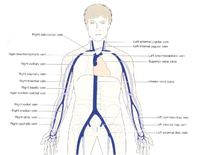
Major Veins of the Body Superior Vena Cava (SVC) • It drains venous blood from head & neck, upper limbs & thorax • It is formed by union of right & left innominate (brachiocephalic) veins • Each innominate vein is formed by the union of subclavian vein (which drains upper limb & thorax) and internal jugular vein (from the head & neck) • The SVC terminates in the right atrium of the heart Inferior Vena Cava (IVC) • It drains the whole body below the diaphragm • It is formed in the abdomen by union of the right & left common iliac veins • It ascends and pierces the diaphragm to enter the thorax • It terminates in the right atrium of the heart
Superficial Veins of the Upper Limb Basilic vein: • On the medial side of the arm • Continues as the axillary vein, which continues as the subclavian vein Cephalic vein • On the lateral side of the arm • Connected to the basilic vein by the median cubital vein • Drains into the axillary vein
Superficial veins of the lower limb Great saphenous vein: • On the medial side • Drains into the femoral vein Small (short) saphenous vein: • On the lateral side • Terminates in the popliteal vein on the back of the knee