The Circulatory System Circulatory system the transport system


 node – a small mass of tissue")


- Slides: 21
The Circulatory System
Circulatory system – the transport system of the body It has 4 main functions: • Transportation of O 2 and CO 2 • Distribution of nutrients and transport of wastes • Maintenance of body temperature • Circulation of hormones
It consists of 3 general components: • A fluid in which materials are transported, such as blood • A system of blood vessels or spaces throughout the body in which the fluid moves • A pump, such as the heart, that pushes the fluid through the blood vessels or spaces Cardiovascular system – part of the circulatory system comprising the heart and blood vessels
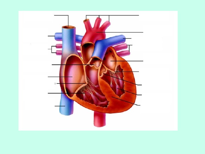
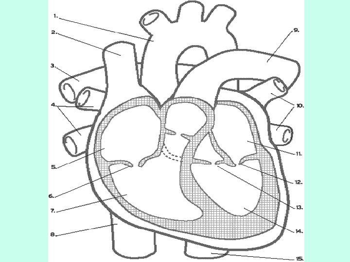
Heart Structure The mammalian heart consists of a double pump separated by a wall of muscle called the septum – separates the right heart pump from the left heart pump Each pump consists of: • A thin-walled atrium – receives blood from veins • A thick-walled ventricle – delivers blood to arteries
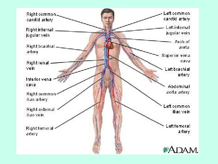
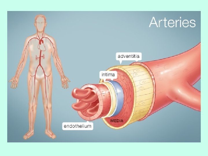
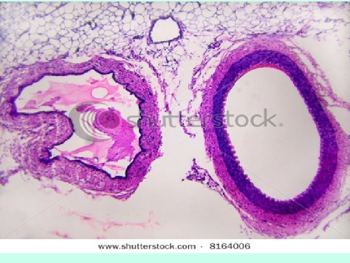
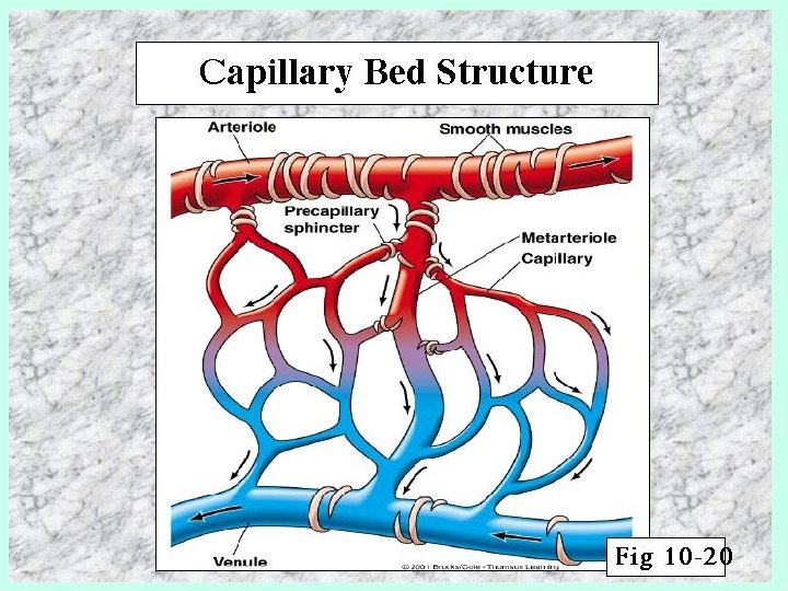
Blood Vessels Arteries carry blood away from the heart • They have thick, muscular walls • Most arteries carry oxygenated blood away from the heart to other parts of the body – Exception: pulmonary artery carries deoxygenated blood from the heart to the lungs • Aorta – largest artery in the body leading directly from the heart and carrying oxygenated blood to the tissues of the body – Blood goes from the aorta, to major arteries, to smaller arterioles, into capillaries
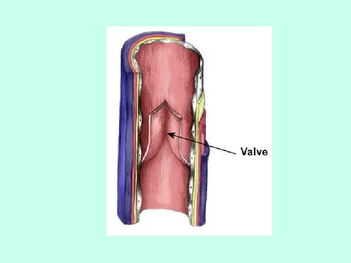
Veins – carry blood back to the heart Have thinner walls, not very elastic • Since veins cannot help with the pumping of blood due to their thin walls, veins have valves that only allow blood flow in one direction. • Also, the contraction of skeletal muscle helps pump blood back to the heart • Most veins carry deoxygenated blood back to the heart – Exception: pulmonary vein that carries oxygenated blood from the lungs to the heart • Vena cava – largest vein in the body that connects directly to the heart – Blood goes from the capillaries, to the venuoles, to the veins, and then to the vena cava
Flow of Blood Through the Heart Deoxygenated blood enters the heart through the superior vena cava or the inferior vena cava and goes into the right atrium The tricuspid atrioventricular (AV) valve opens and the deoxygenated blood enters the right ventricle The pulmonary semilunar valve opens and the blood enters the pulmonary artery which brings the deoxygenated blood to the lungs In the lungs, the blood cells drop off CO 2 and pick up O 2
The oxygenated blood returns to the heart through the pulmonary vein and enters the left atrium The bicuspid atrioventricular (AV) valve opens and the blood enters the left ventricle The aortic semilunar valve opens and the blood enters the aorta where it is pumped to the rest of the body
Heart Rhythms, Sounds, and Pressure Sinoatrial (SA) node – a small mass of tissue in the right atrium that originates the impulses stimulating the heartbeat; acts as a pacemaker Atrioventricular (AV) node – a small mass of tissue through which impulses from the SA node are passed to the ventricles, causing them to contract in unison
Conducting Systems • http: //highered. mcgrawhill. com/sites/0072495855/student_view 0/ch apter 22/animation__conducting_system_of_t he_heart. html
The “lubb-dubb” sound is caused by the closing of the heart valves • Diastole – relaxation of the heart, during which the cavities of the heart fill with blood • Systole – contraction of the ventricles, during which blood is pushed out of the heart – The increase in pressure during systole forces the AV valves shut, causing the “lubb” sound – As the ventricles relax, the pressure inside them decreases and blood flows into them. – This decrease in pressure causes the semilunar valves to close, preventing blood from flowing backwards into the ventricles, causing the “dubb” sound
Heart Sounds • http: //highered. mcgrawhill. com/sites/0072495855/student_view 0/ch apter 22/animation__the_cardiac_cycle__quiz _1_. html
Pulse – change in the diameter of the arteries that can be felt on the body’s surface following heart contractions Blood pressure – the pressure exerted on the walls of the arteries when the ventricles of the heart contract • Normal = 120/80 (systolic/diastolic) • Low blood pressure – reduces the body’s capacity to transport blood • High blood pressure – can weaken an artery, resulting in its rupture
Blood Composition Blood is made of 55% plasma, 45% red blood cells, and less than 1% white blood cells