LECTURE CIRCULATORY SYSTEM CIRCULATORYY SYSTEM The circulatory system


 and atrio-ventricular nodes are composed")


- Slides: 43
LECTURE: CIRCULATORY SYSTEM
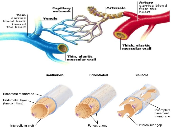
CIRCULATORYY SYSTEM • The circulatory system comprises of two divisions 1. Blood vascular system 2. Lymph vascular system 1. The blood vascular system: This system includes • Heart • arteries • Capillaries • veins
1. THE BLOOD VASCULAR SYSTEM: CAPILLARIES: • Delicate endothelial tubes that connect the arterial and venous sides of circulation are called capillaries. • Arranged in the form of networks. • The capillary wall consists of a single layer of flat endothelial cells which are surrounded externally by a small amount of connective tissue. • Outer to the basal lamina of the endothelium is present a thin layer of delicate collagenous and reticular fibers. Classification of capillaries: 1. Continues capillaries 2. Fenestrated capillaries 3. Sinusoidal capillaries
CAPILLARIES: 1. Continues capillaries: • These capillaries do not have any pores, gaps, or discontinuities in their wall. Location: • They are found in muscles, nervous tissues, connective tissue, lungs and skin. 2. Fenestrated capillaries: This variety of capillaries is characterized by the presence of numerous circular pores(fenestrae) in the lining endothelial cells. Location: • Found in those regions where rapid exchange of substances between the blood and tissues is required • Found in intestinal mucosa, endocrine glands, choroid plexus and ciliary body.
CAPILLARIES: 3. Sinusoidal capillaries: • In some organs the capillaries have luminal diameter much greater than that of an ordinary capillary, these capillaries are known as sinusoidal capillaries or sinusoids. • They exhibit features that are not characteristic of ordinary capillaries. • The sinusoidal capillaries have irregular and tortuous walls. • Wide gaps exist between the endothelial cells which also have an incomplete basal lamina.
• The endothelial cells also show pores. • As indicated by their structural characteristics the sinusoidal capillaries permit a very rapid exchange of fluids between the blood and tissues. Location: • Found in liver, spleen and bone marrows and endocrine organs
GENERAL STRUCTURAL PATTERN OF BLOOD VESSELS • All arteries and veins exhibit a generalized structural pattern. • The basic design remains the same but differences between the blood vessels of different types and sizes exist. • Generally the wall of a blood vessel is composed of three concentric layers 1. tunica intima 2. tunica media 3. tunica adventitia
GENERAL STRUCTURAL PATTERN OF BLOOD VESSELS 1. Tunica intima: • Simply called intima is the innermost layer. • It consist of a single layer of endothelial cells lining the internal surface of the vessel. • Beneath the endothelium is a thin layer of delicate loose connective tissue known as sub endothelial layer. • The tunica intima is separated from tunica media by a layer of elastic fibers called internal elastic lamina.
GENERAL STRUCTURAL PATTERN OF BLOOD VESSELS 2. Tunica media: • It is the middle layer and is chiefly composed of circularly arranged smooth muscle fibers. • Variable amount of collagenous and elastic fibers are found between the smooth muscle fibers. • An external elastic lamina may also be present at the junction of the tunica media and tunica adventitia.
3. Tunica adventitia: • Also called adventitia is the outer most coat which composed of collagenous and elastic fibers coursing mainly in a longitudinal direction. • The adventitia gradually merges with the connective tissue through which the vessel runs.
Tunica adventitia
ARTERIES Depending on their size the arteries are classified into the following three main types: 1. Arterioles and small arteries 2. Medium size arteries 3. Large arteries The structure and relative thickness of the three component tunics vary according to the types of artery
ARTERIES 1 -Arterioles and small arteries: • In these vessels all the three tunics are distinguishable. • The intima consist only of endothelium, the sub endothelial connective tissue is absent. • An internal elastic lamina may be seen in the small arteries. • The media of an arteriole consist only of one or two layers of circularly arranged muscle fibers. • Whereas the media of a small artery may have up to eight layers.
ARTERIES 1 -Arterioles and small arteries………… • The adventitia o f arteriole and small arteries consists of thin layer of longitudinally oriented collagenous and elastic fibers. • Arteriole function to regulate the distribution of blood to different capillary networks by vasoconstriction or vasodilation. • These vessels are considered to be the chief controllers of the systemic blood pressure.
MEDIUM-SIZED ARTERIES • These arteries are also known as muscular arteries because their thick walls contain a lot amount of smooth muscle in the tunica media. • The medium-size arteries perform the function of distribution of blood to various regions in organs of the body according to the functional demands of the region concerned. • Also called distribution arteries. • Most of the named arteries of the body belong to this group e. g. Axillary artery, radial artery, femoral and tibial arteries.
The tunica intima: • The tunica intima of a muscular artery shows all the three typical layers i. e. the endothelial , sub-endothelial connective tissue and internal elastic lamina. • The sub endothelial connective tissue consist of delicate collagenous and elastic fibers. • The internal elastic lamina is very prominent and exist in the form of thick fenestrated band of closely interwoven elastic fibers.
MEDIUM-SIZED ARTERIES Tunica media: • Tunica media of a medium-sized artery is made up of several layers of circular disposed smooth muscle fibers. • Between muscle layers are found small quantities of connective tissue composed mainly of collagenous. • In larger arteries an external elastic lamina may be seen as a network of elastic fibers at the junction of media and adventitia.
The tunica adventitia: • The tunica adventitia of a muscular artery is of sufficient thickness and may be nearly as thick as the media. • It is composed of longitudinally coursing collagenous and elastic fibers.
LARGE ARTERIES • These Arteries have a very large diameter. • Structurally they are characterized by presence of a large amount of elastic tissue in their wall due to which these arteries are also called elastic arteries. • The walls of these vessels are relatively thin and proportion to their diameter. • The aorta and its main branches belong to this variety of arteries. • The large arteries are also referred to as conducting arteries because they conduct blood from the heart to the medium sized distributing arteries.
INTIMA: • The intima of an elastic artery is relatively thick and is lined by endothelial cells. • The sub-endothelial layer consists of collagenous and elastic fibers. • No separate internal elastic lamina can be identified due to abundance of elastic fibers.
LARGE ARTERIES MEDIA: • The media of an elastic artery is composed of a series of concentrically arranged distinct membranes 40 -70 in number. • Between the elastic lamella are present fibroblast and amorphous ground substance and smooth muscle fiber which also form spiral course. • No external elastic lamina is present. ADVENTITIA: • The adventitia of an elastic artery is in thin and consist mainly of collagenous fibers arranged in longitudinal spirals. • The elastic arteries absorb some of the pulse beat by expansion of their walls. • During diastole the large arteries return to normal size.
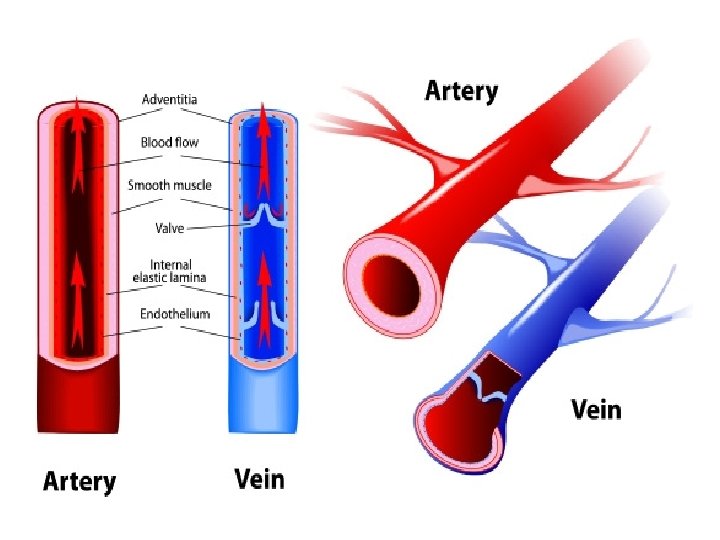
VEINS • The basic structural pattern of veins resemble that of arteries but marked differences are present. • Generally the internal diameter of a vein is larger than an artery of the same size because the veins have thinner walls because of the scarcity of the muscular and elastic components. • Like arteries veins are classified into three types but the classification is not rigid and individual variations may be present within any of the following three groups 1. Venules 2. Medium sized veins 3. Large veins
VEINS 1. VENULES: • Here the intima is composed only of endothelium. • The media is thin and consist of a few layers of smooth muscle fibers. • The adventitia is relatively thick and is made up of collagenous fibers
VEINS 2. MEDIUM-SIZED VEINS: • This group includes the named veins of the body and their principle tributaries. • The tunica intima consist of endothelium and a thin subendothelial layer of delicate collagenous and elastic fibers. • An internal elastic lamina may be distinguishable. • The media is composed of circularly arranged smooth muscle cells and collagenous fibers. • The adventitia is well developed and form bulk of the vein wall, It is composed of thick bundles of collagenous fibers.
VEINS 3. LARGE VEINS: • This group include the superior/inferior venae cava and portal vein. • The intima of a large vein consist of endothelium and a thin layer of sub endothelial connective tissue. • An internal elastic lamina may occasionally be distinguishable.
VEINS 3. LARGE VEINS…………. • The media is thin and poorly developed. It consist mainly of collagenous fibers, the smooth muscle fiber are much reduced or entirely absent. • The adventitia is thickest of the three coats and shows three zones containing dense fibroelsatic connective tissue with coarse collagenous fibers.
THE HEART The heart may be regarded as a blood vessels with very large lumen and extremely thick muscular walls. The walls of the heart consists of the following layers 1. The inner layer (endocardium) 2. The middle layer (myocardium) 3. The outer layer (epicardium)
ENDOCARDIUM • Homologous to the tunica intima of blood vessels • It is lined by a layer of endothelium beneath which is a thin layer of sub endothelial connective tissue composed of collagenous fibers, elastic fibers and some smooth muscle cells. • Deeper to the sub-endothelial connective tissue is present a layer of lose connective tissue called subendocardium , this layer binds the endocardium proper to the myocardium
MYOCARDIUM • The myocardium correspond to the tunica media and consists of the cardiac muscles. • Its thickness varies in different parts of the heart being thinnest in the atria and thickest in the left ventricle.
EPICARDIUM • This is actually the visceral layer of pericardium. • It is lined externally by simple squamous epithelium(mesothelium). • Th epicardium is attached to the myocardium by a sub-epicardial layer of loose areolar connective tissue which contains blood vessels, nerves and fat.
IMPULSE GENERATIN/CONDUCTING SYSTEM OF THE HEART • The Sino-arterial(SA) and atrio-ventricular nodes are composed of small spindle shaped specialized cardiac cells. • The atrio-ventricular bundle and its branches consist of modified cardiac muscle fibers known as Purkinje fibers. • These fibers have a faster rate of conduction than the ordinary cardiac muscle fibers.
Structurally the Purkinje fibers differ from ordinary cardiac muscle fibers in the following aspects: 1: They have a larger diameter 2: They usually have two nuclei located centrally 3: The sarcoplasm of Purkinje fibers is rich in glycogen 4: Purkinje fibers contain relatively more sarcoplasm 5: They have few myofibrils which are usually restricted to the periphery of the fibers.
IMPULSE GENERATIN/CONDUCTING SYSTEM OF THE HEART The pathway of conducting system is made up of 5 elements: • The sino-atrial (SA) node • The atrio-ventricular (AV) node • The bundle of His • The left and right bundle branches • The Purkinje fibres
Bundle of His
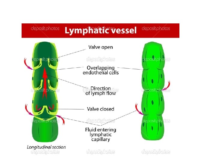
THE LYMPHATIC VASCULAR SYSTEM • This group consist of lymph capillaries, lymphatic vessels and lymphatic ducts. • The lymph vascular system collect fluid from tissue spaces and returns it to the blood: this fluid is called lymph. • Lymph drainage is one way flow (periphery to heart) and is not a circulation in true sense.
THE LYMPHATIC VASCULAR SYSTEM LYMPH CAPILLARIES: • Delicate endothelial tubes that originate in various tissues as blind ended vessels. • They have slightly larger lumen than blood capillaries but their caliber is not uniform. • Endothelial cells do not have pores but adjacent cells show frequent intercellular gaps. • The lymph capillaries branch and anastomose freely to form extensive networks in the spaces between blood capillaries.
THE LYMPHATIC VASCULAR SYSTEM LYMPHATIC VESSELS: • These vessels resemble veins in structure but have much thinner walls. • In the large vessels the three tunics , i. e. intima media and adventitia may be distinguished but usually there is no clear-cut demarcation between the successive coats. • The tunica intima is lined by endothelium, underneath which is present a network of elastic and collagenous fibers. • The tunica media is composed of circular running smooth muscle fibers between which a few delicate elastic fibers may be present. • The tunica adventitia is the thickest coat and consist of longitudinally running collagenous and elastic fibers with a few smooth muscle cells.
THE LYMPHATIC VASCULAR SYSTEM LYMPATIC DUCTS: • These ducts includes the thoracic and right lymphatic ducts and have a structure similar to a vein of equal size except for the presence of a great amount of smooth muscle in the media. • The intima of lymphatic ducts consist of endothelium and sub endothelial layer of delicate connectives tissue containing a few smooth muscle fibers.
THE LYMPHATIC VASCULAR SYSTEM LYMPATIC DUCTS: • The media is the thickest coat. • It is made up of longitudinal and circular bundles of smooth muscle fibers. • Between the muscle bundles is present abundant connective tissue composed principally of collagenous fibers. • The adventitia is poorly defined and consist of longitudinally arranged coarse collagenous fibers with a few smooth muscle cells.