Circulatory system The circulatory system or cardiovascular system

 is an organ system that moves nutrients, gases,")
 that is composed of a liquid")
, 4. 2 to 5. 4 million (female)")


 is a classification of")

![Red blood cell compatibility table[33][34] Recipi ent[1] Donor[1] O- O- Y O+ Y A-](https://slidetodoc.com/presentation_image_h2/b6d4f608ae274bd3428cf29a33e7901c/image-18.jpg "Red blood cell compatibility table[33][34] Recipi ent[1] Donor[1] O- O- Y O+ Y A-")


 of blood. This")
 and is")


 that affects any part")
 of")


, colloquially called \"bad cholesterol\". Many believe that,")


 2. Plaque 3. Artery Wall http: //www. youtube. com/watch? v=gv. Rt.")

, more commonly known as a heart")
- Slides: 53
Circulatory system
The circulatory system (or cardiovascular system) is an organ system that moves nutrients, gases, and wastes to and from cells, helps fight diseases and helps stabilize body temperature and p. H to maintain homeostasis. Humans, as well as other vertebrates, have a closed circulatory system; meaning that the blood never leaves the reticulation of arteries, veins and capillaries.
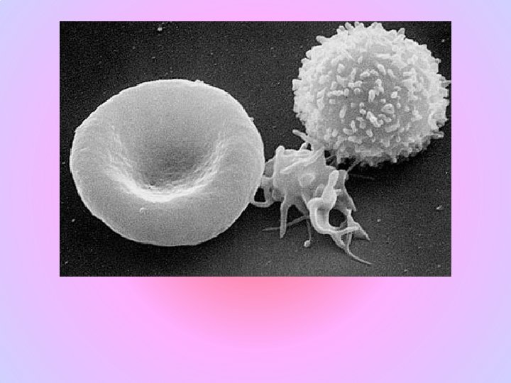
Blood is a specialized fluid (technically a tissue) that is composed of a liquid called blood plasma and blood cells suspended within the plasma. The blood cells present in blood are red blood cells (also called RBCs or erythrocytes), white blood cells (including both leukocytes and lymphocytes) and platelets (also called thrombocytes). Plasma is predominantly water containing dissolved proteins, salts and many other substances; and makes up about 55% of blood by volume. Mammals have red blood, which is bright red when oxygenated, due to hemoglobin. Hemoglobin transports oxygen from the lungs to the rest of the body, such as to the muscles, where it releases the oxygen. It also transport cellular wastes including carbon dioxide which is a bi-product of cellular respiration.
4. 7 to 6. 1 million (male), 4. 2 to 5. 4 million (female) erythrocytes: [3] In mammals, mature red blood cells lack a nucleus and organelles. The proportion of blood occupied by red blood cells is normally about 45%. 4, 000 -11, 000 leukocytes: White blood cells are part of the immune system; they destroy and remove old or aberrant cells and cellular debris, as well as attack infectious agents (pathogens) and foreign substances. The cancer of leukocytes is called leukemia. 200, 000 -500, 000 thrombocytes: Platelets are responsible for blood clotting (coagulation). They change fibrinogen into fibrin. This fibrin creates a mesh onto which red blood cells collect and clot, which then stops more blood from leaving the body and also helps to prevent bacteria from entering the body
Plasma About 55% of whole blood is blood plasma, a fluid that is the blood's liquid medium, which by itself is straw-yellow in color. The blood plasma volume totals of 2. 7 -3. 0 liters in an average human. It is essentially an aqueous solution containing 92% water, 8% blood plasma proteins, and trace amounts of other materials. Plasma circulates dissolved nutrients, such as, glucose, amino acids and fatty acids (dissolved in the blood or bound to plasma proteins), and removes waste products, such as, carbon dioxide, urea and lactic acid. "Dried plasma" was developed and first used in WWII. Prior to the United States' involvement in the war, liquid plasma and "whole blood" were used. The resulting Army-Navy dried plasma package came in two tin cans containing 400 cc bottles. One bottle contained enough distilled to completely reconstitute the dried plasma contained within the other bottle. In about three minutes, the plasma would be ready to use and could stay fresh for around four hours. [.
Blood Vessels Veins The highways with which the blood cells, platelets, nutrients, and wastes travel through are the blood vessels of the human body. Blood vessels come in a variety sizes and directions. With only one exception all veins and venules travel to the heart. Veins and venules are carrying blood that needs to be oxygenated and filtered for waste products. Veins function to return deoxygenated blood to the heart, and are essentially tubes that collapse when their lumens are not filled with blood. The thick, outer-most layer of a vein is made of collagen, wrapped in bands of smooth muscle while the interior is lined with epithelial cells. Most veins have one-way flaps called venous valves that prevent blood from back flowing and pooling in the lower extremities due to the effects of gravity.
Epithelial Cells Endothelial Cells Smooth Muscle Connective Tissue In the picture the left side is a vein and the right is an artery. Both vessels are surrounded with connective tissue, then in the middle smooth muscle and finally in the inner lumen epithelial cells. The difference between the two is that the artery has far more smooth muscle and connective tissue than that of the vein. Why does the artery have a greater thickness of tissue.
Arteries and veins take blood from and to the heart, but the system of vessels start with the large arteries and veins and branch out to include arterioles , venules, and capillaries. Arterioles: An arteriole is a small diameter blood vessel that extends and branches out from an artery and leads to capillaries. Arterioles have thin muscular walls (usually one to two layers of smooth muscle) and are the primary site of vascular resistance. Vascular resistance is the resistance to the flow of blood due to the difference in size of the blood vessel. With arterioles being the first shrinking of vessel diameter there is considerable back pressure that must be overcome by the heart’s push or contraction. An analogy would be trying to drink a milk shake from a thin straw as oppose to an wide straw. Or even better think of a funnel water delays or resists going through a smaller opening.
From arterioles then branch into capillaries. Capillaries are the smallest of a body's blood vessels, measuring 5 -10 um in diameter, which connect arterioles and venules, and enable the interchange of water, oxygen, carbon dioxide, and many other nutrient and waste chemical substances between blood and surrounding tissues. Copy the picture
The walls of capillaries are composed of only a single layer of cells, the endothelium. This layer is so thin that molecules such as oxygen, water and lipids can pass through them by diffusion and enter the tissues. Waste products such as carbon dioxide and urea can diffuse back into the blood to be carried away for removal from the body. Capillaries are so small the red blood cells need to partially fold into bullet-like shapes in order to pass through them in single file.
Through the body all cells are supplied with oxygen and nutrients and deposit waste products through capillary walls.
Arteries are muscular blood vessels that carry blood away from the heart.
lymphatic system The lymphatic system and the cardiovascular system are closely related structures. The lymphatic system is important to the body's defense mechanisms. It filters out organisms that cause disease, produces certain white blood cells and generates antibodies. It is also important for the distribution of fluids and nutrients in the body, because it drains excess fluids and protein so that tissues do not swell up. "Lymph" is a milky body fluid that contains a type of white blood cells, called "lymphocytes, " along with proteins and fats. Lymph seeps outside the blood vessels in spaces of body tissues and is stored in the "lymphatic" system to flow back into the bloodstream. Through the flow of blood in and out of arteries, and into the veins, and through the lymph nodes and into the lymph, the body is able to eliminate the products of cellular breakdown and bacterial invasion.
It is through the actions of this system including the spleen, the thymus, lymph nodes and lymph ducts that our body is able to fight infection and to ward off invasion from foreign invaders. Lymph plays an important role in the immune system and in absorbing fats from the intestines. The lymphatic vessels are present wherever there are blood vessels and transport excess fluid to the end vessels without the assistance of any "pumping" action. There are more than 100 tiny, oval structures (called lymph nodes). These are mainly in the neck, groin and armpits, but are scattered all along the lymph vessels. http: //www. innerbody. com/image/lympov. html
Blood Types A blood type (also called a blood group) is a classification of blood based on the presence or absence of inherited antigenic substances on the surface of red blood cells often called “Markers”. These antigens may be proteins, carbohydrates, glycoproteins or glycolipids, depending on the blood group system, and some of these antigens are also present on the surface of other types of cells of various tissues. Several of these red blood cell surface antigens, that stem from one allele (or very closely linked genes), collectively form a blood group system. Blood types are inherited and represent contributions from both parents. A total of 29 human blood group systems are now recognized by the International Society of Blood Transfusion (ISBT).
Marker molecules or antigens determine blood types and the type of blood that can be received in a transfusion.
Red blood cell compatibility table[33][34] Recipi ent[1] Donor[1] O- O- Y O+ Y A- Y A+ Y B- Y B+ Y AB- Y AB+ Y O+ A- A+ B- B+ AB- AB+ Y Y Y Y Y
The Heart
Facts about the Human Heart • Women hearts beat faster than men. • Three years after a person quits smoking, there chance of having a heart attack is the same as someone who has never smoked before. • The human heart weighs less than a pound. • The human heart can create enough pressure that it could squirt blood at a distance of thirty feet. • The first open heart surgery was performed by Dr. Daniel Hall Williams in 1893. • The human heart beats roughly 35 million times a year. • Olive oil can help in lowering cholesterol levels and decreasing the risk of heart complications. • In a lifetime, the heart pumps about one million barrels of blood.
• People that suffer from gum disease are twice as likely to have a stroke or heart attack. • Most heart attacks occur between the hours of 8 and 9 AM. • The human heart beast roughly 35 million times a year. • During a typical human life span, the human heart will beat approximately 2. 5 billion times. • In one day your heart beats 100, 000 times. • Blood takes about 20 seconds to circulate throughout the entire vascular system. Your system of blood vessels – arteries, veins and capillaries – is over 60, 000 miles long. That's long enough to go around the world more than twice! The adult heart pumps about 5 quarts of blood each minute – approximately 2, 000 gallons of blood each day – throughout the body.
• Your body has about 5. 6 liters (6 quarts) of blood. This 5. 6 liters of blood circulates through the body three times every minute. In one day, the blood travels a total of 19, 000 km (12, 000 miles)-that's four times the distance across the US from coast to coast. • The aorta, the largest artery in the body, is almost the diameter of a garden hose. Capillaries, on the other hand, are so small that it takes ten of them to equal the thickness of a human hair
The heart weighs between 7 and 15 ounces (200 to 425 grams) and is a little larger than the size of your fist. By the end of a long life, a person's heart may have beat (expanded and contracted) more than 3. 5 billion times. In fact, each day, the average heart beats 100, 000 times, pumping about 2, 000 gallons (7, 571 liters) of blood. Your heart is located between your lungs in the middle of your chest, behind and slightly to the left of your breastbone (sternum). A double-layered membrane called the pericardium surrounds your heart like a sac. The outer layer of the pericardium surrounds the roots of your heart's major blood vessels and is attached by ligaments to your spinal column, diaphragm, and other parts of your body. The inner layer of the pericardium is attached to the heart muscle. A coating of fluid separates the two layers of membrane, letting the heart move as it beats, yet still be attached to your body.
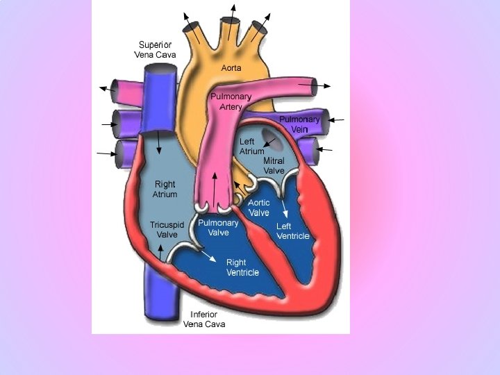
Your heart has 4 chambers. The upper chambers are called the left and right atria, and the lower chambers are called the left and right ventricles. A wall of muscle called the septum separates the left and right atria and the left and right ventricles. The left ventricle is the largest and strongest chamber in your heart. The Heart Valves Four valves regulate Blood flow through your heart: The tricuspid valve regulates blood flow between the right atrium and right ventricle. The pulmonary valve controls blood flow from the right ventricle into the pulmonary arteries, which carry blood to your lungs to pick up oxygen. The mitral valve lets oxygen-rich blood from your lungs pass from the left atrium into the left ventricle. The aortic valve opens the way for oxygen-rich blood to pass from the left ventricle into the aorta, your body's largest artery, where it is delivered to the rest of the body.
The Heart Valves Four valves regulate Blood flow through your heart: The tricuspid valve regulates blood flow between the right atrium and right ventricle. The pulmonary valve controls blood flow from the right ventricle into the pulmonary arteries, which carry blood to your lungs to pick up oxygen. The mitral valve lets oxygen-rich blood from your lungs pass from the left atrium into the left ventricle. The aortic valve opens the way for oxygen-rich blood to pass from the left ventricle into the aorta, your body's largest artery, where it is delivered to the rest of the body.
The Heart Beat A heartbeat is a two-part pumping action that takes about a second. As blood collects in the upper chambers (the right and left atria), the heart's natural pacemaker , which is a specialized bundle of neurons known as the sinoatrial node (SA node), sends out an electrical signal that causes the atria to contract. This contraction pushes blood through the tricuspid and mitral valves into the resting lower chambers (the right and left ventricles). This part of the two-part pumping phase (the longer of the two) is called diastole.
The second part of the pumping phase begins when the ventricles are full of blood. The electrical signals from the SA node travel along a pathway of cells to the ventricles, causing them to contract. This is called systole. As the tricuspid and mitral valves shut tight to prevent a back flow of blood, the pulmonary and aortic valves are pushed open. While blood is pushed from the right ventricle into the lungs to pick up oxygen, oxygen-rich blood flows from the left ventricle to the heart and other parts of the body
After blood moves into the pulmonary artery and the aorta, the ventricles relax, and the pulmonary and aortic valves close. The lower pressure in the ventricles causes the tricuspid and mitral valves to open, and the cycle begins again. This series of contractions is repeated over and over again, increasing during times of exertion and decreasing while you are at rest. The heart normally beats about 60 to 80 times a minute when you are at rest, but this can vary. As you get older, your resting heart rate rises. Also, it is usually lower in people who are physically fit. Your heart does not work alone, though. Your brain tracks the conditions around you—climate, stress, and level of physical activity—and adjusts your cardiovascular system to meet those needs. The human heart is a muscle designed to remain strong and reliable for a hundred years or longer. By reducing your risk factors for cardiovascular disease, you may help your heart stay healthy longer.
The Coronary Arteries The heart muscle, like every other organ or tissue in your body, needs oxygen-rich blood to survive. Blood is supplied to the heart by its own vascular system, called coronary circulation. The aorta branches off into two main coronary arteries. These coronary arteries branch off into smaller arteries, which supply oxygen-rich blood to the entire heart muscle. The right coronary artery supplies blood mainly to the right side of the heart. The right side of the heart is smaller because it pumps blood only to the lungs.
The left coronary artery, which branches into the left anterior descending artery and the circumflex artery, supplies blood to the left side of the heart. The left side of the heart is larger and more muscular because it pumps blood to the rest of the body.
http: //www. nationalgeographic. com/human-heart/index. html
Diseases of the Digestive System and Cardiovascular System Contents: Adenomatous Polyposis Coli - Alagille Syndrome - Anus Diseases Appendicitis - Barrett Esophagus - Biliary Atresia - Biliary Tract Diseases - Budd. Chiari Syndrome - Caroli Disease - Celiac Disease - Cholangitis - Cholecystitis Cholelithiasis - Colitis, Ulcerative - Crohn Disease - Deglutition Disorders Digestive System Diseases - Duodenal Ulcer - Dysentery - Enterocolitis, Pseudomembranous - Esophageal Achalasia - Esophageal Atresia - Esophageal Diseases - Esophagitis - Exocrine Pancreatic Insufficiency - Fatty Liver - Fecal Incontinence - Gastritis, Hypertrophic - Gastroenteritis Gastroesophageal Reflux - Gastrointestinal Diseases - Gastroparesis Hemorrhoids - Hepatitis, Chronic - Hirschsprung Disease Hypertension, Portal - Inflammatory Bowel Diseases - Intestinal Diseases Intestinal Neoplasms - Intestinal Neuronal Dysplasia (not on Me. SH) - Intestinal Obstruction - Irritable Bowel Syndrome - Lactose Intolerance - Liver Cirrhosis Liver Diseases - Meckel Diverticulum - Pancreatic Diseases - Pancreatic Neoplasms - Pancreatitis - Peptic Ulcer - Peutz-Jeghers Syndrome - Proctitis Proctocolitis - Rectal Diseases - Rectal Prolapse - Short Bowel Syndrome Tracheoesophageal Fistula - Whipple Disease - Zollinger-Ellison Syndrome
Hepatocellular carcinoma is arising here in a liver with micronodular cirrhosis as a consequence of chronic alcoholism. Hepatocellular carcinomas can also arise in livers with macronodular cirrhosis following viral hepatitis.
Viral hepatitis of the liver is shown grossly with necrosis, hemorrhage, and collapse of the parenchyma.
Benign gastric ulcer. As much as 80% of ulcers are associated with Helicobacter pylori, a spiral-shaped bacterium that lives in the acidic environment of the stomach, however only 20% of those cases go to a doctor. Ulcers can also be caused or worsened by drugs such as Aspirin and other NSAIDs. Contrary to general belief, more peptic ulcers arise in the duodenum rather than the stomach.
Colitis Signs and symptoms of colitis include pain, tenderness in the abdomen, fever, swelling of the colon tissue, bleeding, erythema (redness) of the surface of the colon, rectal bleeding, blood in stool, rapid weight loss, aches and pains within the joints, and ulcerations of the colon. Common tests which reveal these signs include X-rays of the colon, testing the stool for blood and pus, sigmoidoscopy, and colonoscopy. Additional tests include stool cultures and blood tests, including blood chemistry tests. A high erythrocyte sedimentation rate (ESR) is one typical finding in acute exacerbations of colitis.
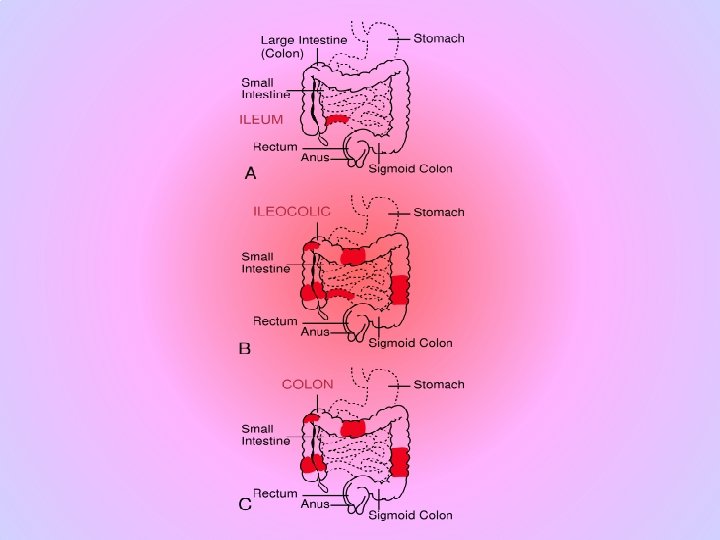
Crohn's disease is a chronic, episodic, inflammatory bowel disease (IBD) that affects any part of the entire wall of the bowel or intestines. Crohn's disease can affect any part of the gastrointestinal tract from mouth to anus; as a result, the symptoms of Crohn's disease vary among afflicted individuals. The disease is characterized by areas of inflammation with areas of normal lining between in a symptom known as skip lesions. The main gastrointestinal symptoms are abdominal pain, diarrhea (which may be bloody, though this may not be visible to the naked eye), constipation, vomiting, weight loss or weight gain. Crohn's disease can also cause complications outside of the gastrointestinal tract such as skin rashes, arthritis, and inflammation of the eye. [1]
Hiatus hernia A hiatus hernia or hiatal hernia is the protrusion (or herniation) of the upper part of the stomach through a tear or weakness in the diaphragm. The symptoms include acid reflux, and pain, similar to heartburn, in the chest and upper stomach. In most patients, hiatus hernias cause no symptoms. Sometimes patients experience heartburn and regurgitation, when stomach acid refluxes back into the esophagus.
The following are possible causes or contributing factors for having a hiatus hernia. Increased pressure within the abdomen caused by: Heavy lifting or frequent bending over Frequent or hard coughing Hard sneezing Violent vomiting Straining with constipation Obesity (extra weight pushes down on the abdomen increasing the pressure) Use of the sitting position for defecation (See epidemiology below) Heredity Smoking Drug use, such as cocaine. Stress
Coronary Diseases http: //yourtotalhealth. ivillage. com/heartattack. html
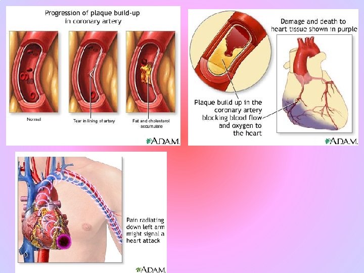
Atherosclerosis is a condition in which patchy deposits of fatty material (atheromas or atherosclerotic plaques) develop in the walls of medium-sized and large arteries, leading to reduced or blocked blood flow.
Atherosclerosis develops from low-density lipoprotein cholesterol (LDL), colloquially called "bad cholesterol". Many believe that, when this lipoprotein gets through the wall of an artery, oxygen free radicals react with it to form oxidized-LDL. [1] The body's immune system responds by sending specialised white blood cells (macrophages and T-lymphocytes) to absorb the oxidised-LDL. Unfortunately, these white blood cells are not able to process the oxidised-LDL, and ultimately grow then rupture, depositing a greater amount of oxidised cholesterol into the artery wall. This triggers more white blood cells, continuing the cycle. http: //www. macneal. com/cardiacriskcalculatorstart. aspx? gclid=CIC 855 i 98 Zg. CFQI Mswodx 2 Mp 1 w http: //www. youtube. com/watch? v=N_Lu. JGobrpw
The two most important oxygen-centered free radicals are superoxide and hydroxyl radical. They are derived from molecular oxygen under reducing conditions. However, because of their reactivity, these same free radicals can participate in unwanted side reactions resulting in cell damage. Many forms of cancer are thought to be the result of reactions between free radicals and DNA, resulting in mutations that can adversely affect the cell cycle and potentially lead to malignancy. Some of the symptoms of aging such as atherosclerosis are also attributed to free-radical induced oxidation of many of the chemicals making up the body. In addition free radicals contribute to alcohol-induced liver damage, perhaps more than alcohol itself. Radicals in cigarette smoke have been implicated in inactivation of alpha 1 -antitrypsin in the lung. This process promotes the development of emphysema.
1. Atherosclerosis is caused by repeated injury to the walls of arteries. 2. Many factors contribute to this injury, including high blood pressure, tobacco smoke, diabetes and high levels of cholesterol in the blood. 3. Often, the first symptom is pain or cramps at times when blood flow cannot keep up with the tissues‘ need for oxygen. 4. To prevent atherosclerosis, people need to stop using tobacco, improve their diet, exercise regularly, and maintain control of their blood pressure and diabetes. 5. If atherosclerosis causes complications, such as a heart attack or stroke, these are treated.
In the United States and most other developed countries, atherosclerosis is the leading cause of illness and death. Estimates for 2005 in the United States alone are that about 16 million people have atherosclerotic heart disease and 5. 8 million have stroke. Cardiovascular disease, primarily coronary and cerebrovascular atherosclerosis, caused almost 870, 000 deaths in 2005—almost twice as many as cancer caused and 9 times as many as injuries caused. This year an estimated 1. 2 million Americans will have a new or recurrent heart attack. http: //yourtotalhealth. ivillage. com/coronary-artery-bypass-surgery. html
To help prevent atherosclerosis, people need to stop tobacco use (see Coronary Artery Disease: Smoking), lower LDL cholesterol levels (see Cholesterol Disorders: Treatment), lower blood pressure (see High Blood Pressure: Drug Therapy), lose weight (see Obesity: Diet and Nutritional Counseling), and exercise (see Exercise and Fitness: Starting an Exercise Program). People who have diabetes must maintain strict control of their blood sugar (glucose). People who are at high risk for atherosclerosis also may benefit from taking certain drugs. Helpful drugs include the statins (even if cholesterol levels are normal or only slightly high) and aspirin. Some Trade Names ECOTRIN /ASPERGUM
1. Lumen (Opening) 2. Plaque 3. Artery Wall http: //www. youtube. com/watch? v=gv. Rt. P 3 wl_AY&feature=related
Atherosclerosis typically begins in early adolescence, and is usually found in most major arteries, yet is asymptomatic and not detected by most diagnostic methods during life. Autopsies of healthy young men that died during the Korean and Vietnam Wars showed evidence of the disease. It most commonly becomes seriously symptomatic when interfering with the coronary circulation supplying the heart or cerebral circulation supplying the brain, and is considered the most important underlying cause of strokes, heart attacks, various heart diseases including congestive heart failure, and most cardiovascular diseases, in general. Atheroma in arm, or, more often, leg arteries, and producing decreased blood flow, is called Peripheral artery occlusive disease (PAOD http: //yourtotalhealth. ivillage. com/plaque-rupture. html
Heart Attack Acute myocardial infarction (AMI or MI), more commonly known as a heart attack, is a medical condition that occurs when the blood supply to a part of the heart is interrupted, most commonly due to rupture of a vulnerable plaque.