
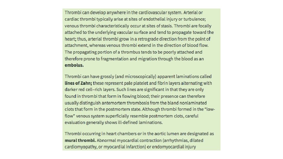
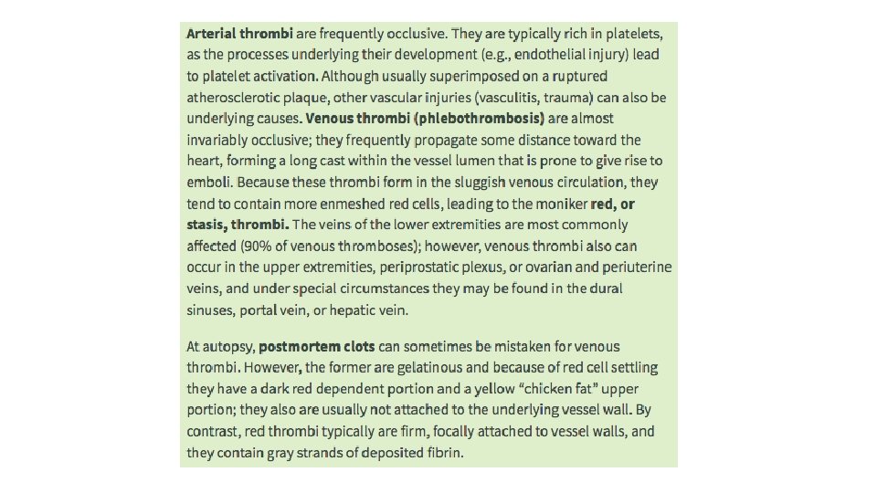
Thromboembolism and Shock Mural thrombi A Thrombus in

Thromboembolism, and Shock

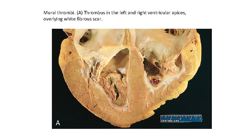
Mural thrombi. (A) Thrombus in the left and right ventricular apices, overlying white fibrous scar.
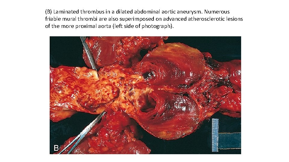
(B) Laminated thrombus in a dilated abdominal aortic aneurysm. Numerous friable mural thrombi are also superimposed on advanced atherosclerotic lesions of the more proximal aorta (left side of photograph).
Fate of the Thrombus If a patient survives an initial thrombotic event, during the ensuing days to weeks the thrombus evolves through some combination of the following four processes: Propagation. The thrombus enlarges through the accretion of additional platelets and fibrin, increasing the odds of vascular occlusion or embolization. Embolization. Part or all of the thrombus is dislodged and transported elsewhere in the vasculature. Dissolution. If a thrombus is newly formed, activation of fibrinolytic factors may lead to its rapid shrinkage and complete dissolution. With older thrombi, extensive fibrin polymerization renders the thrombus substantially more resistant to plasmin-induced proteolysis, and lysis is ineffectual. This acquisition of resistance to lysis has clinical significance, as therapeutic administration of fibrinolytic agents (e. g. , t-PA in the setting of acute coronary thrombosis) generally is not effective unless administered within a few hours of thrombus formation.
Organization and recanalization. Older thrombi become organized by the ingrowth of endothelial cells, smooth muscle cells, and fibroblasts. In time, capillary channels are formed that—to a limited extent—create conduits along the length of the thrombus, thereby reestablishing the continuity of the original lumen. Further recanalization can sometimes convert a thrombus into a vascularized mass of connective tissue that is eventually incorporated into the wall of the remodeled vessel. Occasionally, instead of organizing, the center of a thrombus undergoes enzymatic digestion, presumably because of the release of lysosomal enzymes from entrapped leukocytes. If bacterial seeding occurs, the contents of degraded thrombi serve as an ideal culture medium, and the resulting infection may weaken the vessel wall, leading to the formation of a mycotic aneurysm Clinical Features Thrombi are significant because they cause obstruction of arteries and veins and may give rise to emboli. The effect that is of the greatest clinical importance depends on the site of thrombosis. Thus, although venous thrombi can cause congestion and edema in vascular beds distal to an obstruction, they are most worrisome because of their potential to embolize to the lungs and cause death. Conversely, whereas arterial thrombi can embolize and cause tissue infarction, their tendency to obstruct vessels (e. g. , in coronary and cerebral vessels) is considerably more important.

An organized thrombus. Low-power view of a thrombosed artery stained for elastic tissue. The original lumen is delineated by the internal elastic lamina (arrows) and is completely filled with organized thrombus

Venous Thrombosis (Phlebothrombosis) Most venous thrombi occur in the superficial or the deep veins of the leg. Superficial venous thrombi usually arise in the saphenous system, particularly in the setting of varicosities; these rarely embolize but they can be painful and can cause local congestion and swelling from impaired venous outflow, predisposing the overlying skin to the development of infections and varicose ulcers. Deep venous thromboses (DVTs) in the larger leg veins at or above the knee joint (e. g. , popliteal, femoral, and iliac veins) are more serious because they are prone to embolize. Although such DVTs may cause local pain and edema, collateral channels often circumvent the venous obstruction. Consequently, DVTs are entirely asymptomatic in approximately 50% of patients and are recognized only after they have embolized to the lungs.
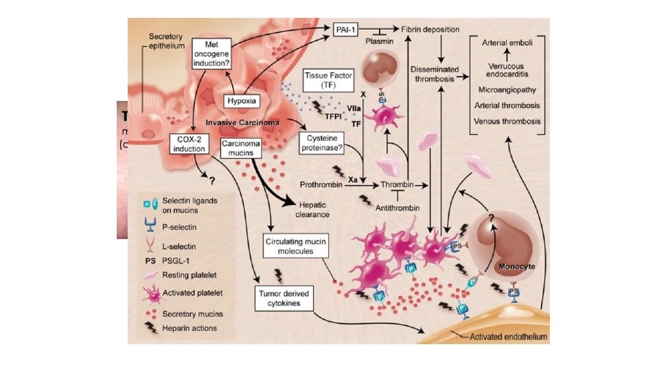
Lower-extremity DVTs are associated with stasis and hypercoagulable states. Thus, common predisposing factors include congestive heart failure, bed rest, and immobilization; the latter two factors reduce the milking action of leg muscles and thus slow venous return. Trauma, surgery, and burns not only immobilize a patient but also are associated with vascular injury, procoagulant release, increased hepatic synthesis of coagulation factors, and reduced t-PA production. Many factors contribute to the thrombotic diathesis of pregnancy; in addition to the potential for amniotic fluid infusion into the circulation at the time of delivery, pressure produced by the enlarging fetus and uterus can produce stasis in the veins of the legs, and late pregnancy and the postpartum period are associated with hypercoagulability. Tumor-associated procoagulant release is largely responsible for the increased risk of thromboembolic phenomena seen in disseminated cancers. These are sometimes referred to as migratory thrombophlebitis, because of the tendency to involve several different venous beds transiently, or as Trousseau syndrome, for the physician who both described the disorder and suffered from it. Regardless of the specific clinical setting, the risk of DVT is increased in persons older than 50 years.
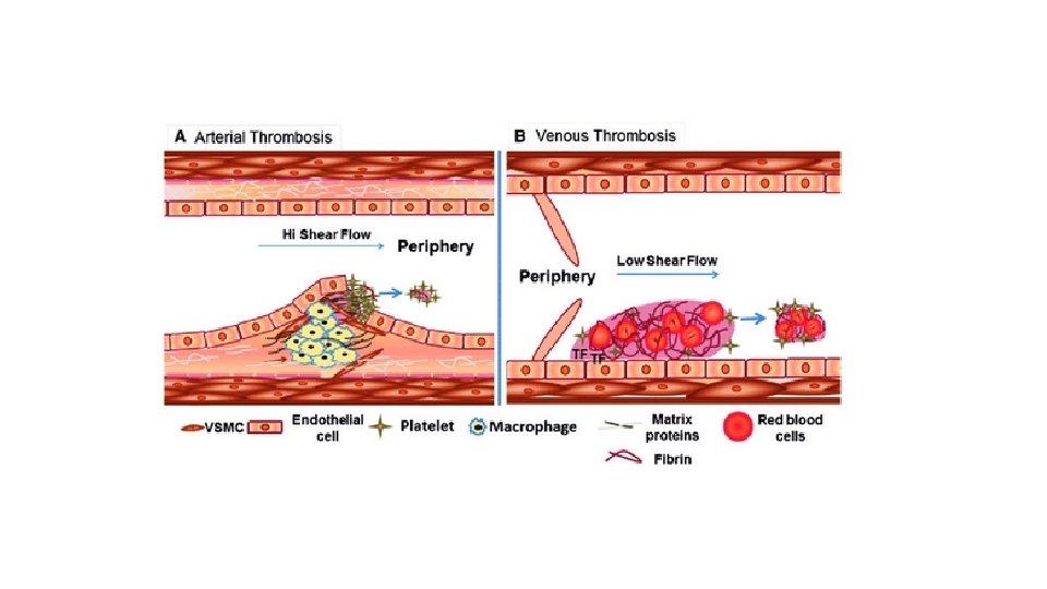
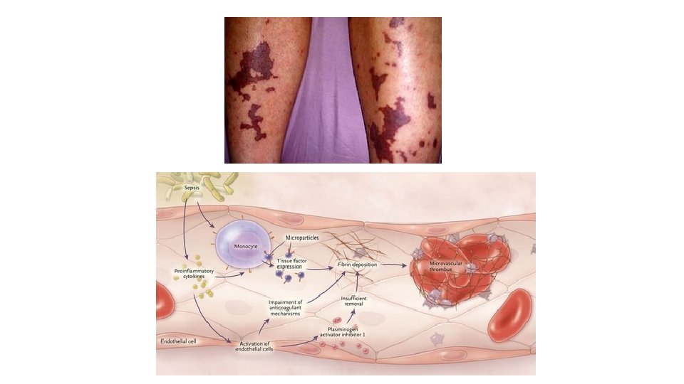
Arterial and Cardiac Thrombosis Atherosclerosis is a major cause of arterial thromboses because it is associated with the loss of endothelial integrity and with abnormal blood flow. Myocardial infarction can predispose to cardiac mural thrombi by causing dyskinetic myocardial contraction and endocardial injury, and rheumatic heart disease may engender atrial mural thrombi by causing atrial dilation and fibrillation. Both cardiac and aortic mural thrombi are prone to embolization. Although any tissue can be affected, the brain, kidneys, and spleen are particularly likely targets because of their rich blood supply. Disseminated Intravascular Coagulation (DIC) DIC is widespread thrombosis within the microcirculation that may be of sudden or insidious onset. It may be seen in disorders ranging from obstetric complications to advanced malignancy. To complicate matters, the widespread microvascular thrombosis consumes platelets and coagulation proteins (hence the synonym consumptive coagulopathy), and at the same time, fibrinolytic mechanisms are activated. The net result is that excessive clotting and bleeding may co-exist in the same patient.
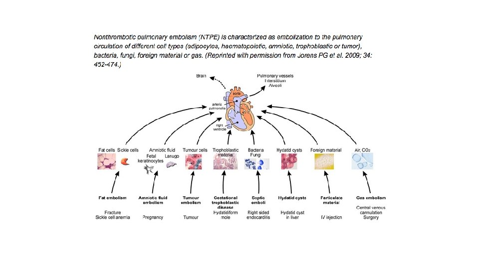
Embolism An embolus is a detached intravascular solid, liquid, or gaseous mass that is carried by the blood from its point of origin to a distant site, where it often causes tissue dysfunction or infarction. Pulmonary emboli originate from deep venous thromboses and are responsible for the most common form of thromboembolic disease. The incidence of pulmonary embolism (PE) is 2 to 4 per 1000 hospitalized patients. In more than 95% of cases, venous emboli originate from thrombi within deep leg veins proximal to the popliteal fossa; embolization from lower leg thrombi is uncommon. Fragmented thrombi from DVTs are carried through progressively larger channels and usually pass through the right side of the heart before arresting in the pulmonary vasculature. Depending on size, a PE can occlude the main pulmonary artery, lodge at the bifurcation of the right and left pulmonary arteries (saddle embolus), or pass into the smaller, branching arterioles. Frequently, multiple emboli occur, either sequentially or as a shower of smaller emboli from a single large thrombus; a patient who has had one pulmonary embolus is at increased risk for having more. Rarely, an embolus passes through an atrial or ventricular defect and enters the systemic circulation (paradoxical embolism).
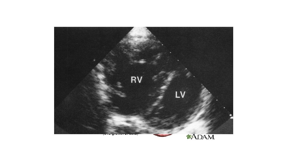
Most pulmonary emboli (60%– 80%) are small and clinically silent. With time, they undergo organization and become incorporated into the vascular wall; in some cases, organization of thromboemboli leaves behind bridging fibrous webs. At the other end of the spectrum, a large embolus that blocks a major pulmonary artery can cause sudden death. Embolic obstruction of medium-sized arteries and subsequent rupture of downstream capillaries rendered anoxic can cause pulmonary hemorrhage. Such emboli do not usually cause pulmonary infarction because the area also receives blood through an intact bronchial circulation (dual circulation). However, a similar embolus in the setting of left-sided cardiac failure (and diminished bronchial artery perfusion) can lead to a pulmonary infarct. Embolism to small end-arteriolar pulmonary branches usually causes infarction. Multiple emboli occurring through time can cause pulmonary hypertension and right ventricular failure (cor pulmonale).
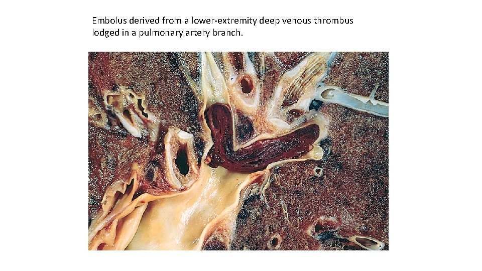
Embolus derived from a lower-extremity deep venous thrombus lodged in a pulmonary artery branch.
Systemic Thromboembolism Most systemic emboli (80%) arise from intracardiac mural thrombi; twothirds of these are associated with left ventricular infarcts and another 25% with dilated left atria (e. g. , secondary to mitral valve disease). The remainder originate from aortic aneurysms, thrombi overlying ulcerated atherosclerotic plaques, fragmented valvular vegetations , or the venous system (paradoxical emboli); 10% to 15% of systemic emboli are of unknown origin. By contrast with venous emboli, which lodge primarily in the lung, arterial emboli can travel virtually anywhere; their final resting place understandably depends on their point of origin and the relative flow rates of blood to the downstream tissues. Common arteriolar embolization sites include the lower extremities (75%) and central nervous system (10%); intestines, kidneys, and spleen are less common targets. The consequences of embolization depend on the caliber of the occluded vessel, the collateral supply, and the affected tissue’s vulnerability to anoxia; arterial emboli often lodge in end arteries and cause infarction.
Fat Embolism Soft tissue crush injury or rupture of marrow vascular sinusoids (eg, due to a long bone fracture) release microscopic fat globules into the circulation. Fat and marrow emboli are common incidental findings after vigorous cardiopulmonary resuscitation but probably are of little clinical significance. Similarly, although fat and marrow embolism occurs in some 90% of individuals with severe skeletal injuries, less than 10% show any clinical findings. However, a minority of patients develop a symptomatic fat embolism syndrome characterized by pulmonary insufficiency, neurologic symptoms, anemia, thrombocytopenia, and a diffuse petechial rash that is fatal in 10% of cases. Clinical signs and symptoms appear 1 to 3 days after injury as the sudden onset of tachypnea, dyspnea, tachycardia, irritability, and restlessness, which can progress rapidly to delirium or coma. Thrombocytopenia is attributed to platelet adhesion to fat globules and subsequent aggregation or splenic sequestration; anemia can result from similar red cell aggregation and/or hemolysis. A diffuse petechial rash (seen in 20%– 50% of cases) is related to rapid onset of thrombocytopenia and can be a useful diagnostic feature.
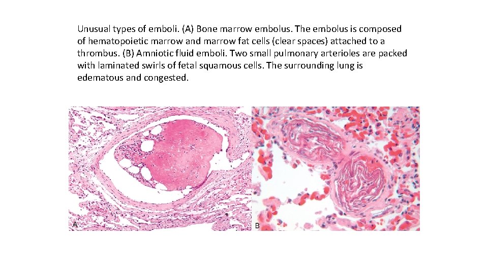
Unusual types of emboli. (A) Bone marrow embolus. The embolus is composed of hematopoietic marrow and marrow fat cells (clear spaces) attached to a thrombus. (B) Amniotic fluid emboli. Two small pulmonary arterioles are packed with laminated swirls of fetal squamous cells. The surrounding lung is edematous and congested.
Amniotic Fluid Embolism Amniotic fluid embolism is an uncommon, grave complication of labor and the immediate postpartum period occurring in 1 in 40, 000 deliveries. The mortality rate approaches 80%, making it the most common cause of maternal death in the developed world and the fifth most common cause of maternal death in the United States, accounting for 10% of maternal deaths in this country; 85% of survivors suffer some form of permanent neurologic deficit. Onset is characterized by sudden severe dyspnea, cyanosis, and hypotensive shock, followed by seizures and coma. If the patient survives the initial crisis, pulmonary edema typically develops, along with (in about half the patients) disseminated intravascular coagulation secondary to release of thrombogenic substances from amniotic fluid. Indeed it is thought that morbidity and mortality in such cases results not from mechanical obstruction of pulmonary vessels but from biochemical activation of the coagulation system and the innate immune system caused by substances in the amniotic fluid. The underlying cause is the entry of amniotic fluid (and its contents) into the maternal circulation via tears in the placental membranes and/or uterine vein rupture. Histologic analysis shows squamous cells shed from fetal skin, lanugo hair, fat from vernix caseosa, and mucin derived from the fetal respiratory or gastrointestinal tracts in the maternal pulmonary microcirculation. Other findings include marked pulmonary edema, diffuse alveolar damage, and systemic fibrin thrombi generated by disseminated intravascular coagulation.
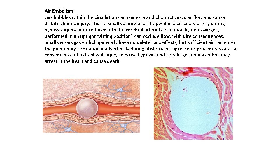
Air Embolism Gas bubbles within the circulation can coalesce and obstruct vascular flow and cause distal ischemic injury. Thus, a small volume of air trapped in a coronary artery during bypass surgery or introduced into the cerebral arterial circulation by neurosurgery performed in an upright “sitting position” can occlude flow, with dire consequences. Small venous gas emboli generally have no deleterious effects, but sufficient air can enter the pulmonary circulation inadvertently during obstetric or laproscopic procedures or as a consequence of a chest wall injury to cause hypoxia, and very large venous emboli may arrest in the heart and cause death.
A particular form of gas embolism called decompression sickness is caused by sudden changes in atmospheric pressure. Scuba divers, underwater construction workers, and persons in unpressurized aircraft who undergo rapid ascent are at risk. When air is breathed at high pressure (e. g. , during a deep sea dive), increased amounts of gas (particularly nitrogen) become dissolved in the blood and tissues. If the diver then ascends (depressurizes) too rapidly, the nitrogen expands in the tissues and bubbles out of solution in the blood to form gas emboli, which cause tissue ischemia. Rapid formation of gas bubbles within skeletal muscles and supporting tissues in and about joints is responsible for the painful condition called the bends (so named in the 1880 s because the afflicted person arches the back in a manner reminiscent of a then -popular women’s fashion pose called the Grecian bend). Gas bubbles in the pulmonary vasculature cause edema, hemorrhages, and focal atelectasis or emphysema, leading to respiratory distress, the so-called “chokes”. Bubbles in the central nervous system can cause mental impairment and even sudden onset of coma. A more chronic form of decompression sickness is called caisson disease (named for pressurized underwater vessels used during bridge construction), in which recurrent or persistent gas emboli in the bones lead to multifocal ischemic necrosis; the heads of the femurs, tibiae, and humeri are most commonly affected.
Infarction An infarct is an area of ischemic necrosis caused by occlusion of the vascular supply to the affected tissue. Infarction primarily affecting the heart and the brain is a common and extremely important cause of clinical illness. Roughly 40% of all deaths in the United States are a consequence of cardiovascular disease, with most of these deaths stemming from myocardial or cerebral infarction. Pulmonary infarction is a common clinical complication, bowel infarction often is fatal, and ischemic necrosis of distal extremities (gangrene) causes substantial morbidity in the diabetic population. Arterial thrombosis or arterial embolism underlies the vast majority of infarctions. Less common causes of arterial obstruction include vasospasm, expansion of an atheroma secondary to intraplaque hemorrhage, and extrinsic compression of a vessel, such as by tumor, a dissecting aortic aneurysm, or edema within a confined space (e. g. , in anterior tibial compartment syndrome). Other uncommon causes of tissue infarction include vessel twisting (e. g. , in testicular torsion or bowel volvulus), traumatic vascular rupture, and entrapment in a hernia sac. Although venous thrombosis can cause infarction, the more common outcome is simply congestion; typically, bypass channels rapidly open to provide sufficient outflow to restore the arterial inflow. Infarcts caused by venous thrombosis thus usually occur only in organs with a single efferent vein (e. g. , testis or ovary).
Infarcts are classified based on their color (reflecting the amount of hemorrhage) and the presence or absence of microbial infection. Thus, infarcts may be either red (hemorrhagic) or white (anemic) and may be either septic or bland. Red infarcts occur (1) as a result of venous occlusions (such as in ovarian torsion); (2) in loose tissues (e. g. , lung) where blood can collect in infarcted zones; (3) in tissues with dual circulations such as lung and small intestine, where partial, albeit inadequate perfusion by collateral arterial supplies is typical; (4) in previously congested tissues (as a consequence of sluggish venous outflow); and (5) when flow is reestablished after infarction has occurred (e. g. , after angioplasty of an arterial obstruction).
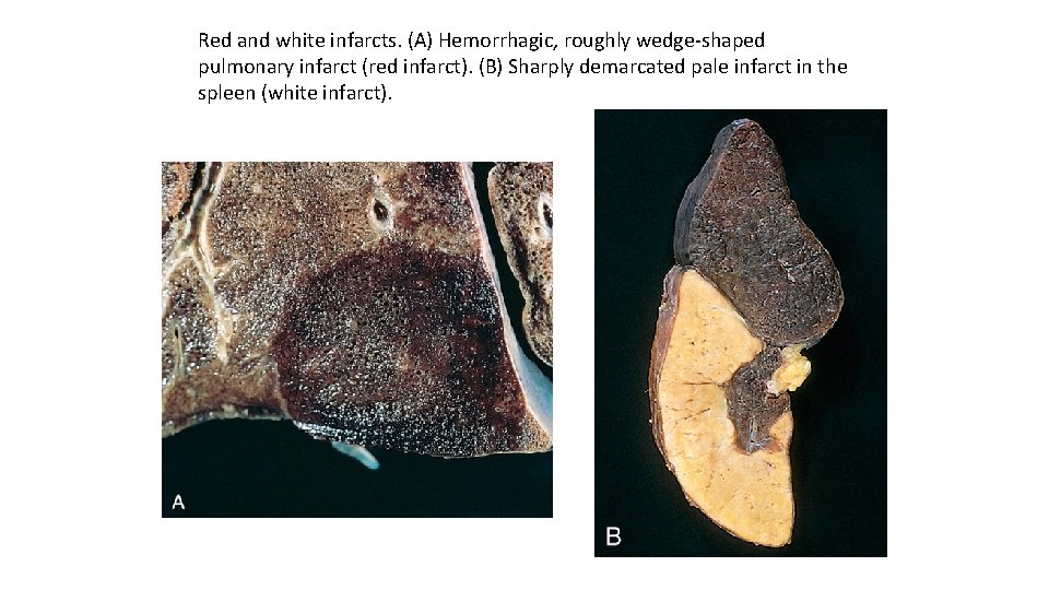
Red and white infarcts. (A) Hemorrhagic, roughly wedge-shaped pulmonary infarct (red infarct). (B) Sharply demarcated pale infarct in the spleen (white infarct).
White infarcts occur with arterial occlusions in solid organs with end-arterial circulations (e. g. , heart, spleen, and kidney), and where tissue density limits the seepage of blood from adjoining patent vascular beds. Infarcts tend to be wedge -shaped, with the occluded vessel at the apex and the organ periphery forming the base; when the base is a serosal surface, there is often an overlying fibrinous exudate. Lateral margins may be irregular, reflecting flow from adjacent vessels. The margins of acute infarcts typically are indistinct and slightly hemorrhagic; with time, the edges become better defined by a narrow rim of hyperemia attributable to inflammation. Infarcts resulting from arterial occlusions in organs without a dual circulation typically become progressively paler and more sharply defined with time. By comparison, hemorrhagic infarcts are the rule in the lung and other spongy organs. Extravasated red cells in hemorrhagic infarcts are phagocytosed by macrophages, and the heme iron is converted to intracellular hemosiderin. Small amounts do not impart any appreciable color to the tissue, but extensive hemorrhages leave a firm, brown residue.
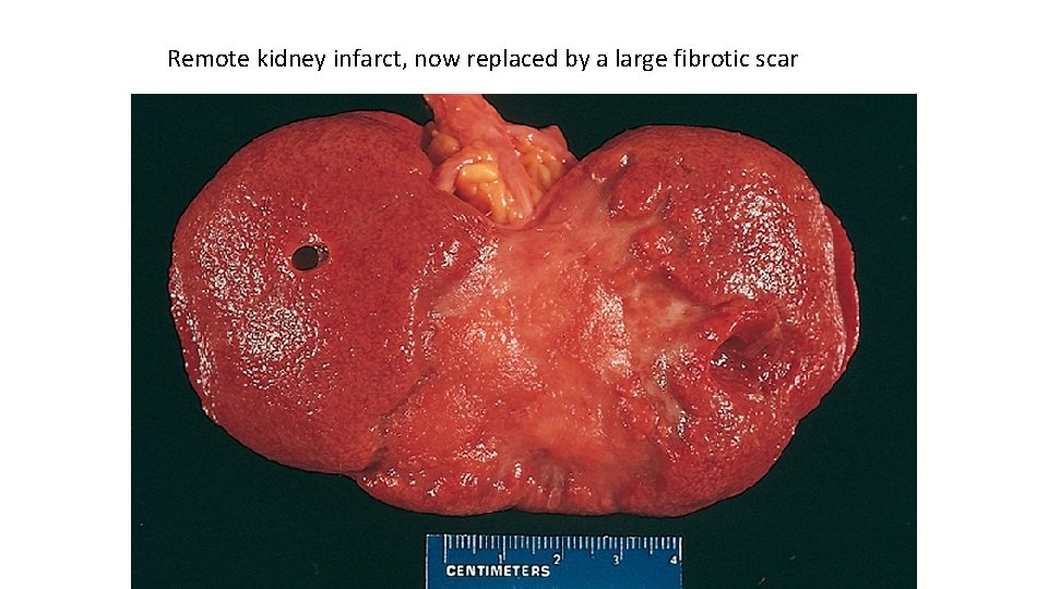
Remote kidney infarct, now replaced by a large fibrotic scar
In most tissues, the main histologic finding associated with infarcts is ischemic coagulative necrosis. An inflammatory response begins to develop along the margins of infarcts within a few hours and usually is well defined within 1 to 2 days. Eventually, inflammation is followed by repair, beginning in the preserved margins. In some tissues, parenchymal regeneration can occur at the periphery of the infarct, where the underlying stromal architecture has been spared. Most infarcts, however, are ultimately replaced by scar. The brain is an exception to these generalizations; ischemic tissue injury in the central nervous system results in liquefactive necrosis Septic infarctis occur when infected cardiac valve vegetations embolize, or when microbes seed necrotic tissue. In these cases the infarct is converted into an abscess, with a correspondingly greater inflammatory response and healing by organization and fibrosis The effects of vascular occlusion range from inconsequential to tissue necrosis leading to organ dysfunction and sometimes death. The range of outcomes is influenced by the following three variables:
Anatomy of the vascular supply The presence or absence of an alternative blood supply is the most important factor in determining whether occlusion of an individual vessel causes damage. The dual supply of the lung by the pulmonary and bronchial arteries means that obstruction of the pulmonary arterioles does not cause lung infarction unless the bronchial circulation also is compromised. Similarly, the liver, which receives blood from the hepatic artery and the portal vein, and the hand forearm, with its parallel radial and ulnar arterial supply, are resistant to infarction. By contrast, the kidney and the spleen both have end-arterial circulations, and arterial obstruction generally leads to infarction in these tissues.
Rate of occlusion. Slowly developing occlusions are less likely to cause infarction because they allow time for the development of collateral blood supplies. For example, small interarteriolar anastomoses, which normally carry minimal blood flow, interconnect the three major coronary arteries. If one coronary artery is slowly occluded (e. g. , by encroaching atherosclerotic plaque), flow in this collateral circulation may increase sufficiently to prevent infarction—even if the original artery becomes completely occluded. Tissue vulnerability to hypoxia. Neurons undergo irreversible damage when deprived of their blood supply for only 3 to 4 minutes. Myocardial cells, although hardier than neurons, still die after only 20 to 30 minutes of ischemia. By contrast, fibroblasts within myocardium remain viable after many hours of ischemia.
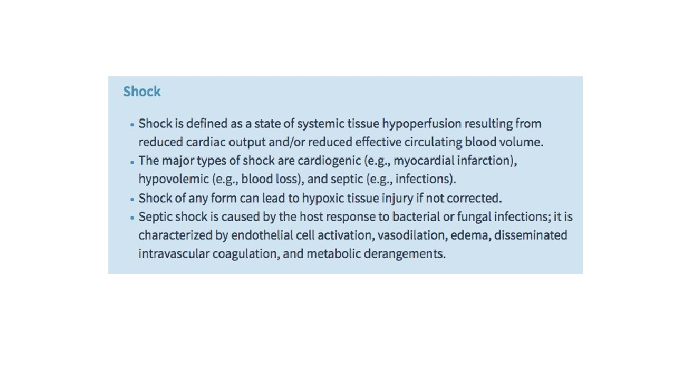
Shock is a state in which diminished cardiac output or reduced effective circulating blood volume impairs tissue perfusion and leads to cellular hypoxia. At the outset, the cellular injury is reversible; however, prolonged shock eventually leads to irreversible tissue injury and is often fatal. Shock may complicate severe hemorrhage, extensive trauma or burns, myocardial infarction, pulmonary embolism, and microbial sepsis. Cardiogenic shock results from low cardiac output as a result of myocardial pump failure. It may be caused by myocardial damage (infarction), ventricular arrhythmias, extrinsic compression (cardiac tamponade) , or outflow obstruction (e. g. , pulmonary embolism).
Hypovolemic shock results from low cardiac output due to loss of blood or plasma volume (e. g. , resulting from hemorrhage or fluid loss from severe burns). Septic shock is triggered by microbial infections and is associated with severe systemic inflammatory response syndrome (SIRS). In addition to microbes, SIRS may be triggered by a variety of insults, including burns, trauma, and/or pancreatitis. The common pathogenic mechanism is a massive outpouring of inflammatory mediators from innate and adaptive immune cells that produce arterial vasodilation, vascular leakage, and venous blood pooling. These cardiovascular abnormalities result in tissue hypoperfusion, cellular hypoxia, and metabolic derangements that lead to organ dysfunction and, if severe and persistent, organ failure and death.
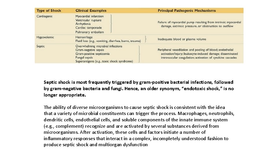
Septic shock is most frequently triggered by gram-positive bacterial infections, followed by gram-negative bacteria and fungi. Hence, an older synonym, “endotoxic shock, ” is no longer appropriate. The ability of diverse microorganisms to cause septic shock is consistent with the idea that a variety of microbial constituents can trigger the process. Macrophages, neutrophils, dendritic cells, endothelial cells, and soluble components of the innate immune system (e. g. , complement) recognize and are activated by several substances derived from microorganisms. After activation, these cells and factors initiate a number of inflammatory responses that interact in a complex, incompletely understood fashion to produce septic shock and multiorgan dysfunction
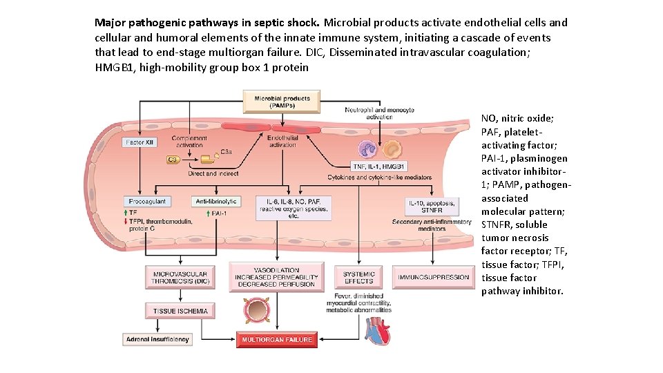
Major pathogenic pathways in septic shock. Microbial products activate endothelial cells and cellular and humoral elements of the innate immune system, initiating a cascade of events that lead to end-stage multiorgan failure. DIC, Disseminated intravascular coagulation; HMGB 1, high-mobility group box 1 protein NO, nitric oxide; PAF, plateletactivating factor; PAI-1, plasminogen activator inhibitor 1; PAMP, pathogenassociated molecular pattern; STNFR, soluble tumor necrosis factor receptor; TF, tissue factor; TFPI, tissue factor pathway inhibitor.
Inflammatory and counterinflammatory responses. In sepsis, various microbial cell wall constituents engage receptors on cells of the innate immune system, triggering proinflammatory responses. Likely initiators of inflammation in sepsis are signaling pathways that lie downstream of Toll-like receptors (TLRs), which recognize a host of microbe-derived substances containing socalled “pathogen-associated molecular patterns” (PAMPs), as well as G-protein–coupled receptors that detect bacterial peptides, and C-type lectin receptors such as Dectins. On activation, innate immune cells produce numerous cytokines, including TNF, IL-1, IFN-γ, IL-12, and IL-18, as well as other inflammatory mediators such as high-mobility group box 1 protein (HMGB 1). Markers of acute inflammation such as C-reactive protein and procalcitonin are also elevated. The latter is a clinically useful indicator of septic shock.
Reactive oxygen species and lipid mediators such as prostaglandins and platelet-activating factor (PAF) are also elaborated. These effector molecules induce endothelial cells (and other cell types) to upregulate adhesion molecule expression and further stimulate cytokine and chemokine production. The complement cascade is also activated by microbial components, both directly and through the proteolytic activity of plasmin resulting in the production of anaphylotoxins (C 3 a, C 5 a), chemotactic fragments (C 5 a), and opsonins (C 3 b), all of which contribute to the proinflammatory state. In addition, microbial components can activate coagulation directly through factor XII and indirectly through altered endothelial function. The accompanying widespread activation of thrombin may further augment inflammation by triggering protease activated receptors on inflammatory cells.
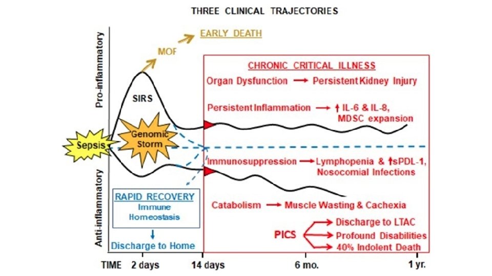
The hyperinflammatory state, initiated by sepsis, triggers counterregulatory immunosuppressive mechanisms, which may involve both innate and adaptive immune cells. As a result, septic patients may oscillate between hyperinflammatory and immunosuppressed states during their clinical course. Proposed mechanisms for the immune suppression include a shift from proinflammatory (TH 1) to antiinflammatory (TH 2) cytokines, production of anti-inflammatory mediators (e. g. , soluble TNF receptor, IL-1 receptor antagonist, and IL-10), lymphocyte apoptosis, the immunosuppressive effects of apoptotic cells, and the induction of cellular anergy. In some patients the counterregulatory mechanisms overshoot the inflammatory responses and the resultant immune suppression renders such patients susceptible to superinfections.
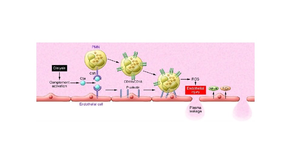
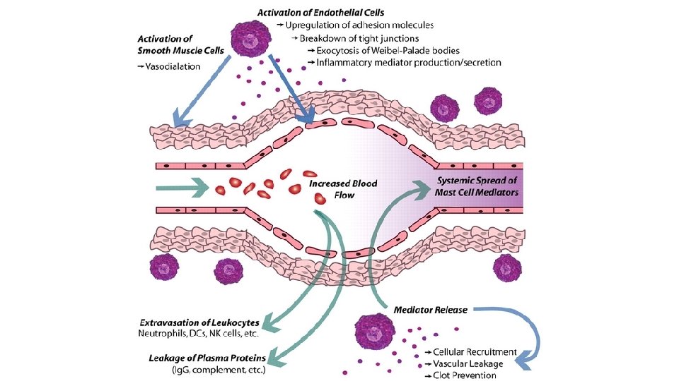
Endothelial activation and injury The proinflammatory state and endothelial cell activation associated with sepsis lead to widespread vascular leakage and tissue edema, which have deleterious effects on both nutrient delivery and waste removal. One effect of inflammatory cytokines is to loosen endothelial cell tight junctions, making vessels leaky and resulting in the accumulation of protein-rich edema fluid throughout the body. This alteration impedes tissue perfusion and may be exacerbated by attempts to support the patient with intravenous fluids. Activated endothelium also upregulates production of nitric oxide (NO) and other vasoactive inflammatory mediators (e. g. , C 3 a, C 5 a, and PAF), which may contribute to vascular smooth muscle relaxation and systemic hypotension.
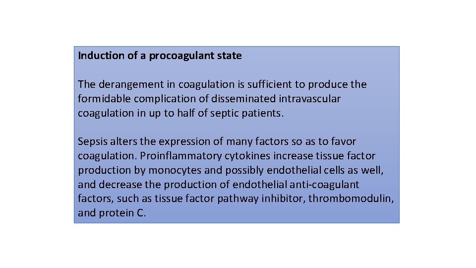
Induction of a procoagulant state The derangement in coagulation is sufficient to produce the formidable complication of disseminated intravascular coagulation in up to half of septic patients. Sepsis alters the expression of many factors so as to favor coagulation. Proinflammatory cytokines increase tissue factor production by monocytes and possibly endothelial cells as well, and decrease the production of endothelial anti-coagulant factors, such as tissue factor pathway inhibitor, thrombomodulin, and protein C.
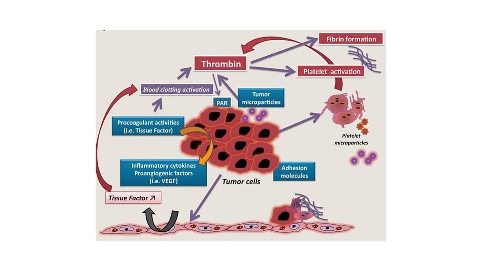
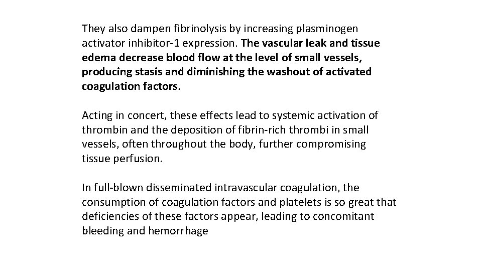
They also dampen fibrinolysis by increasing plasminogen activator inhibitor-1 expression. The vascular leak and tissue edema decrease blood flow at the level of small vessels, producing stasis and diminishing the washout of activated coagulation factors. Acting in concert, these effects lead to systemic activation of thrombin and the deposition of fibrin-rich thrombi in small vessels, often throughout the body, further compromising tissue perfusion. In full-blown disseminated intravascular coagulation, the consumption of coagulation factors and platelets is so great that deficiencies of these factors appear, leading to concomitant bleeding and hemorrhage
Metabolic abnormalities Septic patients exhibit insulin resistance and hyperglycemia. Cytokines such as TNF and IL-1, stress-induced hormones (such as glucagon, growth hormone, and glucocorticoids), and catecholamines all drive gluconeogenesis. At the same time, the proinflammatory cytokines suppress insulin release while simultaneously promoting insulin resistance in the liver and other tissues, likely by impairing the surface expression of GLUT-4, a glucose transporter. Hyperglycemia decreases neutrophil function—thereby suppressing bactericidal activity—and causes increased adhesion molecule expression on endothelial cells. Although sepsis is initially associated with an acute surge in glucocorticoid production, this phase may be followed by adrenal insufficiency and a functional deficit of glucocorticoids. This may stem from depression of the synthetic capacity of intact adrenal glands or frank adrenal necrosis resulting from disseminated intravascular dissemination (Waterhouse-Friderichsen syndrome). Finally, cellular hypoxia and diminished oxidative phosphorylation lead to increased lactate production and lactic acidosis.
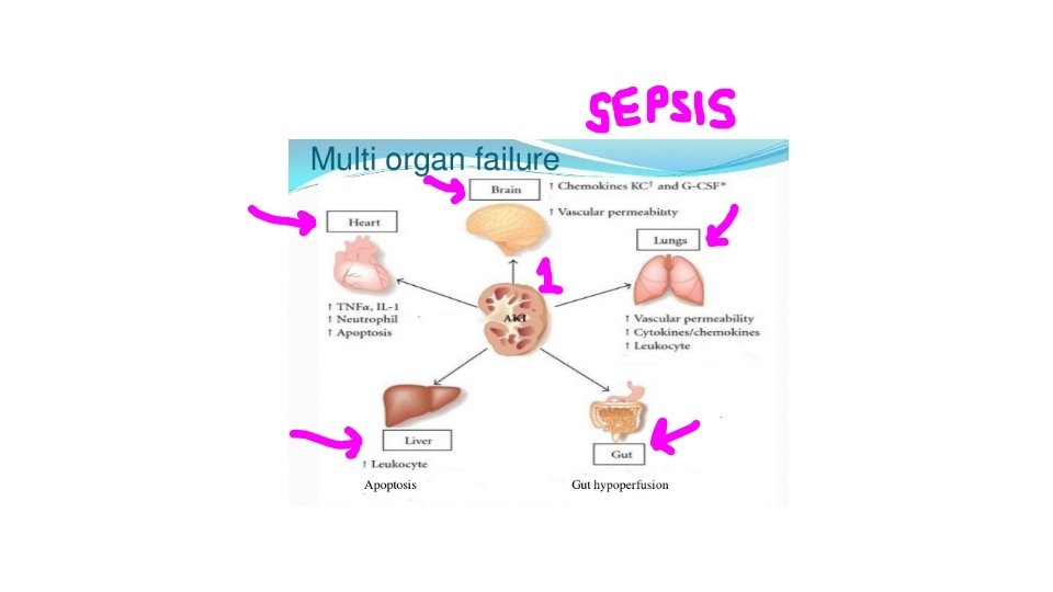
Organ dysfunction Systemic hypotension, interstitial edema, and small vessel thrombosis all decrease the delivery of oxygen and nutrients to the tissues that, because of cellular hypoxia, fail to properly use those nutrients that are delivered. Mitochondrial damage resulting from oxidative stress impairs oxygen use. High levels of cytokines and secondary mediators diminish myocardial contractility and cardiac output, and increased vascular permeability and endothelial injury can lead to the acute respiratory distress syndrome. Ultimately, these factors may conspire to cause the failure of multiple organs, particularly the kidneys, liver, lungs, and heart, culminating in death.
Stages of Shock is a progressive disorder that leads to death if the underlying problems are not corrected. The exact mechanisms of sepsis-related death are still unclear; aside from increased lymphocyte and enterocyte apoptosis, cellular necrosis is minimal. Death typically follows the failure of multiple organs, which usually offer no morphological clues to explain their dysfunction. For hypovolemic and cardiogenic shock, however, the pathways leading to a patient’s demise are reasonably well understood. Unless the insult is massive and rapidly lethal (e. g. , exsanguination from a ruptured aortic aneurysm), shock tends to evolve through three general (albeit somewhat artificial) stages. These stages have been documented most clearly in hypovolemic shock but are common to other forms as well
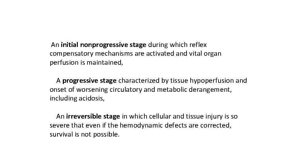
An initial nonprogressive stage during which reflex compensatory mechanisms are activated and vital organ perfusion is maintained, A progressive stage characterized by tissue hypoperfusion and onset of worsening circulatory and metabolic derangement, including acidosis, An irreversible stage in which cellular and tissue injury is so severe that even if the hemodynamic defects are corrected, survival is not possible.
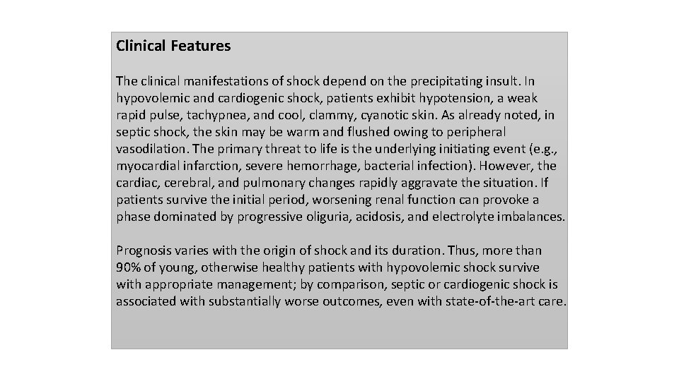
Clinical Features The clinical manifestations of shock depend on the precipitating insult. In hypovolemic and cardiogenic shock, patients exhibit hypotension, a weak rapid pulse, tachypnea, and cool, clammy, cyanotic skin. As already noted, in septic shock, the skin may be warm and flushed owing to peripheral vasodilation. The primary threat to life is the underlying initiating event (e. g. , myocardial infarction, severe hemorrhage, bacterial infection). However, the cardiac, cerebral, and pulmonary changes rapidly aggravate the situation. If patients survive the initial period, worsening renal function can provoke a phase dominated by progressive oliguria, acidosis, and electrolyte imbalances. Prognosis varies with the origin of shock and its duration. Thus, more than 90% of young, otherwise healthy patients with hypovolemic shock survive with appropriate management; by comparison, septic or cardiogenic shock is associated with substantially worse outcomes, even with state-of-the-art care.
- Slides: 55