Jugular Venous Pulse Dr Latchumanadhas Madras medical mission


 is due to atrial relaxation during")


 • It begins and")


: Early rapid ventricular")


 • Apply firm pressure to")


- Slides: 53
Jugular Venous Pulse Dr. Latchumanadhas Madras medical mission
History of JVP • • • Lancis : Venous pulse of EJV Chauvea, marey : Graphic recording of JVP Pontain : Wave pattern in JVP James Mackenzie : Nomenclature of JVP Paul wood : Hemodynamics of JVP
Jugular Venous System • Venous system contains 70 -80% of the circulating blood volumes. • Right atrial and right ventricular filling produce pulsations in the central veins that are transmitted to jugular veins. • An accurate assessment of the jugular venous pulse reflects the hemodynamics of the right sided heart. • Right atrial pressure during systole and right ventricular filling pressure during diastole are producing pulsations and pressure waves in jugular veins.
JVP Inspection
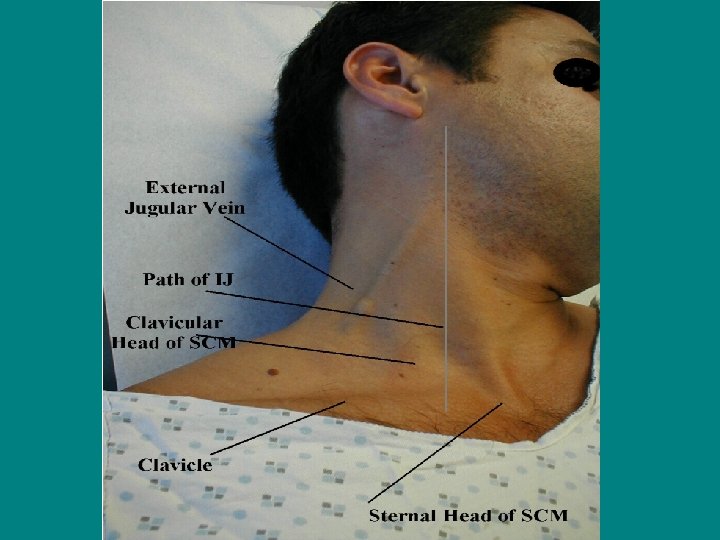
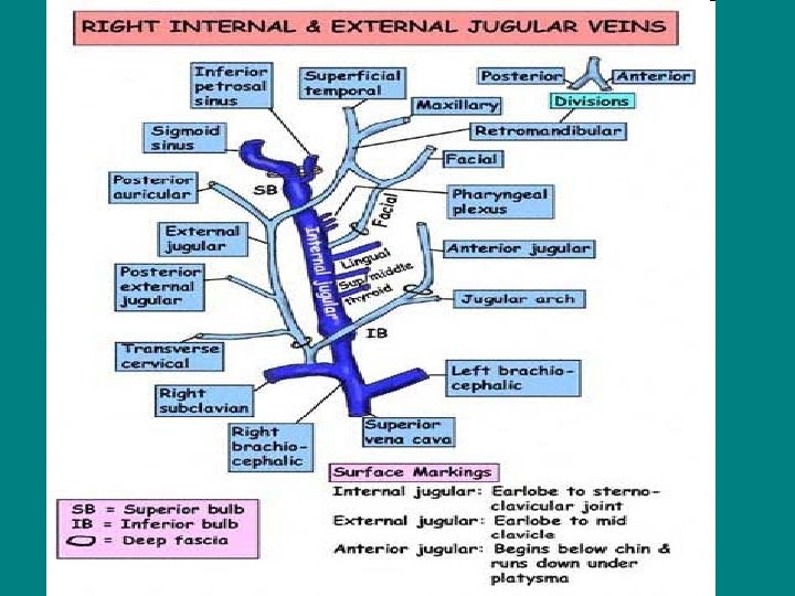
Exam of JVP • Right IJV is usually assessed both for waveform and estimation of CVP • Internal jugular venous pulsation located between two heads of sternocleidomastoid muscle are transmitted to overlying skin. • Right IJV is in direct continuity with SVC and right atrium • Left IJV drains into left innominate vein, which is not in straight line from RA • Right IJV and innominate vein is not compressed by adjacent structures
Right IJV Preferred : Why? • Direct continuation of right atrium • Straight line course through innominate vein to the svc and right atrium • IJV is less likely affected by extrinsic compression from other structures in neck • There are no or less numbers of valves in IJV than EJV • Less impact of vasoconstriction on IJV due to sympathetic activity than EJV
Differences between IJV and Carotid pulses • Superficial and lateral in the neck Deeper and medial in the neck • Better seen than felt Better felt than seen • Has two peaks and two troughs Has single upstroke only • Descents >obvious than crests Upstroke brisker and visible • Digital compression abolishes venous pulse Digital compression has no effect • Jugular venous pressure falls during inspiration Abdominal compression elevates jugular pressure Mean jugular venous pressure falls during standing Do not change with respiration • • Abdominal compression has no effect on carotid pulse Carotid pulse do not change when standing.
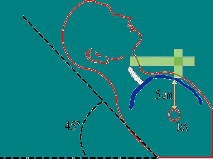
Position of Patient • Patient should lie comfortably and trunk is inclined by 45 degree position • Elevate chin and slightly rotate head to the left • Inclination angle should be subtended between trunk and bed , while neck and trunk should be in same line • When neck muscles are relaxed , shining the light tangentially over the skin and see pulsations • • In patients with low jugular pressure , a lesser (<30‘) inclination is desirable • In patients with high jugular pressure , a greater (60 -90‘) inclination is required to obtain visible pulsations • Simultaneous palpation of the left carotid artery and or apical impulse aids in timing of the venous pulsations in cardiac cycle.
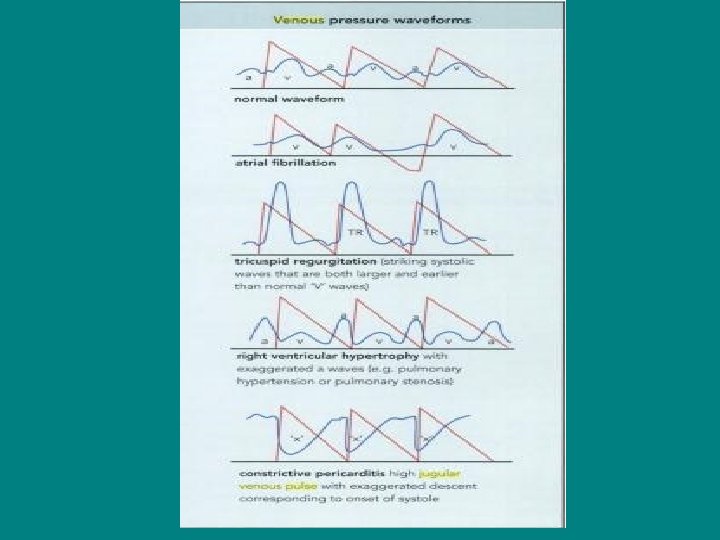
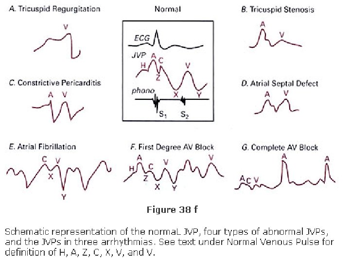
Normal JVP • Normal JVP reflects phasic pressure changes in RA during systole and RV during diastole • Two visible positive waves ( a and v) and two negative troughs ( x and y) • Two additional positive waves can be recorded. C wave interrupts x descent and h wave precedes the next a wave
Normal JVP Waveform • Consists of 3 positive waves – a, c & v • And 3 descents – x, x'(x prime) and y
a Wave • First positive presystolic a wave is due to right atrial contraction results in retrograde blood flow in to svc and jugulars • Effective RA contraction is needed for visible a wave • Dominant wave in JVP and larger than v • It precedes upstroke of the carotid pulse and S 1, but follow the P wave in ECG
x Descent • Systolic x descent (systolic collapse) is due to atrial relaxation during atrial diastole • X descent is most prominent motion of normal JVP which begins during systole and ends just before S 2 • It is larger than y descent • X descent more prominent during inspiration
c WAVE • Second positive wave recorded in JVP which interrupts the x descent • Produced by carotid artery impact on JVP upward bulging of closed TV into RA during isovolumic contraction
x` Descent • x`descent is systolic trough after c wave • Due to fall of right atrial pressure during early RV systole downward pulling of the TV by contracting right ventricle descent of RA floor by contracting RV
v Wave • Third positive wave in JVP which begins in late systole and ends in early diastole • Rise in RA pressure due to continued RA filling during ventricular systole when tricuspid valve closed • It is roughly synchronous with carotid upstroke and corresponds S 2.
y Descent • Diastolic collapse wave (down slope v wave) • It begins and ends during diastole well after S 2 • Decline of RA pressure due to RA emptying during early diastole when tricuspid valve opens • Initial y descent is corresponds to rapid RV filling and later part of y wave is produced by continued diastolic inflow in to RV.
h wave • Small brief positive wave following y descent just prior to a wave during period of diastasis • Described by Hieschfelder in 1907 • It usually seen when diastole is long (as in slow heart rates) • With increasing heart rate, y descent immediately followed by next a wave.
Prominent a Wave • Forceful atrial contraction when there is resistance to RA emptying or increased resistance to ventricular filling • RV inflow obstruction: Tricuspid stenosis or atresia RA mxyoma Decreased ventricular compliance: Pulmonary stenosis Pulmonary hypertension of any cause RV infarction RV cardiomyopathy (including HOCM) Acute pulmonary embolism
Cannon Waves • Whenever RA contracts against closed TV valve during RV systole : Systolic cannon wave. • Venous corrigan by paul wood • Regular cannon waves: Junctional rhythm VT with 1: 1 retrograde conduction Isorhythmic AV dissociation • Irregular cannon waves : Complete heart block Classic AV dissociation Ventricular pacing or ventricular ectopics.
Absent a Wave • When no effective atrial contraction as in atrial fibrillation • In atrial fibrillation , both atrial contraction as well as relaxation become in effective • In sinus tachycardia , when a wave may fuse with preceding v wave , especially when the PR interval is prolonged.
Abnormal x descent • Prominent x descent : Presence of atrial relaxation with intact tricuspid valve and good RV contraction is needed for prominent x descent. • Causes : Constrictive pericarditis Atrial septal defect
Absent x descent • Moderate to severe TR: EARLY SIGN • Obliteration of x descent with early build up of large systolic wave ( cv wave) • RV dysfunction due to PHT with RV failure severe RV infarction • Chronic atrial fibrrilation : loss of atrial relaxation
Prominent v wave • Increased RA volume during ventricular systole produce prominent v wave • Severe TR : giant v wave (Lanci sign) – ventricularization of atrial / jugular pressure • Giant v wave sometimes causes : systolic movement of ear lobe head bobbing with each systole systolic pulsation of liver pulsatile exophthalmos pistol shots heard over IJV
PROMINENT V WAVE • • • Tricuspid regurgitation ASD with mitral regurgitation VSD of LV to RA shunt (Gerbode's defect) Severe congestive heart failure Cor pumonale Atrial fibrillation
Rapid y Descent • Severe TR • C. Pericarditis (Friedreich's sign): Early rapid ventricular filling • PHT with RV dysfunction • Severe RV failure • ASD with mitral regurgitation • Severe RV infarction
Slow y Descent • When RA emptying and RV filling are impaired , y descent is slow and gradual Tricuspid stenosis Right atrial myxoma Pericardial tamponade( y descent may even be absent).
DD of Double Descents • Costrictive pericarditis • RV infarction with mild RV dysfunction • PHT with early RV decompensation DD of single Descent x descent only : cardiac tamponade y descent only : severe TR with RV dysfunction chronic AF with CCF EMF with TR
Estimation of Venous Pressure • Measuring jugular venous pressure • Hepatojugular reflux • Examining the veins on the dorsum of the hand • Examining the veins of the undersurface of the tongue • Assessment of jugular venous pressure at bed side reflect mean right atrial pressure
Measurement of JVP • Sternal angle or angle of Louis is a surface anatomical mark , reference point used for JVP measurement (Paul wood) • It is found approximately 5 cm above the center of the right atrium • Distance between sternal angle and center of right atrium remains relatively constant regardless of position of the thorax • Use sternal angle as zero reference point • Examine the patient in an optimal position(45`recline) and relaxed neck muscles , shining the light tangentially across the skin overlying IJV and see the top of oscillating venous column
Measurement of JVP • Two scale method is commonly used • A horizontal scale at the top of the oscillating venous column in IJV cuts the vertical scale at the sternal angle gives JV pressure in cm of water • Normally JV pressure does not exceed 4 cm above the sternal angle • Since RA is approximately 5 cm below the sternal angle , the jugular venous pressure (RA mean pressure) is corresponds to 9 cm • By way of conversion , normal mean JV pressure does not exceed 7 mm Hg (9 cm column of water / 1. 3 =6. 9) • Elevated JVP : JVP of >4 cm above sternal angle.
Elevated JVP • Increased RVP and reduced compliance: Pulmonary stenosis Pulmonary hypertension Right ventricular failure RV infarction • RV inflow impedance: Tricuspid stenosis / atresia RA myxoma Constrictive pericarditis
Elevated JVP • Circulatory overload : Renal failure Cirrhosis liver Excessive fluid administration • SVC obstruction • COPD
Kussmual's sign • Mean jugular venous pressure increases during inspiration as a result of impaired RV compliance. • Constrictive pericarditis • Severe right heart failure • RV infarction • Restrictive cardiomyopathy
Abdominal -Jugular Reflux • Hepatojugular reflux – Rondot (1898) • Apply firm pressure to periumbilical region for 10 -30 sec with patient lying comfortably and breathing quietly while observe JVP • Normally JV pressure rises transiently(15 sec) to < 3 cm while abdominal pressure is continued • If JV pressure remains elevated until abdominal pressure is continued: called as Positive AJR.
Positive AJR • • Incipient and or compensated RVF LVF with volume overload Tricuspid regurgitation False Positive AJR ( without CCF) COPD Systemic vasoconstriction Increased sympathetic tone Severe anaemia
Gaertner's method • Measurement of JVP by examining the veins on the dorsum of the hand • When patient sitting or lying at a 45‘ elevation , arm slowly and passively raised from dependant position until the vein collapses • Height of the limb above the level of sternal angle at which vein collapses represents the venous pressure • When venous pressure is normal , veins of hand collapse at the level of sternal angle
Cardiac tamponade • • • JVP is usually elevated a wave never prominent v and x wave is normal y descent is diminished or absent Kussmaul's sign usually negative
Constrictive pericarditis • • • JVP is elevated a wave is usually normal v wave is usually equal to a wave x descent –prominent y descent – rapid descent Kussmauls sign is usually positive
Endomyocardial fibrosis • • • JVP is usually elevated a wave is prominent v wave is prominent due to TR x descent is obliterated due to TR Rapid y descent is due to TR Kussmaul's sign is negative
Primary Pulmonary Hypertension • Normal RV compliance : normal JVP • Early RV decompensation : JVP may be elevated a wave is prominent and larger than v wave x and y descent seen and equal Decompensated RVF: JVP is always elevated a and v wave prominent , v wave larger than a wave x descent is diminished or absent rapid y descent due to TR
JVP in ASD • JVP is normal and equal a and v waves. x descent is more prominent. • Elevated JVP may seen in severe PAH and in RVF • Prominent a wave with PS and MS • Prominent v wave with PAH and in RVF with TR • Rapid y descent with RVF or TR
JVP in VSD • • Prominent a wave with severe PS Elevated JVP with CHF Prominent v wave with Gerbode's shunt In Eisenmenger complex : JVPressure usually normal Normal a and v waves CHF and TR is rare
Ebstein Anomaly • JVP is usually normal • Prominent a waves are seen only occasionally • Attenuated x descent and systolic v wave are not reflected in jugular pulse despite appreciable TR • Unimpressive JVP is attributed to damping effect of large capacitance RA and thin, toneless atrialized RV (Hypokinetic TR) • Prominent a and v / elevated JVP with advent of right ventricular failure.
Cyanosis with prominent a wave • It usually indicates intact IVS • Severe PS with intact IVS and Right to Left shunt • Tricuspid atresia • TAPVC with restrictive ASD • Eisenmenger syndrome with PDA • PPH with right to left shunt via PFO
Cyanosis with Elevated JVP • Elevated JVP in cyanotic CHD usually indicates intact IVS or increased PBF or both • Common in TGA or TAPVC with increased PBF • Severe PS with intact IVS and Right to Left shunt • Tricuspid atresia with restrictive ASD • Eisenmenger syndrome with ASD and PDA , JVP may be elevated • PPH with RVF and right to left shunt • Adult TOF
Cyanosis with normal JVP • It usually indicates cyanotic CHD with VSD and reduced PBF • Tetralogy of Fallot • TOF like physiology • VSD with right to left shunt • Pulmonary AV Fistula