Circulation and Blood vessels The Circulatory System Small

Circulation and Blood vessels

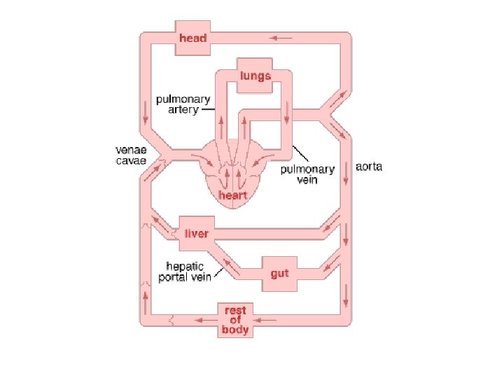
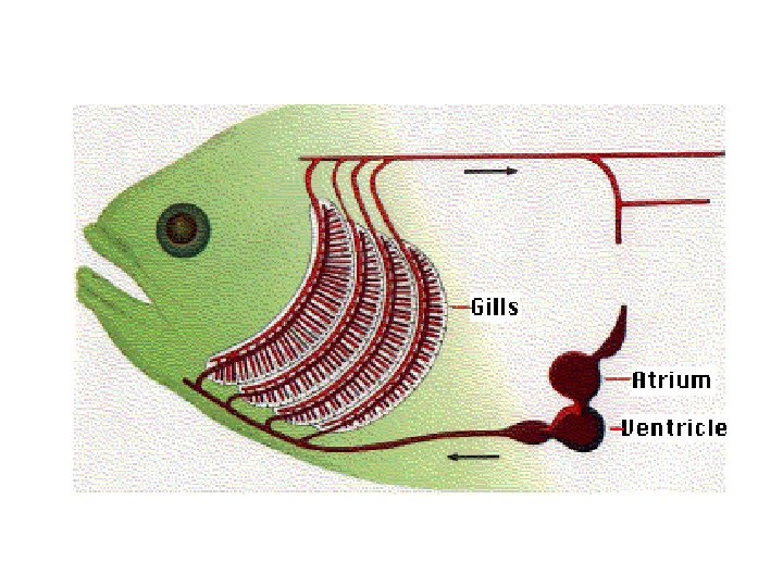
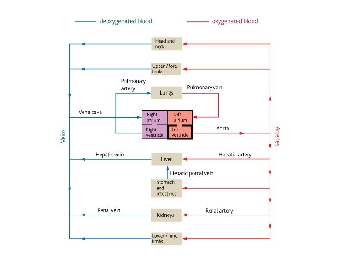
The Circulatory System • Small organisms don’t have a bloodstream, but instead rely on the simple diffusion of materials for transport around their cells. This is OK for single cells, but it would take days for molecules to diffuse through a large animal, so most animals have a circulatory system with a pump to transport materials quickly around their bodies. This is an example of a mass flow system, which means the transport of substances in the flow of a fluid (as opposed to diffusion, which is the random motion of molecules in a stationary fluid). The transport of materials in the xylem and phloem of plants is another example of mass flow. Mass flow systems work together with the specialised exchange systems (such as lungs). • Humans have a double circulatory system with a 4 -chambered heart. In humans the right side of the heart pumps blood to the lungs only and is called the pulmonary circulation, while the left side of the heart pumps blood to the rest of the body – the systemic circulation. • The circulation of blood round the body was discovered by William Harvey in 1628. Until then people assumed that blood ebbed and flowed through the same tubes, because they hadn't seen capillaries.
; Carotid (neck); Renal;")
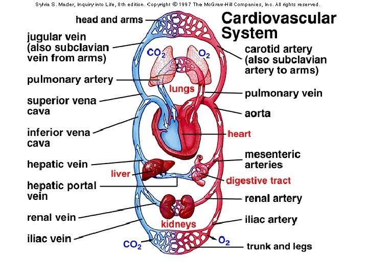
Major blood vessels • Arteries: Aorta; Pulmonary (↑CO 2, ↓O 2); Carotid (neck); Renal; Hepatic • Veins: Vena Cava; Pulmonary (↓CO 2, ↑O 2); Renal; Jugular; Hepatic Portal;

Insects don't have veins or arteries, but they do have circulatory systems. When blood is moved without the aid of vessels, the organism has an open circulatory system. Insect blood, properly called hemolymph, flows freely through the body cavity and makes direct contact with organs and tissues.
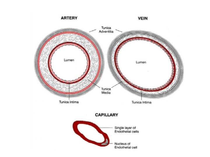

Blood Vessels Artery versus Vein • The wall of the artery is thicker: thicker connective tissue layer, thicker mixed layer of muscle and elastic tissue. • The lumen of the artery is much narrower. • Arteries do not have valves along their length, veins do. • Valves in the veins prevent the backflow of blood so the flow is in one correct direction towards the heart. • Blood flows away from the heart in arteries; blood flows towards the heart in veins. • Blood pressure in arteries is higher and so also the speed of blood flow • Pulsed flow in an artery, steady flow in a vein. • Many tissues, thus both are organs

Vein Artery Capillary For each blood vessel, label each layer and relate the structure to its function in the space underneath
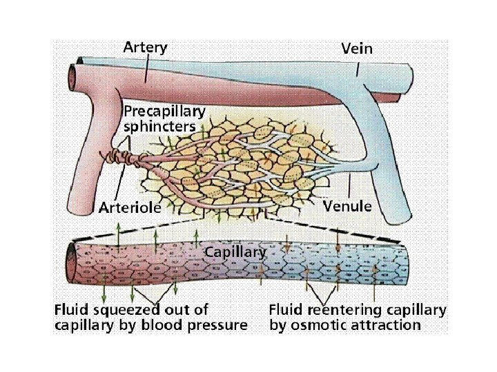

Formation of Tissue Fluid • As the blood enters the capillaries the high hydrostatic pressure forces some of the plasma out through the wall. • The escaped fluid (tissue fluid) is similar in composition to plasma, but lower in protein. • Therefore the remaining blood has a lower water potential • And a lower hydrostatic pressure • At the venule end the hydrostatic pressure is lower • Thus water returns to blood down water potential gradient. • The 10% excess tissue fluid must be drained away – by the lymphatic system.
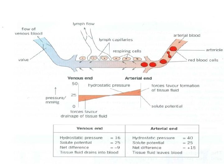
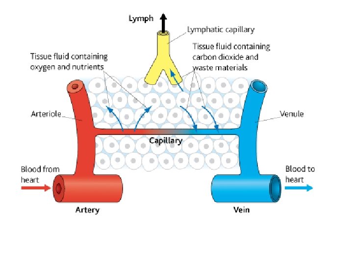

The Lymphatic System – • A collection of special drainage vessels receiving excess tissue fluid. – • Once the tissue fluid enters the lymphatic capillaries it is called lymph. – • Lymph nodes (e. g. tonsils) filter the lymph and produce lymphocytes. – • The lymph vessels have many valves, but low pressure. – • The lymph is moved along by the squeezing action of: • o the skeletal muscles, • o pressure changes in the thorax during breathing and • o by the rhythmic contraction of the lymph vessel walls. – • Lymph re-enters the blood just before the right atrium.

Functions of the Lymphatic System Circulatory role • Return the excess tissue fluid to the blood: this maintains blood volume, pressure and concentration. • Collect and deliver the absorbed lipids from the small intestine to the blood Defence role • The lymph nodes filter out pathogens in the lymph. • Production and ‘export’ of lymphocytes to the blood system for general distribution. • Detection of antigens and production of specific antibodies.
- Slides: 18