
Veins of lower limb and lymphatics By Prof
Veins of lower limb and lymphatics By Prof Dr Zubair/ Dr Kamran /Dr Sobia

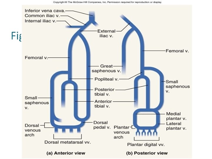
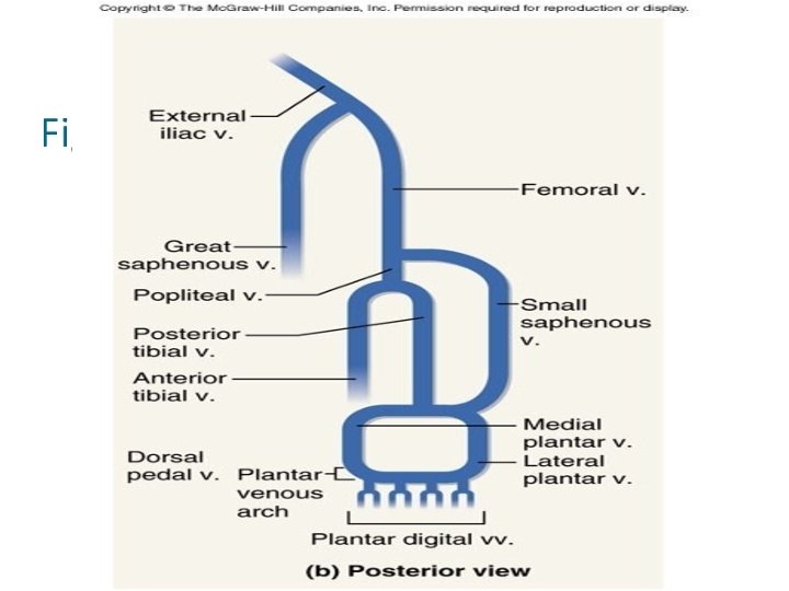
VEINS OF THE LOWER LIMBS � Blood from the lower limbs is drained by both superficial and deep veins. � The superficial veins often anastomose with one another and with deep veins along their length. Deep veins have the same names as corresponding arteries. � All veins of the lower limbs have valves, which are more numerous than in veins of the upper limbs.
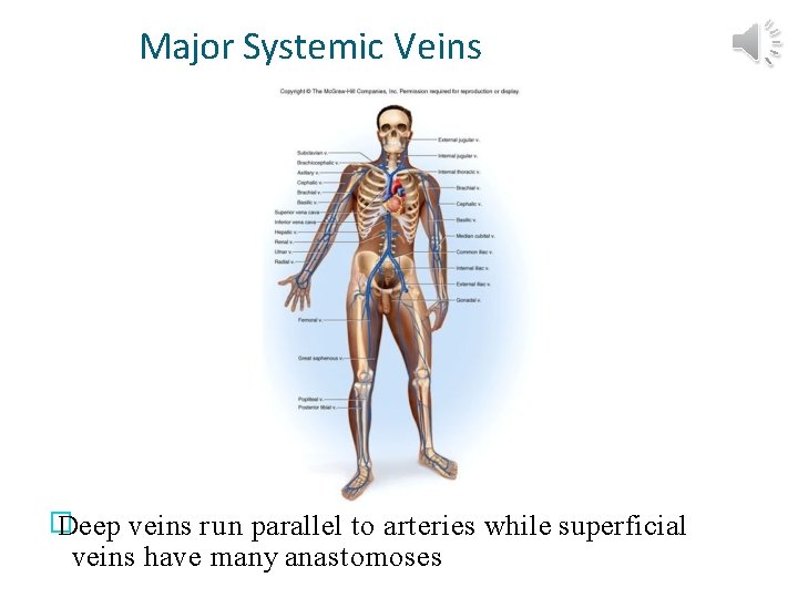
Major Systemic Veins � Deep veins run parallel to arteries while superficial veins have many anastomoses
SUPERFICIAL VEINS Small saphenous veins Great saphenous veins
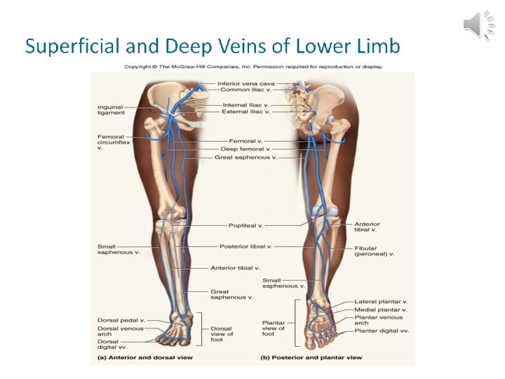
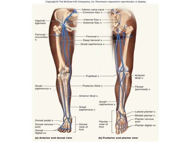
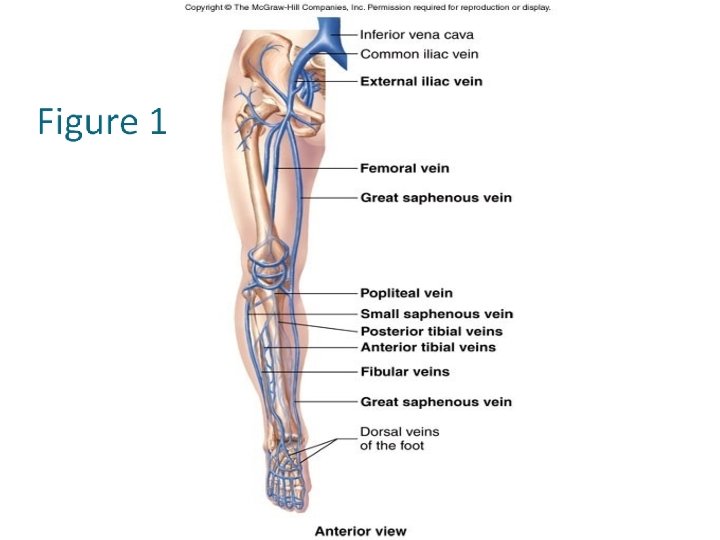
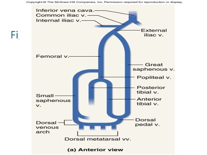
Great (long) saphenous veins • � the longest veins in the body, ascend from the foot the groin in the subcutaneous layer. • � Begin at the medial end of the dorsal venous arches of the foot. • � The dorsal venous arches are networks of veins on the dorsum of the foot formed by the dorsal digital veins, which collect blood from the toes and then unite in pairs to form the dorsal metatarsal veins, which parallel the metatarsals.
Great (long) saphenous veins � As the dorsal metatarsal veins approach the foot, they combine to form the dorsal venous arches. � The great saphenous veins pass anterior to the medial malleolus of the tibia and then superiorly along the medial aspect of the leg and thigh just deep to the skin. � They receives tributaries from superficial tissues and connect with the deep veins as well. � They empty into the femoral veins at the groin. � Drain mainly the medial side of the leg and thigh, the groin, external genital and abdominal wall.
Great (long) saphenous veins • � Along their length, the great saphenous veins have from 10 – 20 valves, with more located in the leg than the thigh. • � These veins are more likely to be subject to varicosities than other veins in the lower limbs because they must support a long column of blood are not well supported by skeletal muscles. • � Often used for prolonged administration of intravenous fluids –important in very young children and in patient of any age who are in shock and whose veins are collapsed.
Small saphenous veins � Begin at the lateral aspect of the dorsal venous arches of the foot. � They pass posterior to the lateral malleolus of the fibula and ascend deep to the skin along the posterior aspect of the leg. � They empty into the popliteal fossa, posterior to the knee. � Along their length, the small saphenous veins have from 9 – 12 valves. � Drain the foot and posterior aspect of the leg. They may communicate with the great saphenous veins in the proximal thigh.
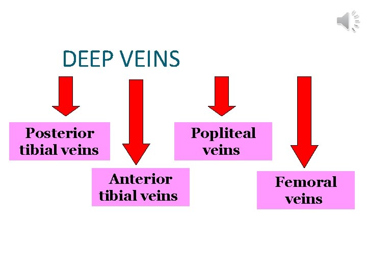
DEEP VEINS Posterior tibial veins Anterior tibial veins Popliteal veins Femoral veins
Posterior tibial veins � The plantar digital veins on the plantar surfaces of the toes unite to form the plantar metatarsal veins, which parallel the metatarsals. � Unite to form the deep plantar venous arches. � From each arch emerges the medial and lateral plantar veins. � Posterior to the medial malleolus of the tibia, form the paired posterior tibial veins, which sometimes merge into a single vessel. They accompany the posterior tibial artery through the leg.
Posterior tibial veins � They ascend deep to the muscles in the posterior aspect of the leg and drain the foot and posterior compartment muscles. � About two-third of the way up the leg, the posterior tibial veins drain blood from the fibular (peroneal) veins, which drain the lateral and posterior leg muscles. � They unite with the anterior tibial veins just inferior to the popliteal fossa to form the popliteal veins.
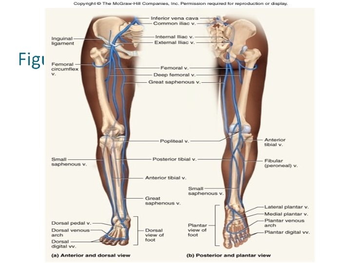
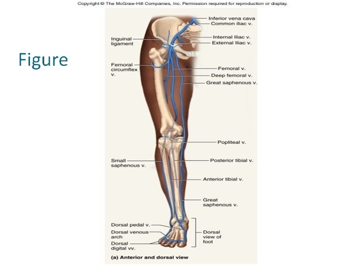
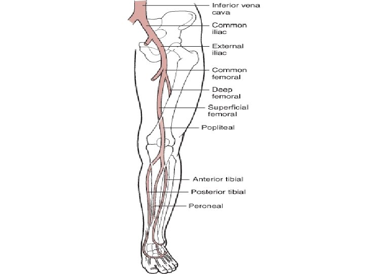
Anterior tibial veins � The paired anterior tibial veins arise in the dorsal venous arch and accompany the anterior tibial artery. � Ascend in the interosseous membrane and unite with the posterior tibial veins to form the popliteal veins. � Drain the ankle joint, knee joint, tibiofibular joint and anterior portion of the leg.
Popliteal veins • � Formed by the union of the anterior and posterior tibial veins. • � Receive blood from the small saphenous veins and tributaries that correspond to branches of the popliteal artery. • � Drain the knee joint and the skin, muscles and bones of portion of the calf and thigh around the knee joint.
Femoral veins � Accompany the femoral arteries and are continuations of the popliteal veins just superior to the knee. � The femoral veins extend up the posterior surface of the thighs and drain the muscles of the thighs, femurs, external genitals and superficial lymph nodes. � The largest tributaries of the femoral veins are the deep veins of the thigh (deep femoral veins). � Just before penetrating the abdominal wall, the femoral veins receive the deep femoral veins and the great saphenous veins.
Femoral veins � The veins formed from this union penetrate the body wall and enter the pelvic cavity. � Here they are known as the external iliac veins.
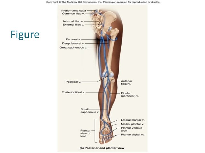
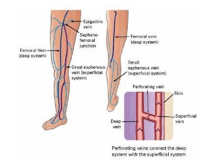
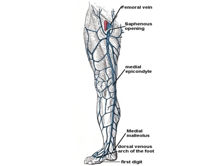
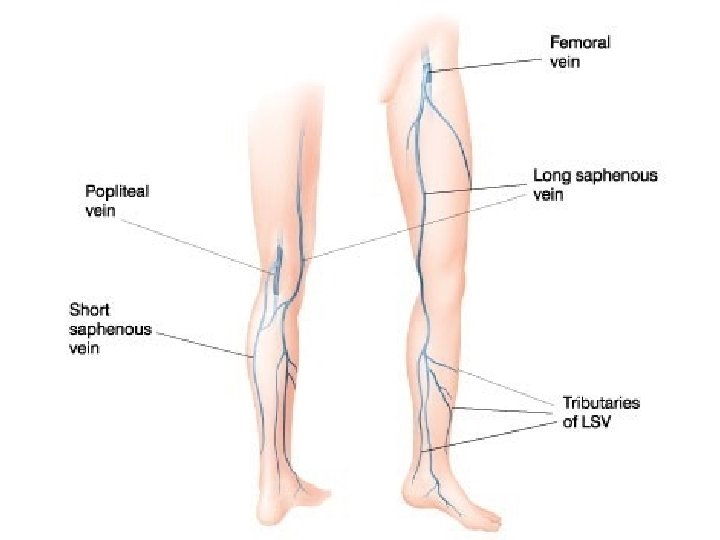
Venous drainage • � The chief superficial veins are the great and small saphenous veins. • � The great (or long) saphenous vein begins on the medial side of the dorsal venous network of the foot. • � It ascends about a finger breadth anterior to the medial malleolus, where it can be found for catheterization.
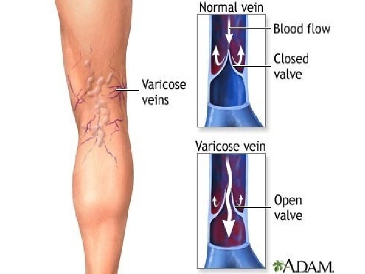
Venous drainage � It crosses the medial surface of the tibia obliquely, passes posterior to the medial condyles of the tibia and femur, and then ascends along the medial side of the thigh. � In the femoral triangle, it pierces the cribriform fascia (at the saphenous opening) and the femoral sheath, ending in the femoral vein � A varicose vein is one that has permanently lost its valvular efficiency, a condition that is not uncommon in the great saphenous vein, which is frequently tortuous and dilated when a subject is in the erect position.
Venous drainage • � The great saphenous vein receives many tributaries, including the superficial epigastric vein. • � Communications occur between the superficial epigastric vein and lateral thoracic veins (by way of the thoraco-epigastric vein). Therefore, there is a communication between the femoral and the axillary veins. • � These communications are important, and they become enlarged in the event of obstruction of either the superior or inferior vena cava.
Venous drainage � The chief deep veins of the lower limb are the femoral and the popliteal veins. � The deep veins begin on the plantar aspect of the foot and accompany the anterior and posterior tibial arteries and the fibular artery. � Many valves are present.
Venous drainage � The superficial and deep veins are connected by perforating veins. �An important series is found in the leg, where valves direct blood from superficial to deep veins. � Muscular action, combined with the arrangement of the valves, is important in returning blood from the lower limb, and venous return is much reduced during quiet standing, when blood pooling may result in hypotension.
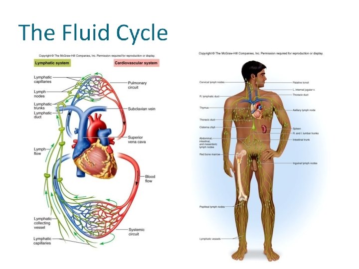
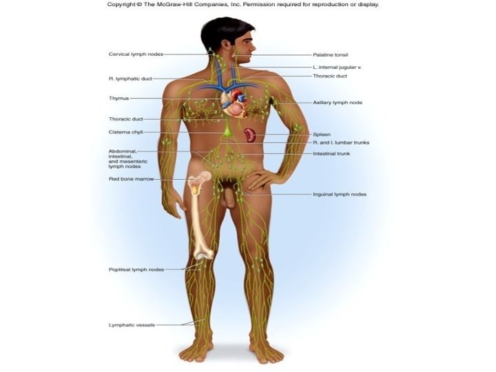
Lymphatic drainage � The superficial lymphatic vessels of the lower limb are arranged as medial trunks, which end in the inguinal nodes, and as a lateral set, which either joins the medial trunks superior to the knee or ends in the popliteal nodes. �The popliteal nodes receive the deep lymphatic trunks that accompany the tibial blood vessels and the lateral group of superficial trunks.
Lymphatic drainage � Their efferents accompany the femoral blood vessels to the deep inguinal nodes. � The inguinal nodes are mostly subcutaneous and are frequently palpable in vivo. � The superficial inguinal nodes lie both longitudinally along the great saphenous vein and transversely, just inferior to the inguinal ligament, whereas the deep inguinal nodes (into which the superficial drain) lie medial to the femoral vein.
Lymphatic drainage � When the inguinal nodes are found to be enlarged, their territory of drainage, i. e. , the trunk inferior to the level of the umbilicus (including perineum, external genitalia, and anus) as well as the entire lower limb should be examined. � The efferent lymphatic vessels from the inguinal nodes proceed to the external iliac nodes and drain ultimately into the lumbar (aortic) nodes.

- Slides: 41