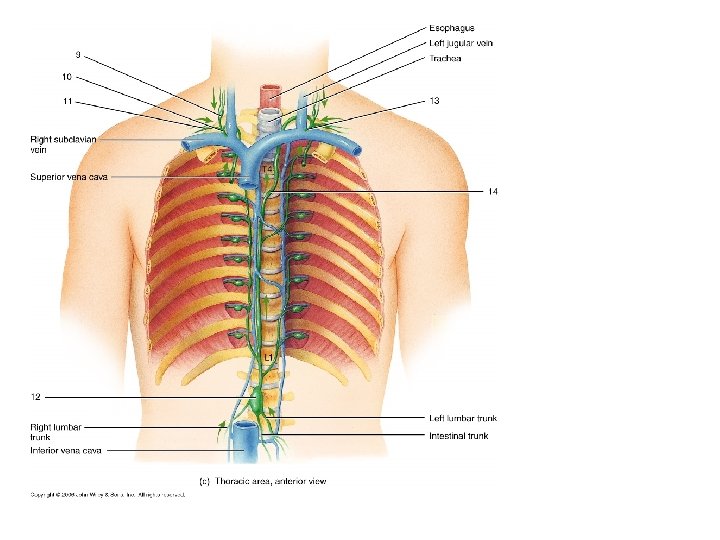
Right internal jugular vein Right jugular trunk Right

 Lymph drainage")


 Gross structure")

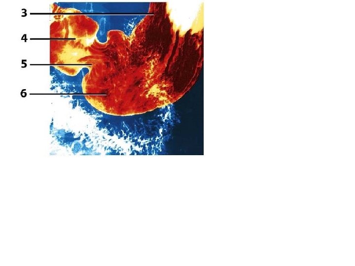
 Mature T cell 2 _____ 9")


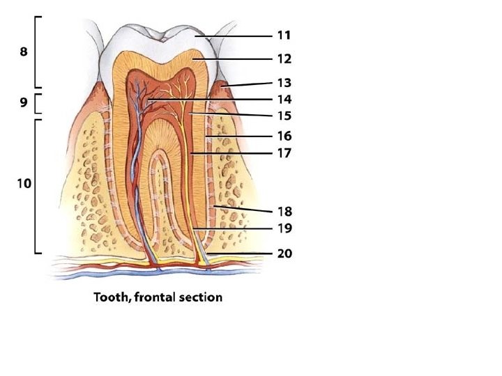
 Frontal section")
 Pharyngeal structure")
 Pharyngeal")
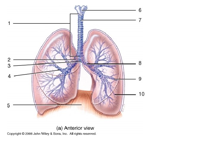

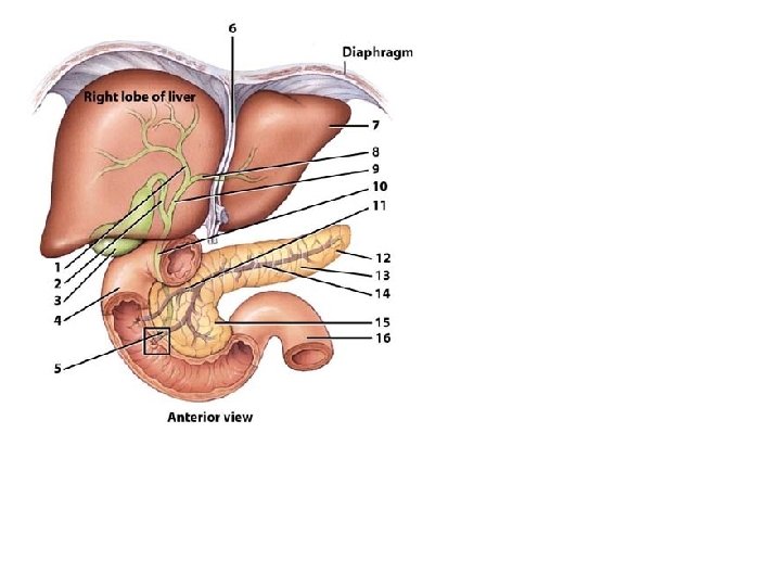
 3 4 (a) Anterior view")

 (a) Transverse section")
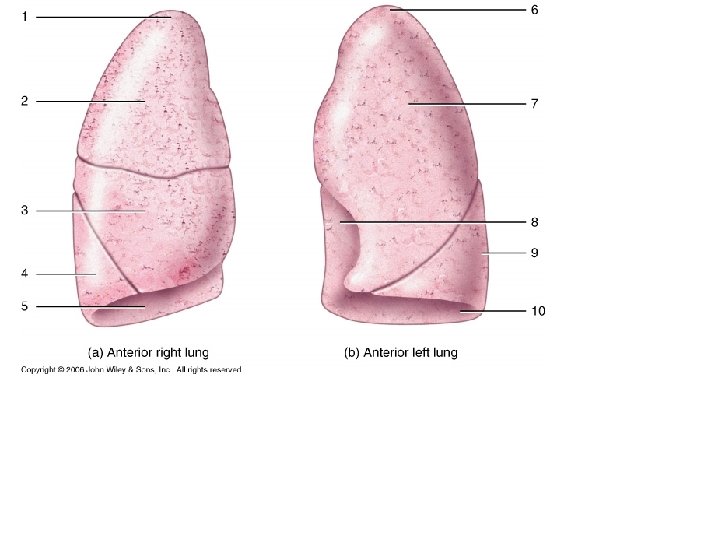
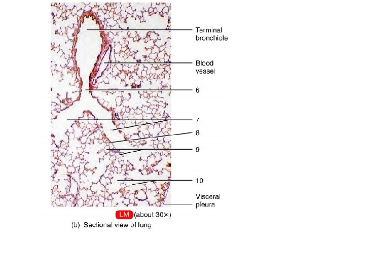
 Medial view of right lung")


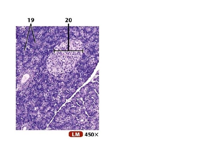
 Hepatopancreatic ampulla 20")
- Slides: 56
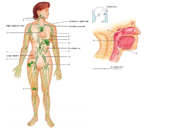
Right internal jugular vein Right jugular trunk Right subclavian trunk Left jugular trunk 1 2 3 4 Intestinal and lumber trunks 6 Left subclavian vein 5 (a) Overview of lymphatic vessels
7 8 (b) Lymph drainage
4 Left internal jugular vein 5 Right subclavian vein Left subclavian vein 10 11 12 13 Small intestine 6 7 Large intestine 14 8 15 16 9 (b) Anterior view
Thyroid gland Brachiocephalic veins Trachea Thymus Right common carotid artery Parietal pericardium Superior vena cava Right lung Left lung Diaphragm (a) Thymus of adolescent
SUPERIOR 2 Colic impression 1 Gastric impression 3 Renal impression (a) Gross structure
1 Palatine tonsil 2 Submandibular node 3 Cervical node 4 Thymus gland Axillary node 8 5 Intestinal node 6 Appendix Spleen 9 Peyer's patches 10 Iliac node 11 Inguinal node 12 7 Red bone marrow (b) Location of immune system cells
Red Bone Marrow 1 _____ 7 _____ (clone) Mature T cell 2 _____ 9 _____ 10 _____ 3 _____ (clone) 4 _____ (clone) 6 _____ 5 _____ 8 _____
4 5 1 6 7 1 2 2 3 4 3 5 6 7 8 9 10 Anterior view
2 3 4 Frontal sinus Frontal bone Sphenoid bone 5 Sphenoidal sinus 6 7 1 8 9 (a) Sagittal section
Frontal sinus Ethmoidal sinus 13 10 11 14 12 Maxillary sinus (b) Frontal section
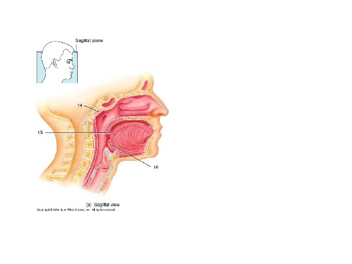
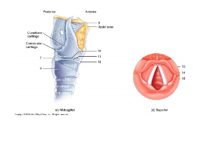
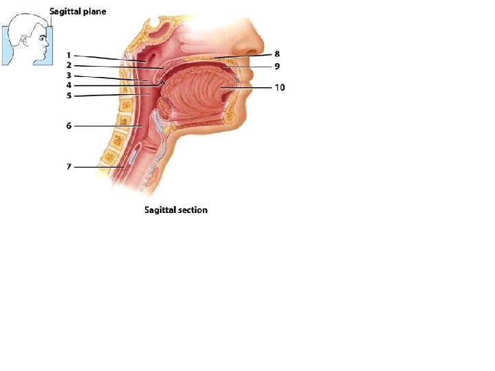
1 2 3 (a) Pharyngeal structure
Sagittal plane 10 4 5 6 7 11 12 8 13 9 (b) Pharyngeal structure
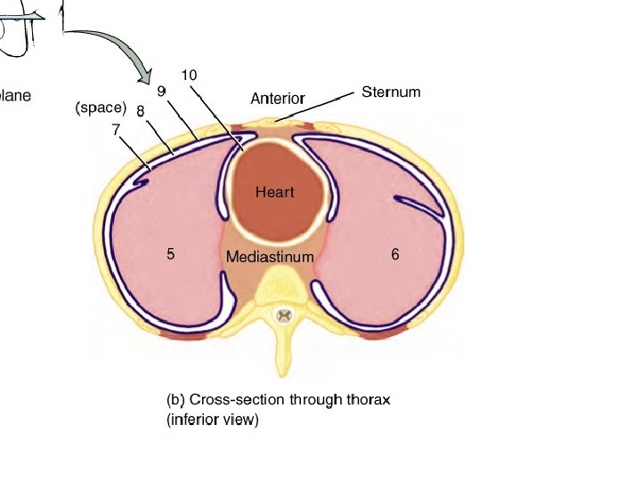
1 2 (space) 3 4 (a) Anterior view
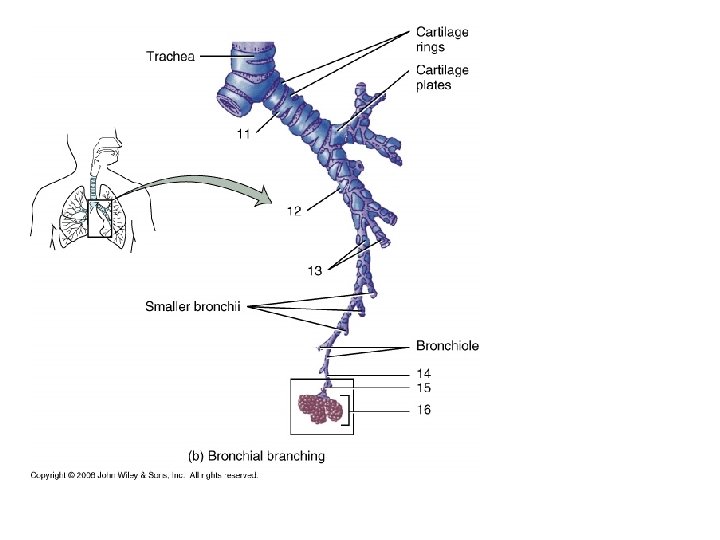
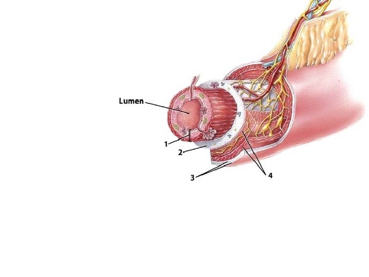
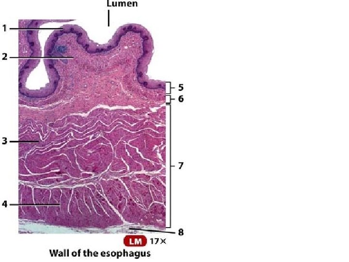
Lumen Esophagus Trachea Epithelial lining Trachealis muscle Transverse plane
1 POSTERIOR 2 3 4 5 ANTERIOR LM (2. 6 x) (a) Transverse section of trachea and esophagus
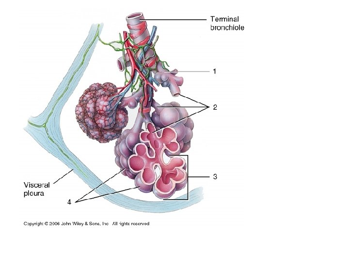
15 11 16 12 17 13 18 14 (c) Medial view of right lung (d) Medial view of left lung
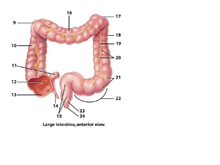
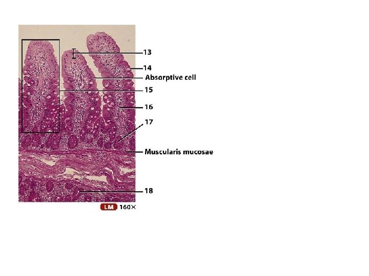
12. Small intestine: Primary site of nutrient absorption
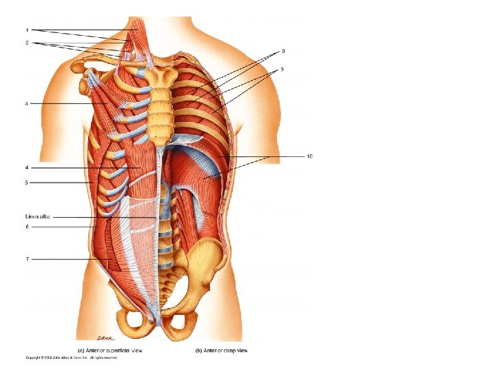
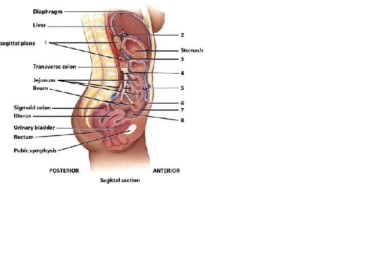
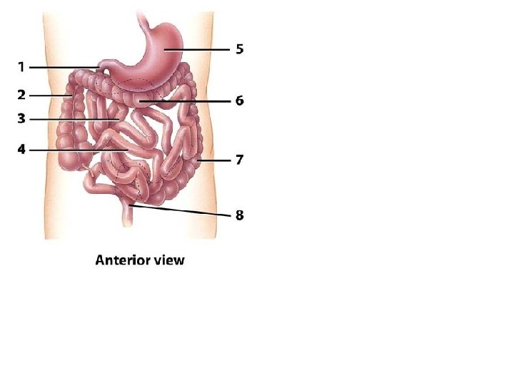
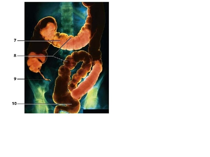
11. Lessor omentum Gallbladder 11 Liver Stomach Duodenum Transverse colon Descending colon Ascending colon Sigmoid colon (c) Anterior view with liver and gallbladder lifted
12 12. Greater omentum Transverse colon 13. Mesocolon 13 14. Mesentery Jejunum 14 Descending colon Ileum Sigmoid colon (d) Anterior view with greater omentum lifted and small intestine reflected to right side
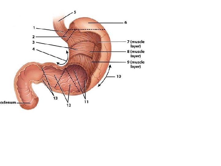
Duodenum Esophagus Pyloric canal Pyloric sphincter Pyloric antrum Cardia Lower esophageal sphincter Fundus Lesser curvature Rugae of mucosa Body Greater curvature
19 Mucosa of duodenum 18 17 (b) Hepatopancreatic ampulla 20