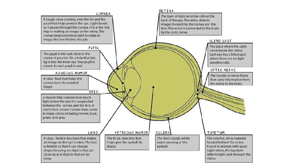
2 Cornea Tough is clear covering over the


- Slides: 13
2. Cornea Tough, is clear covering over the iris and pupil that helps protect the eye. Light bend as it passes through the cornea. This is the first step in making an image on the retina. The lens finishes making the image.
5. Pupil – a dark circle in the center of your iris. It’s the hole that lets light into the inner eye.
3. Aqueous Humor – clear fluid that helps the cornea keep its rounded shape.
4. Iris- a muscle that controls how much light enters the eye. It is suspended between the cornea and the lens. Cow’s iris is brown. Human iris’s come in many colors
6. Lens – a clear flexible structure that makes an image on the eye’s retina. It is so flexible it can change shape, focusing on objects close and far away.
7. Vitreous humor – the thick, clear jelly that helps give the eyeball its shape.
1. Sclera – the thick, tough, white outer covering of the eyeball.
Tapetum – colorful shiny material located behind the retina. Found in animals with good night vision. Reflects light back thru the retina.
9. Optic Nerve – the bundle of nerve fibers that carry info from the retina to the brain.
Blind spot – place where the optic nerve leave the retina. No light sensitive cells.
8. Retina – layer of light sensitive cells at the back of the eye. The retina detects images focused by the cornea and the lens. The retina is connected to the brain by the optic nerve.