
STRESS ECHO Dr Ajay Nair Senior Resident Dept



- Slides: 67
STRESS ECHO Dr Ajay Nair Senior. Resident Dept of Cardiology Calicut Medical. College
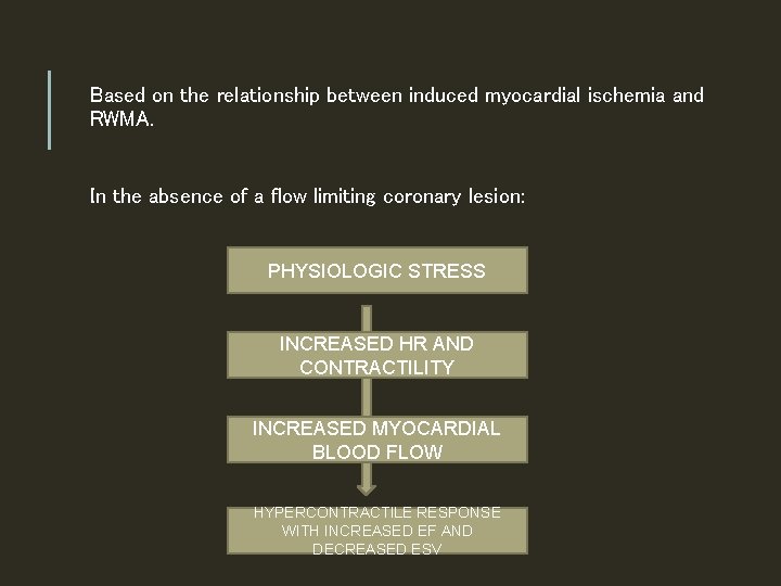
Based on the relationship between induced myocardial ischemia and RWMA. In the absence of a flow limiting coronary lesion: PHYSIOLOGIC STRESS INCREASED HR AND CONTRACTILITY INCREASED MYOCARDIAL BLOOD FLOW HYPERCONTRACTILE RESPONSE WITH INCREASED EF AND DECREASED ESV
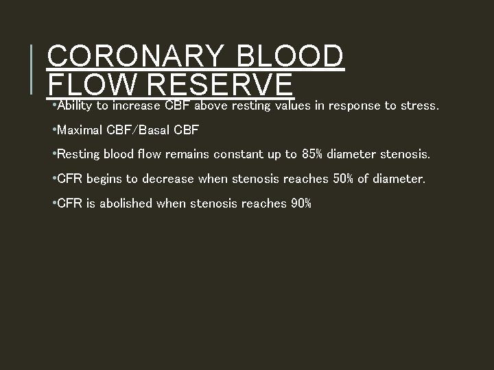
CORONARY BLOOD FLOW RESERVE • Ability to increase CBF above resting values in response to stress. • Maximal CBF/Basal CBF • Resting blood flow remains constant up to 85% diameter stenosis. • CFR begins to decrease when stenosis reaches 50% of diameter. • CFR is abolished when stenosis reaches 90%
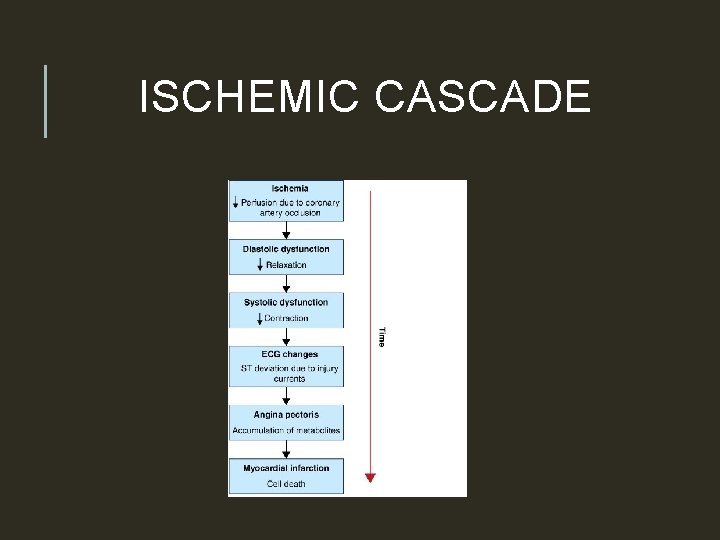
ISCHEMIC CASCADE
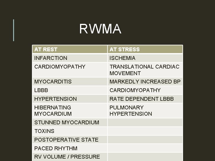
RWMA AT REST AT STRESS INFARCTION ISCHEMIA CARDIOMYOPATHY TRANSLATIONAL CARDIAC MOVEMENT MYOCARDITIS MARKEDLY INCREASED BP LBBB CARDIOMYOPATHY HYPERTENSION RATE DEPENDENT LBBB HIBERNATING MYOCARDIUM PULMONARY HYPERTENSION STUNNED MYOCARDIUM TOXINS POSTOPERATIVE STATE PACED RHYTHM RV VOLUME / PRESSURE
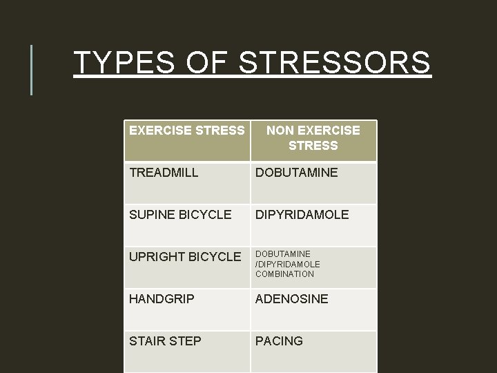
TYPES OF STRESSORS EXERCISE STRESS NON EXERCISE STRESS TREADMILL DOBUTAMINE SUPINE BICYCLE DIPYRIDAMOLE UPRIGHT BICYCLE DOBUTAMINE /DIPYRIDAMOLE COMBINATION HANDGRIP ADENOSINE STAIR STEP PACING
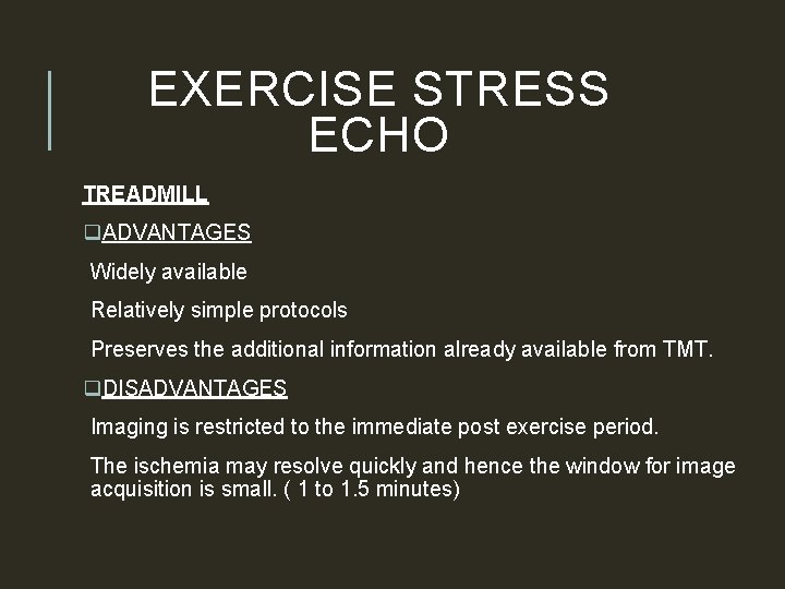
EXERCISE STRESS ECHO TREADMILL q. ADVANTAGES Widely available Relatively simple protocols Preserves the additional information already available from TMT. q. DISADVANTAGES Imaging is restricted to the immediate post exercise period. The ischemia may resolve quickly and hence the window for image acquisition is small. ( 1 to 1. 5 minutes)
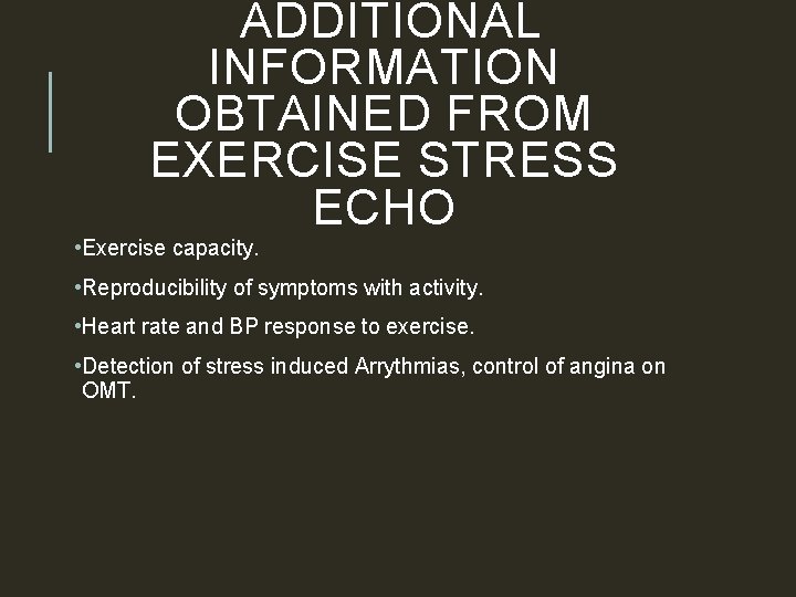
ADDITIONAL INFORMATION OBTAINED FROM EXERCISE STRESS ECHO • Exercise capacity. • Reproducibility of symptoms with activity. • Heart rate and BP response to exercise. • Detection of stress induced Arrythmias, control of angina on OMT.
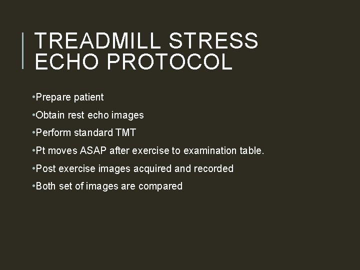
TREADMILL STRESS ECHO PROTOCOL • Prepare patient • Obtain rest echo images • Perform standard TMT • Pt moves ASAP after exercise to examination table. • Post exercise images acquired and recorded • Both set of images are compared
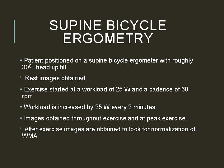
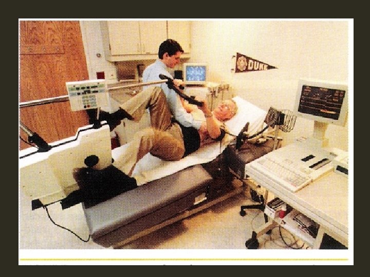
SUPINE BICYCLE ERGOMETRY • Patient positioned on a supine bicycle ergometer with roughly 300 head up tilt. • Rest images obtained • Exercise started at a workload of 25 W and a cadence of 60 rpm. • Workload is increased by 25 W every 2 minutes • Images obtained throughout exercise and at peak exercise. • After exercise images are obtained to look for normalization of WMA
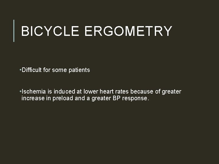
BICYCLE ERGOMETRY • Difficult for some patients • Ischemia is induced at lower heart rates because of greater increase in preload and a greater BP response.
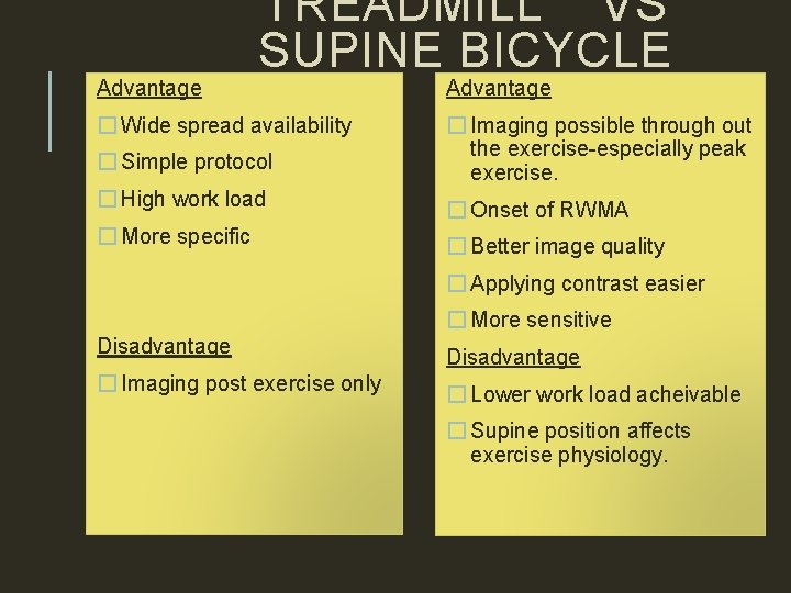
Advantage TREADMILL VS SUPINE BICYCLE � Wide spread availability Advantage � Simple protocol � Imaging possible through out the exercise-especially peak exercise. � High work load � Onset of RWMA � More specific � Better image quality � Applying contrast easier � More sensitive Disadvantage � Imaging post exercise only � Lower work load acheivable � Supine position affects exercise physiology.
PHARMACOLOGIC STRESS TESTING
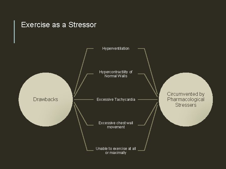
Exercise as a Stressor Hyperventilation Hypercontractility of Normal Walls Drawbacks Excessive Tachycardia Excessive chest wall movement Unable to exercise at all or maximally Circumvented by Pharmacological Stressers
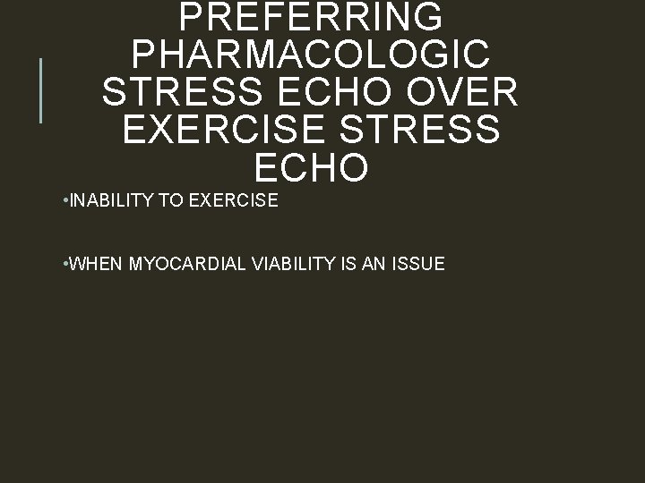
PREFERRING PHARMACOLOGIC STRESS ECHO OVER EXERCISE STRESS ECHO • INABILITY TO EXERCISE • WHEN MYOCARDIAL VIABILITY IS AN ISSUE
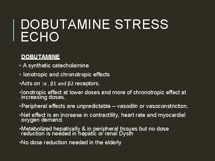
DOBUTAMINE STRESS ECHO DOBUTAMINE • A synthetic catecholamine • Ionotropic and chronotropic effects • Acts on : α , β 1 and β 2 receptors. • Ionotropic effect at lower doses and more of chronotropic effect at increasing doses. • Peripheral effects are unpredictable – vasodiln or vasoconstriction. • Net effect is an increase in contractility, heart rate and myocardial oxygen demand. • Metabolized hepatically & in peripheral tissues but no dose reduction is needed in hepatic or renal Dysfn • No dose reduction needed in the elderly
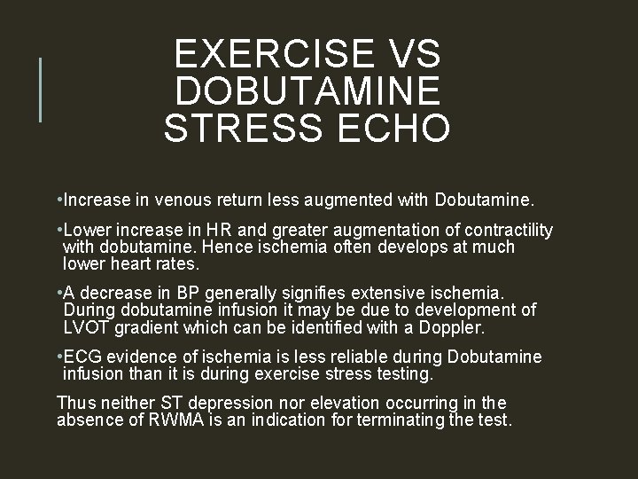
EXERCISE VS DOBUTAMINE STRESS ECHO • Increase in venous return less augmented with Dobutamine. • Lower increase in HR and greater augmentation of contractility with dobutamine. Hence ischemia often develops at much lower heart rates. • A decrease in BP generally signifies extensive ischemia. During dobutamine infusion it may be due to development of LVOT gradient which can be identified with a Doppler. • ECG evidence of ischemia is less reliable during Dobutamine infusion than it is during exercise stress testing. Thus neither ST depression nor elevation occurring in the absence of RWMA is an indication for terminating the test.
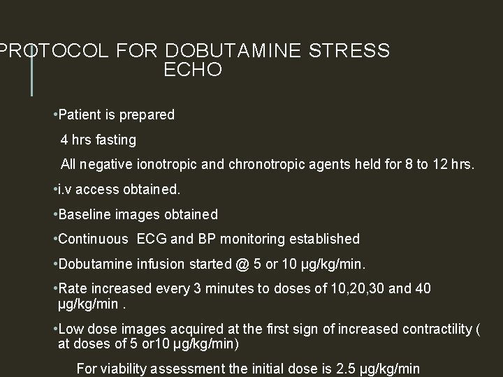
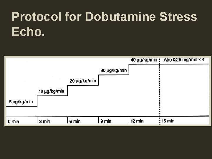
PROTOCOL FOR DOBUTAMINE STRESS ECHO • Patient is prepared 4 hrs fasting All negative ionotropic and chronotropic agents held for 8 to 12 hrs. • i. v access obtained. • Baseline images obtained • Continuous ECG and BP monitoring established • Dobutamine infusion started @ 5 or 10 µg/kg/min. • Rate increased every 3 minutes to doses of 10, 20, 30 and 40 µg/kg/min. • Low dose images acquired at the first sign of increased contractility ( at doses of 5 or 10 µg/kg/min) For viability assessment the initial dose is 2. 5 µg/kg/min
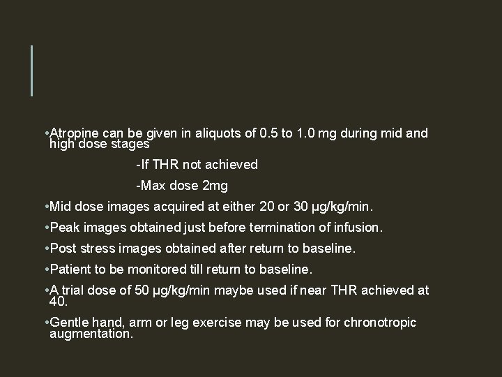
• Atropine can be given in aliquots of 0. 5 to 1. 0 mg during mid and high dose stages -If THR not achieved -Max dose 2 mg • Mid dose images acquired at either 20 or 30 µg/kg/min. • Peak images obtained just before termination of infusion. • Post stress images obtained after return to baseline. • Patient to be monitored till return to baseline. • A trial dose of 50 µg/kg/min maybe used if near THR achieved at 40. • Gentle hand, arm or leg exercise may be used for chronotropic augmentation.
Protocol for Dobutamine Stress Echo.
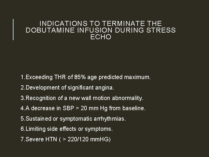
INDICATIONS TO TERMINATE THE DOBUTAMINE INFUSION DURING STRESS ECHO 1. Exceeding THR of 85% age predicted maximum. 2. Development of significant angina. 3. Recognition of a new wall motion abnormality. 4. A decrease in SBP > 20 mm Hg from baseline. 5. Sustained or symptomatic arrhythmias. 6. Limiting side effects or symptoms. 7. Severe HTN ( > 220/120 mm. HG)
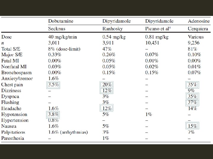
SAFETY • Safety has been studied extensively • Short t 1/2 (2 minutes) , hence induced ischemia readily reversed by termination of infusion. • Severe cases - short acting i. v β Blocker, Esmolol or Metoprolol can be used. • Most common side effects – Minor arrythmias like VPCs or Atrial arrythmias.
� In one series of 1, 118 patients referred for dobutamine stress echocardiography, there were no incidents of death, myocardial infarction, or sustained ventricular tachycardia or fibrillation (Mertes et al. , 1993).
DIPYRIDAMOLE Potent coronary vasodilator Provokes anginal attack in angina patients Vasodilatory action: Inhibition of reuptake of adenosine by endothelial cells CBF increases 4 to 5 times in normal vessel Reduction of subendocardial blood flow in stenotic coronary arteries
DIPYRIDAMOLE Coronary steal phenomenon Standard protocol: 0. 54 mg/kg for 4 min High dose protocol: 0. 84 mg/kg Antidote: theophylline
DIPYRIDAMOLE Contraindication active wheezing high degree AV block hypotension(SBP<90 mm. Hg) recent use of dipyridamole(<24 hours) Relative contraindication Hx of reactive airway disease sick sinus syndrome severe sinus bradycardia
ADENOSINE • Naturally occurring agent • Receptors A 1: slows HR and conduction A 2 a: c-AMP Decreases calcium uptake by SR Smooth muscle relax vasodilation • Half life: 2 seconds-Needs constant IV infusion • Rapidly removed by RBCs and endothelial cells.
ADENOSINE – SIDE EFFECT Flushing: 37% Dyspnea: 35% GI discomfort: 15% Headache: 14% Light-headedness 9% Most side effect – short-lived and mild
CHOOSING BETWEEN PHARMACOLOGIC AGENTS Dobutamine is more likely to cause true ischemia rather than a mere flow mismatch and thus a WMA is more likely to be detectable on Echo. For inducing perfusion abnormality both Dobutamine and vasodilators are equivalent. Thus Dobutamine is considered the agent of choice for stress Echo.
INTERPRETATION OF STRESS ECHO HYPERKINESIS – The normal response to stress. Lack of Hyperkinesis: - Myocardial ischemia - Non –ischemic CMP. - Beta blocker therapy - Severe hypertension - Delay in image acquisition - Elderly patients especially women
VOLUME RESPONSE Decrease in ESV and EDV – Normal response 25 -30% decrease in ESV and EDV is the normal response. An abnormal volume response is defined as an increase in volume from rest to stress of > 17%. However with Supine bicycle ergometry, due to increased venous return, an increase is LV volume may be a normal response.
USE OF CONTRAST AGENTS: When endocardial borders can’t be properly identified in 2 or more LV segments contrast may be used.
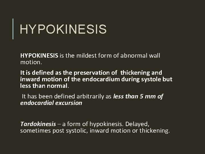
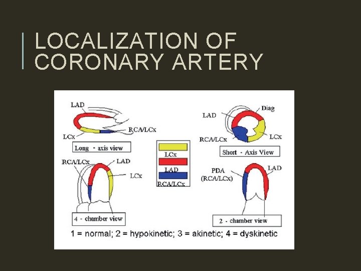
HYPOKINESIS is the mildest form of abnormal wall motion. It is defined as the preservation of thickening and inward motion of the endocardium during systole but less than normal. It has been defined arbitrarily as less than 5 mm of endocardial excursion Tardokinesis – a form of hypokinesis. Delayed, sometimes post systolic, inward motion or thickening.
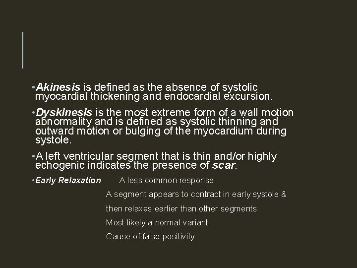
• Akinesis is defined as the absence of systolic myocardial thickening and endocardial excursion. • Dyskinesis is the most extreme form of a wall motion abnormality and is defined as systolic thinning and outward motion or bulging of the myocardium during systole. • A left ventricular segment that is thin and/or highly echogenic indicates the presence of scar. • Early Relaxation: A less common response A segment appears to contract in early systole & then relaxes earlier than other segments. Most likely a normal variant Cause of false positivity.
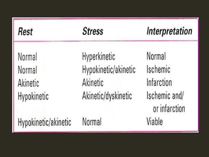
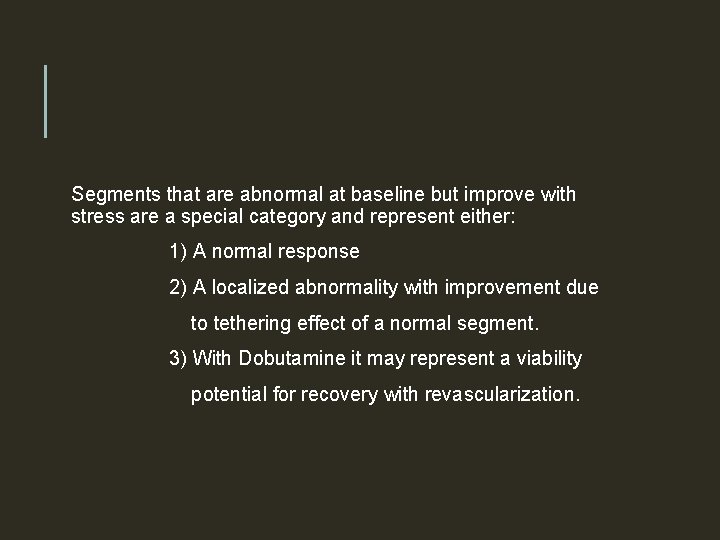
Segments that are abnormal at baseline but improve with stress are a special category and represent either: 1) A normal response 2) A localized abnormality with improvement due to tethering effect of a normal segment. 3) With Dobutamine it may represent a viability potential for recovery with revascularization.
LOCALIZATION OF CORONARY ARTERY
CORRELATION WITH SYMPTOMS AND ECG • Usually concordant CHANGES • When discordant, rely on Echo changes. • In fact one of the most common indications for stress echo are situations when Stress ECG may be false negative: -LVH -WOMEN -LBBB • In a study using bicycle ergometry by Ryan et al. precise concordance between ECG and Echo occurred in nearly 50% cases and when disagreement did occur Echo was mostly correct. • However a strongly positive exercise stress test should not be ignored.
DETECTION OF CAD • Using CAG as the standard the overall sensitivity of Exercise Stress Echo has ranged from 71 to 94%. • Specificity ranges between 80 to 90%. • Studies on Dobutamine stress echo also gave similar results. • Stress ECG – Sensitivity – 77% - Specificity – 68% • Factors affecting sensitivity- Degree of coronary stenosis - Location of stenosis - Presence of LVH
LBBB: - Most common cause of false positive result -Abnormal septal motion -Myocardial thickening is however preserved. -Trimming the loops to avoid the first few frames of systole can be helpful.
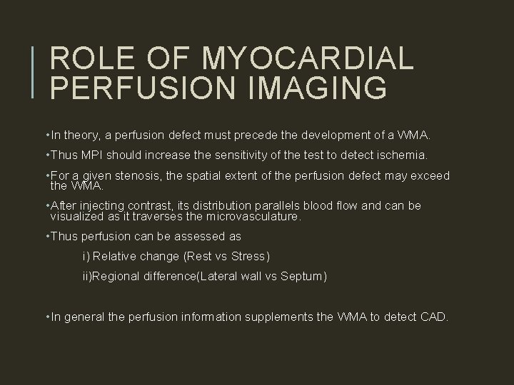
ROLE OF MYOCARDIAL PERFUSION IMAGING • In theory, a perfusion defect must precede the development of a WMA. • Thus MPI should increase the sensitivity of the test to detect ischemia. • For a given stenosis, the spatial extent of the perfusion defect may exceed the WMA. • After injecting contrast, its distribution parallels blood flow and can be visualized as it traverses the microvasculature. • Thus perfusion can be assessed as i) Relative change (Rest vs Stress) ii)Regional difference(Lateral wall vs Septum) • In general the perfusion information supplements the WMA to detect CAD.
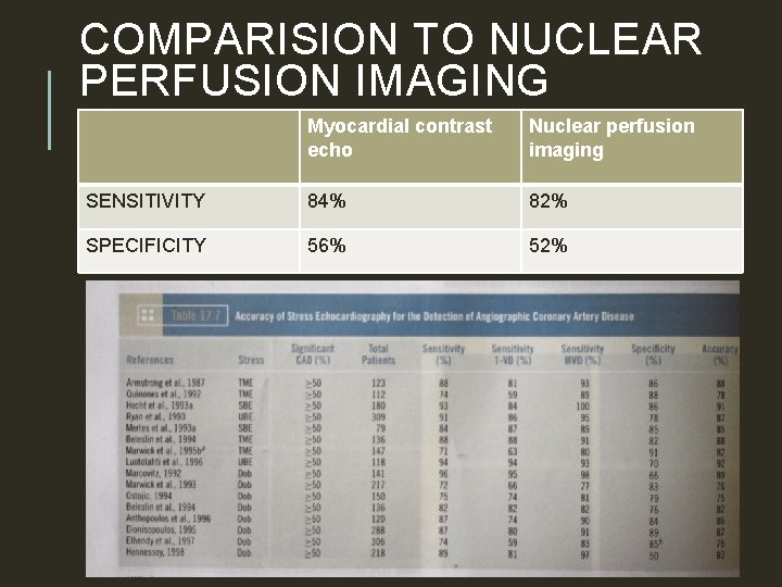
COMPARISION TO NUCLEAR PERFUSION IMAGING Myocardial contrast echo Nuclear perfusion imaging SENSITIVITY 84% 82% SPECIFICITY 56% 52%
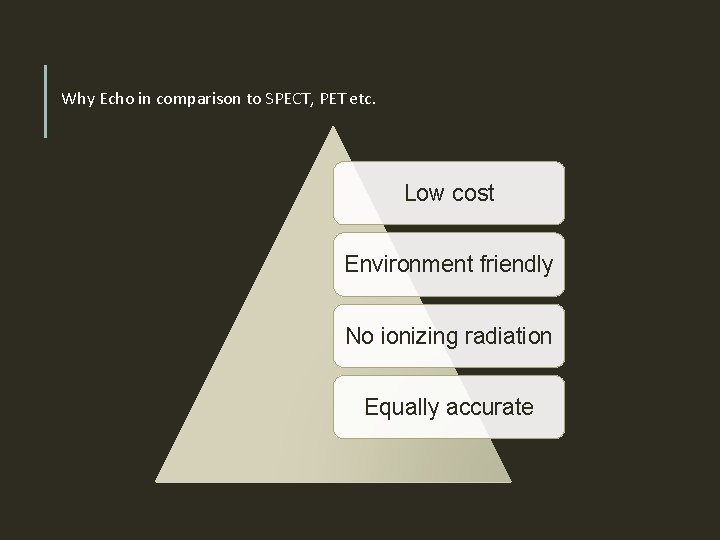
Why Echo in comparison to SPECT, PET etc. Low cost Environment friendly No ionizing radiation Equally accurate
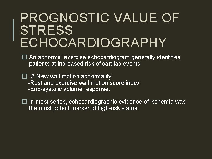
PROGNOSTIC VALUE OF STRESS ECHOCARDIOGRAPHY � An abnormal exercise echocardiogram generally identifies patients at increased risk of cardiac events. � -A New wall motion abnormality -Rest and exercise wall motion score index -End-systolic volume response. � In most series, echocardiographic evidence of ischemia was the most potent marker of high-risk status
STRESS ECHO AFTER MI §Used both to identify high and low risk subsets and to predict the location and extent of CAD. §The goal is to identify ischemia at a distance §Positive finding would be detection of a new WMA remote from the site of previous infarction. §Inducible ischemia is a powerful indicator of high risk and suggests the need for further evaluation.
PREOPERATIVE RISK ASSESSMENT �Most studies used dobutamine stress. �Mainly before major peripheral vascular surgery and therefore included patients who frequently are unable to exercise. �In this high-risk subset, the presence or absence of an inducible WMA has been the most potent determinant of relative risk. �The absence of an inducible wall motion abnormality confers a very favorable prognosis, with a negative predictive value of 93% to 100%.
In a Meta-analysis examining the value of dipyridamole thallium and dobutamine echocardiography before vascular surgery (Shaw et al. , 1996), the presence of an inducible wall motion abnormality on echocardiography provided the greatest ability to discriminate between high- and low-risk status
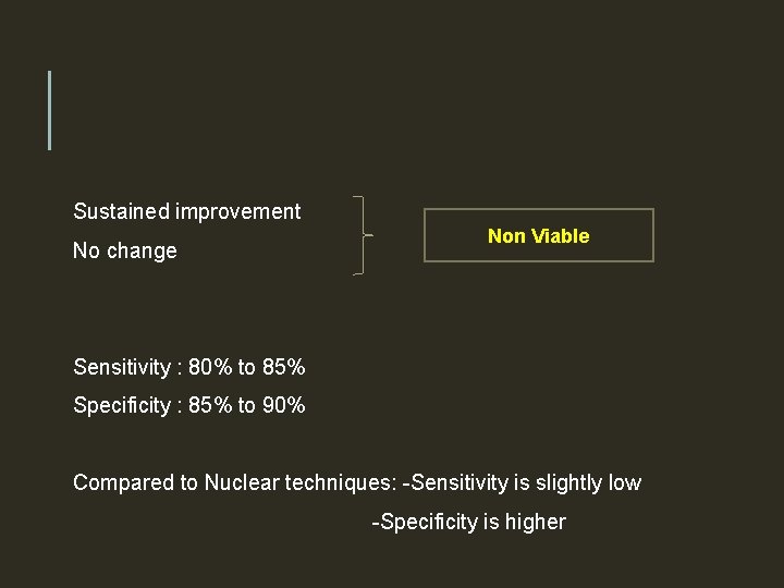
ASSESMENT OF MYOCARDIAL VIABILITY • The term viable refers to myocardium that has the potential for functional recovery. • The resting Echo is neither sensitive nor specific for this purpose. • The use of Dobutamine echo is basd on the observation that viable myocardium will augment in response to β adrenergic stimulation whereas non viable myocardium will not. • Dobutamine is infused at incremental rates and the response is carefully noted. • The BIPHASIC RESPONSE, i. e augmentation at low dose followed by deterioration at higher doses is most predictive of the capacity for functional recovery.
Sustained improvement No change Non Viable Sensitivity : 80% to 85% Specificity : 85% to 90% Compared to Nuclear techniques: -Sensitivity is slightly low -Specificity is higher
STRESS ECHO IN VALVULAR HEART DISEASE AS � No role in patients with definite cardiac symptoms. � The principal role of exercise testing is to unmask symptoms or abnormal blood pressure responses in patients with AS who claim to be asymptomatic.
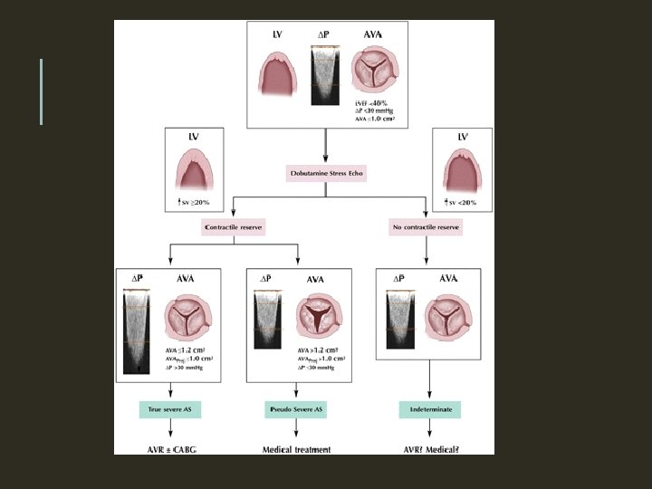
AS WITH LOW-FLOW, LOW-GRADIENT, AND LV DYSFUNCTION Anatomically severe AS and LV systolic dysfunction (EF<40%) often presents with a relatively low-pressure gradient, such as a mean gradient of 30 to 40 mm Hg or less. Difficult to differentiate them from a primary cardiomyopathic process and a thickened but nonstenotic aortic valve producing an outflow murmur. PSEUDOSEVERE AS
TRUE SEVERE AS- The small and relatively fixed AVA contributes to an increase in afterload, a decrease in ejection fraction, and a reduction stroke volume. PSEUDOSEVERE AS-The predominant factor is myocardial disease The severity of AS is overestimated on the basis of AVA because there is incomplete opening of the valve.
In both situations, the low-flow state and low-pressure gradient contribute to a calculated AVA that meets criteria for severe AS at rest (1. 0 cm 2) The resting echocardiogram does not distinguish between these 2 situations Distinction is essential because patients with true severe AS and poor LV function will generally benefit significantly from AVR. Patients with pseudosevere AS will not and may also have a higher risk of perioperative mortality.
AR • Exercise testing may elicit symptoms in patients with AR who are apparently asymptomatic based on the medical history, thus identifying candidates for surgery. • In addition, pre-operative exercise capacity is helpful in predicting survival and recovery of function after AVR
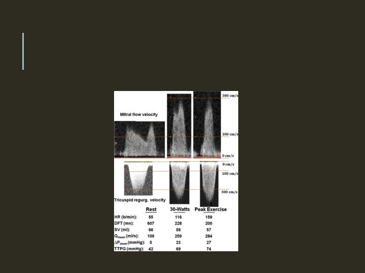
MS • In asymptomatic patients with severe MS (mean gradient >10 mm Hg and mitral valve area [MVA] <1. 0 cm 2), or • Symptomatic patients with moderate MS (mean gradient of 5 to 10 mm Hg and MVA of 1. 0 to 1. 5 cm 2) • The measurement of PAP (measured from the TR velocity) during exercise or dobutamine stress echo can help distinguish those who could benefit from valvuloplasty or valve replacement.
• Patients with reduced atrioventricular compliance show a more pronounced increase in PAP during exercise or dobutamine than those with normal compliance • In some patients with Moderate MS at rest, the physiologic effects of heart rate sensitivity and atrioventricular compliance can produce exercise-induced pulmonary hypertension and exertional dyspnea. • The resting values of transmitral gradient and pulmonary arterial pressure do not necessarily reflect the actual severity of the disease • Stress echocardiography is useful for assessing the severity of MS, assessing its hemodynamic impact, and explaining exerciseinduced symptoms.
• The current ACC/AHA guidelines have given a Class I recommendation for stress echocardiography in patients with MS and discordance between symptoms and stenosis severity. • The threshold values proposed by the ACC/AHA guidelines for consideration for intervention are a mean transmitral pressure gradient >15 mm Hg during exercise or a peak pulmonary artery systolic pressure >60 mm Hg during exercise. • In these patients, BMV or Sx is recommended, even with apparently moderate MS at rest.
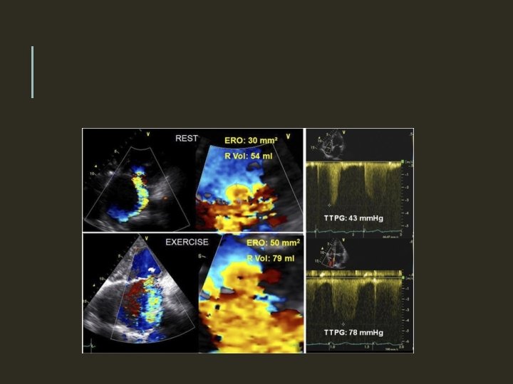
MR • Asymptomatic severe MR : Exercise stress echocardiography may help identify patients with unrecognized symptoms or subclinical latent LV dysfunction. • In symptomatic patients in whom the severity of MR is estimated to be only mild at rest, exercise echocardiography may be useful in elucidating the cause.
• Worsening of MR severity • A marked increase in PAP • Impaired exercise capacity • Occurrence of symptoms during exercise echocardiography • Useful findings for identifying a subset of apparently asymptomatic patients at higher risk who may benefit from early surgery. • A pulmonary artery systolic pressure >60 mm Hg during exercise has been suggested as a threshold value above which asymptomatic patients with severe MR might be referred for surgical valve repair (Class IIa recommendation )
PROSTHETIC HEART VALVES Stress echocardiography -valuable in confirming or excluding the presence of hemodynamically significant prosthetic valve stenosis or PPM, especially when there is discordance between the patient's symptomatic status and the prosthetic valve hemodynamics measured at rest In contrast to a normally functioning and well-matched prosthesis a stenotic prosthetic valve is generally associated with a marked increase in gradient with exercise
A disproportionate increase in transvalvular gradient (>20 mm Hg for aortic prostheses or >12 mm Hg for mitral prostheses) generally indicates severe prosthesis dysfunction or PPM High resting and stress gradients occur more often with smaller (21 for aortic and 25 for mitral) rather than larger -sized prostheses, and mismatched rather than non mismatched prostheses.
THANK YOU