EYE BANKING Dr Ajai Agrawal Additional Professor Department

EYE BANKING Dr. Ajai Agrawal Additional Professor Department of Ophthalmology A. I. I. M. S. RISHIKESH

Acknowledgment • Photographs in this presentation are courtesy of Dr. Samar. K. Basak

Learning Objectives At the end of the class, students shall be able to • Understand the importance and need of eye banking • Have basic knowledge of structure and functions of eye banks • Understand the various surgical procedures for
 performed 1 st successful human corneal transplantation. • 1935:")
History • 1903: E. Zirm(Czechoslovakia) performed 1 st successful human corneal transplantation. • 1935: V P Filatov (Russia): Father of keratoplasty and modern eye banking. • 1944: Dr. R. Townley Paton established the first eye bank in New York City. • 1945: The first eye bank in India was

• 1960 : 1 st successful corneal transplantation in India by Dr. Dhanda (Indore). • 1974: Mc. Karey and Kaufman developed M K medium which allowed the excised corneo-scleral rim to be preserved for up to 4 days at 4°C.

Why do we need an Eye Bank ?


Infectious keratititis

Injuries • Open globe injuries • Chemical injuries Can be devastating Need early intervention

What is an Eye Bank ?

Eye Bank is a non profit organization which deals with the collection, storage and distribution of the donor cornea for the purpose of corneal grafting, research and supply of eye tissues to other eye banks for ophthalmic purposes.

Structure of Eye Bank • Medical section : Medical Director ( A qualified Corneal Surgeon), Trained technicians • Administrative Section: Eye Donation Counsellor / Social Worker / Health Educator / Clerk

Functions of the Administrative Section The administrative section is responsible for - Public awareness programmes - Liaison with government, local voluntary and other health care agencies - Fund raising

Functions of the Medical Section Medical section deals with the entire technical operation of the eye bank: -Tissue harvesting, evaluation, preservation and distribution (maintaining medical quality of highest

Functions of the Eye Bank Networking of eye banks under the umbrella of a national organisation ( e. g. Eye Bank Association of India) allows -Public education programmes - Institution of newer eye banking procedures - Training programmes and

Eye Banking System
 • affiliated to a registered eye bank")
Eye Banking System Eye Donation Center (EDC) • affiliated to a registered eye bank (1) public and professional awareness about eye donation (2) co-ordinate with donor families and hospitals to motivate eye donation (3) to harvest corneal tissue and collect blood for serology (4) to ensure safe transportation of tissue to the parent eye bank.
 • Provide a round-the-clock public response system over the telephone and")
Eye Bank (EB) • Provide a round-the-clock public response system over the telephone and conduct public awareness programs on eye donation. • Co-ordinate with donor families and hospitals to motivate eye donation/Hospital Cornea Retrieval Programs – (HCRP) • To harvest corneal tissue • To process, preserve and evaluate the collected tissue • To distribute tissue in an equitable manner for Keratoplasty • To ensure safe transportation of tissue Eye Bank Training Centre (EBTC) • All of the eye bank functions plus training for all levels of

Equipments EQUIPMENT S Slit lamp EBTC EB EDC Required Not required Refrigerators Required Preferable Serology Required Not required Specular microscope Required Instruments for corneal excision Autoclave Required Required Should have access Laminar flow Required if Not required collection is ˃ 200/yr

How It Works ? Recovery or retrieval Cornea Processing Distribution

Deceased family calls Eye Bank Grief counselor motivates and obtains consent Retrieval/ Recovery of tissue

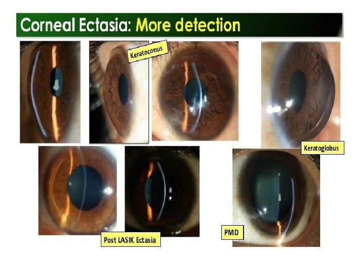
Tissue Retrieval • Contraindications: Systemic: • AIDS • Rabies • Active viral hepatitis • Creutzfeldt-Jakob disease • SSPE • Reye’s syndrome • Death from unknown causes • Congenital Rubella • Active septicemia • Leukemia (blast form) • Lymphoma/lymphosarco Ocular: • Intrinsic eye diseases ü Retinoblastoma ü Active conjuctivitis, iritis, uv eitis, vitritis, retinitis ü Congenital abnormalities (keratoconus) ü Central opacities, pterygium • Prior refractive procedures (radial

Preliminary preparations • Obtain legal permission. • Go through the donor’s medical records for any contraindications. • Wash hands and be prepared with aseptic dressing. • Identify the donor. • Collection of postmortem blood: 10 ml ü Femoral vein ü Subclavian vein ü Heart ü Jugular vein

Enucleation

Corneo-scleral button excision

Serological testing • • HIV HBV HCV Syphilis

Evaluation of donor tissue • Gross examination ü Whole globe: eyes with excessive stromal hydration should be discarded unless specular microscopy can be done for endothelial cell count. ü Corneoscleral button: Colour of the tissue storage media is to be noted to rule out contamination.

Evaluation of donor tissue • Slit Lamp Biomicroscopic examination

Endothelial cell count AGE Average Endothelial cell count 10 -19 2, 900 -3, 500 20 -29 2, 600 -3, 400 30 -39 2, 400 -3, 200 40 -49 2, 300 -3, 100 50 -59 2, 100 -2, 900 60 -69 2, 000 -2, 800 70 -79 1, 800 -2, 600 80 -89 1, 500 -2, 300 Critical cell density: 300 -500 cells/mm 2 Functional cell density: 1500 -2200 cells/mm 2 * Philips C, Laing R, Yee R. Specular Microscopy. In: Krachmer JH, Mannis MJ, Holland EJ (eds). Cornea, 2 nd ed. Philadelphia: Elsevier Mosby, 2005: 261 -77.

Exclusion Criteria for penetrating keratoplasty* • Cell density less than 2000 cells per square millimeter. (Corneas with cell density less than 2000 cells / sq. mm may be suitable for lamellar procedures). • Extreme polymegathism or pleomorphism. • Presence of significant guttata. • Presence of many non-hexagonal or abnormally shaped cells. • Presence of inflammatory cells, bacteria, or debris on endothelial surface. • Numerous vacuolated cells. *Standards of Eye banking in India 2009; NPCB; Director General of Health & Family Welfare, Govt. of India
")
Storage of donor tissue Storage Short term 2 -3 days Moist chamber (24 hrs) M-K medium Intermediate 7 -10 days K-sol, Dexsol, Optisol GS Long term 30 days Organ culture medium, MEM Very long term 1 year Cryopreservat ion

Preservation of cornea • Moist chamber storage ü Storage of whole globe ü 4◦ C ü 24 hours • Advantage: Simple • Disadvantage: Corneal stromal edema.

Preservation of cornea • o o o o o Tissue Media Cornea storage Dextran Media Chondroitin sulphate MK Electrolytes K-SOL p. H buffer system Antibiotics CSM Essential amino acids DEXSOL Antioxidants, ATP precursors OPTISOL Insulin Epidermal growth factor PROCELL Antiprotease, anticoagulants Storage time (days) 4 7 7 10 14 14

M-K medium: • Described by Mc-Carey & Kauffman. • Mixture of tissue culture medium (TC-199) and Dextran (5%, 40, 000 MW) • Buffer: HEPES (N hydroxyethyl- piperazine-Nethane Sulphonic acid) • Antibiotics: Penicillin, Gentamicin, Polymyxin • Storage period- 96 hrs. K-Sol: • Purified chondroitin sulphate in tissue culture medium (TC 199). • Storage: 7 -10 days in 40 C.

Preservation of cornea • Long term Organ Culture storage system ü MEM media(minimum essential media) ü Developed by Harry Eagle. ü 34 degree C ü Incubated at room temperature in nutrient medium ü Storage period : 30 days ü Advantage: Enables HLA matching • Very long time preservation: ü Cryopreservation

CORNEAL TRANSPLANTATION

Cornea as transplant • Immune privilege of cornea ü Absence of blood and lymphatic channel in the graft and its bed ü Absence of MHC class II APCs in the graft ü Reduced expression of MHC coded alloantigen on graft cells ü Immunosupressive microenvironment of aqueous humor. ü Anterior chamber associated immune deviation.

Corneal Transplantation • Corneal transplantation refers to surgical replacement of a full-thickness or lamellar portion of the host cornea with that of a donor eye. • Allograft/autograft • Full-thickness( Penetrating)/ Partial thickness (

Corneal Transplantation : Schematic

Types of Keratoplasty • Optical – to improve vision • Tectonic- to restore or preserve corneal integrity • Therapeutic- to remove infected corneal tissue

Keratoplasty : Schematic Diagram
 • Keratoconus • Post- cataract surgery edema • Corneal")
Indications of Penetrating Keratoplasty( PK) • Keratoconus • Post- cataract surgery edema • Corneal dystrophies and degenerations • Mechanical or chemical trauma • Microbial/postmicrobial keratitis • Congenital opacity

Lamellar keratoplasty • Lamellar keratoplasty refers to replacement of only a portion of the corneal layers of the host cornea with the graft. • Indications: -Opacification of superficial corneal stroma -Marginal thinning or infiltration -Localised thinning / descemetocele formation
 • Descemet")
Types of Lamellar Keratoplasty • Superficial/ Deep anterior lamellar keratoplasty ( SALK/DALK) • Descemet stripping automated endothelial keratoplasty (DSAEK) • Descemet membrane endothelial
")
LEGAL ASPECTS IN INDIA • Under the Transplantation of Human Organs Act, 1994 (THOA) 1. The qualification of doctors permitted to perform enucleation (surgical eye removal) has been reduced from MS (Ophth. ) to MBBS. 2. Eye donation in India is always decided by the donor’s surviving relatives and not by the actual donor. 3. Enucleating doctors always have to legally obtain a written consent from the relatives of the deceased before they actually

- Slides: 56