GNB Pathology lecture 2018 Pathology and mechanisms of

GNB Pathology lecture 2018 Pathology and mechanisms of malabsorption Dr. Maha Arafah Dr. Ahmed Al Humaidi

Objectives Upon completion of this lecture the students will: 1. Understand that the malabsorption is caused by either abnormal digestion or small intestinal mucosal disease 2. Know that malabsorption can affect many organ systems ( alimentary tract, hematopoietic system, musculoskeletal system, endocrine system, epidermis, nervous system) 3. Know the following aspects of celiac disease: a. definition b. pathogenesis c. clinical features d. pathology (gross and microscopic features) e. complications (T-cell lymphoma and GI tract carcinoma) 4. Know the cause and types of Lactose intolerance

Definition of malabsorption Malabsorption Syndrome Inability of the intestine to absorb nutrients adequately into the blood stream Impairment can be of single or multiple nutrients depending on the abnormality

Causes of malabsorption Physiology – The main purpose of the gastrointestinal tract is to digests and absorbs nutrients (fat, carbohydrate, and protein), micronutrients (vitamins and trace minerals), water, minerals and electrolytes.

Causes of malabsorption Mechanisms and Causes of Malabsorption Syndrome 1. Inadequate digestion 3. Primary mucosal abnormalities 4. Inadequate small intestine 2. Deficient bile salt 5. Lymphatic obstruction

Causes of malabsorption Mechanisms and Causes of Malabsorption Syndrome Primary mucosal abnormalities Celiac disease Tropical sprue Whipple's disease Amyloidosis Radiation enteritis Abetalipoproteinemia Giardiasis Inadequate small intestine Intestinal resection Crohn's disease Mesenteric vascular disease with infarction Jejunoileal bypass Lymphatic obstruction Intestinal lymphangiectasia Malignant lymphoma Macroglobulinemia M an y ca us es Inadequate digestion Postgastrectomy Deficiency of pancreatic lipase Chronic pancreatitis Cystic fibrosis Pancreatic resection Zollinger-Ellison syndrome Deficient bile salt Obstructive jaundice Bacterial overgrowth Stasis in blind loops, diverticula Fistulas Hypomotility states (diabetes) Terminal ileal resection Crohns' disease Precipitation of bile salts (neomycin)

Causes of malabsorption Pathophysiology Inadequate digestion Or Small intestine abnormalities = Malabsorption

Causes of malabsorption Pathophysiology Inadequate digestion Stomach Pancreas Bile Small intestine abnormalities mucosa Inadequate small intestine Lymphatic obstruction Postgastrectomy Deficiency of pancreatic lipase Chronic pancreatitis Cystic fibrosis Pancreatic resection Obstructive jaundice Terminal ileal resection

Causes of malabsorption Pathophysiology Inadequate digestion Stomach Pancrease Bile Small intestine abnormalities mucosa Inadequate small intestine Lymphatic obstruction Celiac disease Tropical sprue Whipple's disease Giardiasis Intestinal resection Crohn's disease Intestinal lymphangiectasia Malignant lymphoma

Know that malabsorption can affect many organ systems Malabsorption Syndrome Clinical features 1. There is increased fecal excretion of fat (steatorrhea) and the systemic effects of deficiency of vitamins, minerals, protein and carbohydrates. Steatorrhea is passage of soft, yellowish, greasy stools containing an increased amount of fat 2. Growth retardation and failure to thrive in children 3. Weight loss despite increased oral intake of nutrients

Know that malabsorption can affect many organ systems Systemic effects of the malabsorption syndromes • Weight loss and anorexia • Abdominal distension and borborygmi (increased bowel sounds) • Diarrhoea (loose, bulky stools) • Steatorrhoea—malabsorption of fat, producing pale, foul-smelling stools that characteristically float in water • Muscle wasting

Know that malabsorption can affect many organ systems Malabsorption Syndrome Clinical features Depend on the deficient nutrient Protein Swelling or oedema Anaemias B 12, folic acid and iron deficiency vitamin D, calcium Muscle cramp vitamin K and other coagulation factor (fatigue and weakness) Osteomalacia and osteoporosis Bleeding tendencies

Know that malabsorption can affect many organ systems Clinical features

Diagnosis of malabsorption Diagnosis There is no specific test for malabsorption. Investigation is guided by symptoms and signs. 1. Fecal fat study to diagnose steatorrhoea 2. Blood tests 3. Endoscopy Biopsy of small bowel

Celiac disease Malabsorption Syndrome Celiac disease An immune reaction to gliadin fraction of the wheat protein gluten Usually diagnosed in childhood – mid adult. Patients have raised antibodies to gluten autoantibodies Highly specific association with class II HLA-DQ 2 or HLA-DQ 8 alleles.

Celiac disease a 33 -amino acid peptide resistant to degradation by gastric, pancreatic, and small intestinal proteases

Celiac disease Clinical features Celiac disease Typical presentation GI symptoms that characteristically appear at age 9 -24 months. Symptoms begin at various times after the introduction of foods that contain gluten. A relationship between the age of onset and the type of presentation; Infants and toddlers…. GI symptoms and failure to thrive Childhood…………………minor GI symptoms, inadequate rate of weight gain, Young adults……………anemia is the most common form of presentation. Adults and elderly…. . . GI symptoms are more prevalent

Celiac disease Endoscopy Normal Celiac disease

Celiac disease Celiac Disease Histology • Mucosa is flattened with marked villous atrophy. • Increased • Crypt Subtotal villous atrophy intraepithelial lymphocytosis elongation Normal Total villous atrophy

Celiac disease Celiac Disease Diagnosis Clinical documentations of malabsorption. Stool ………. Fat Serology is +ve for Ig. A to tissue transglutaminase or Ig. G to deamidated gliadin or anti-endomysial antibodies Small intestine biopsy demonstrate villous atrophy. Improvement of symptom and mucosal histology on gluten withdrawal from diet. wheat, barley, flour Other grains, such as rice and corn flour, do not have such an effect.

Celiac disease Celiac Disease Complications Osteopenia , osteoporosis Infertility in women Short stature, delayed puberty, anemia, Malignancies: [intestinal T-cell lymphoma] 10 to 15% risk of developing GI lymphoma.

Lactose Intolerance

Lactose Intolerance Pathophysiology lactase Lactose At the brush border of enterocytes Lactose Intolerance glucose + galactose Low or absent activity of the enzyme lactase

Lactose Intolerance causes Inherited lactase deficiency Congenital lactase deficiency Childhood-onset and adult-onset lactase deficiency extremely rare common Genetically programmed progressive loss of the activity of the small intestinal enzyme lactase. Gastroenteritis: Infectious diarrhea, particularly viral gastroenteritis in younger children, may damage the intestinal mucosa enough to reduce the quantity of the lactase enzyme Acquired lactase deficiency Transient Secondary lactase deficiency due to intestinal mucosal injury by an infectious, allergic, or inflammatory process

Lactose Intolerance Clinical Bloating, abdominal discomfort, and flatulence …………… 1 hour to a few hours after ingestion of milk products

Lactose Intolerance

Lactose Intolerance Diagnosis Empirical treatment with a lactose-free diet, which results in resolution of symptoms; Hydrogen breath test

Lactose Intolerance Hydrogen breath test. • An oral dose of lactose is administered • The sole source of H 2 is bacterial fermentation; • Unabsorbed lactose makes its way to colonic bacteria, resulting in excess breath H 2. • Increased exhaled H 2 after lactose ingestion suggests lactose malabsorption.

Lactose Intolerance A 3 -week trial of a diet that is free of milk and milk products is a satisfactory trial to diagnose lactose intolerance

Lactose Intolerance summary • Deficiency/absence of the enzyme lactase in the brush border of the intestinal mucosa → maldigestion and malabsorption of lactose • Unabsorbed lactose draws water in the intestinal lumen • In the colon, lactose is metabolized by bacteria to organic acid, CO 2 and H 2; acid is an irritant and exerts an osmotic effect • Causes diarrhea, gaseousness, bloating and abdominal cramps


Questions A 33 -year-old man with a 5 year history of chronic diarrhea has recently lost 6 g and become chronically fatigued. A biopsy specimen of his jejunum (top) is compared with a normal jejunal biopsy specimen. After a change in diet, he improves.

Q 1. What is your diagnosis? The flattening with atrophy of the villi, the loss of goblet cells, and the chronic inflammation are features of celiac disease (nontropical sprue or gluten-sensitive enteropathy). Q 2. What serologic tests can aid in diagnosis? Testing for antiendomysial antibodies (directed against transglutaminase in epithelial cells), antigliadin antibodies, and antireticulin antibodies may be helpful. Q 3. What was the dietary change? Gluten (found in wheat, rye, or barley flour) contains the gliadin protein that binds to epithelial cells; cytotoxic T cell injury directed against gliadin-modified cells leads to cell loss. The removal of gluten (gliadin) from the diet removes the antigenic stimulus to the T cells, and the epithelium is eventually restored. Q 4. What skin condition can accompany this disease? Dermatitis herpetiformis, with sub-basilar skin blistering and associated Ig. A deposition, may occur with this condition.

Question • A 23 -year-old woman from Hong Kong who is living in Zurich has had bouts of explosive, watery diarrhea with abdominal bloating since her arrival 6 months ago. Her stool has no occult blood, ova, parasites, or bacterial pathogens. Her laboratory findings are shown.
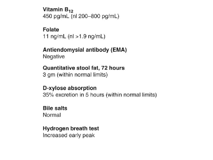

1. What is your diagnosis? lactase deficiency. Disaccharidases are secreted from apical enterocytes, with infants expressing more than adults. Europeans typically have more lactase expression than Asians do. 2. What is the pathogenesis? When disaccharides are not absorbed, gut bacteria have increasing substrate to ferment, which produces hydrogen that is absorbed and exhaled. It is the bacterial overgrowth and osmotic diarrhea that cause the unpleasant symptoms. 3. How can this be treated? Avoiding foods containing lactose sugar include dairy products will reduce the symptoms.
- Slides: 37