
Connective Tissue Components Dr Jack L Haar I

- Slides: 55
Connective Tissue Components Dr. Jack L. Haar I. Introduction – A. Distribution and role – B. Embryonic origin 1. Mesoderm 2. Neural crest
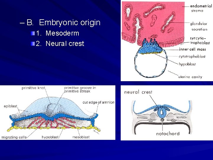
– B. Embryonic origin 1. Mesoderm 2. Neural crest
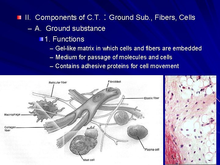
II. Components of C. T. : Ground Sub. , Fibers, Cells – A. Ground substance 1. Functions – Gel-like matrix in which cells and fibers are embedded – Medium for passage of molecules and cells – Contains adhesive proteins for cell movement
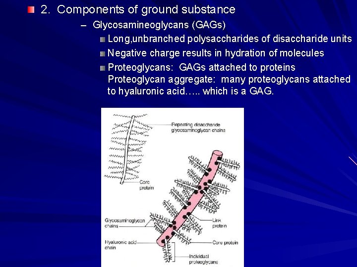
2. Components of ground substance – Glycosamineoglycans (GAGs) Long, unbranched polysaccharides of disaccharide units Negative charge results in hydration of molecules Proteoglycans: GAGs attached to proteins Proteoglycan aggregate: many proteoglycans attached to hyaluronic acid…. . which is a GAG.
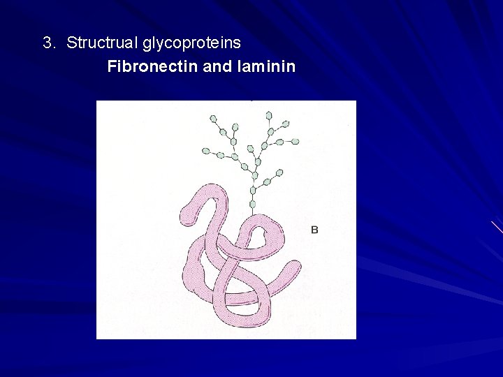
3. Structrual glycoproteins Fibronectin and laminin
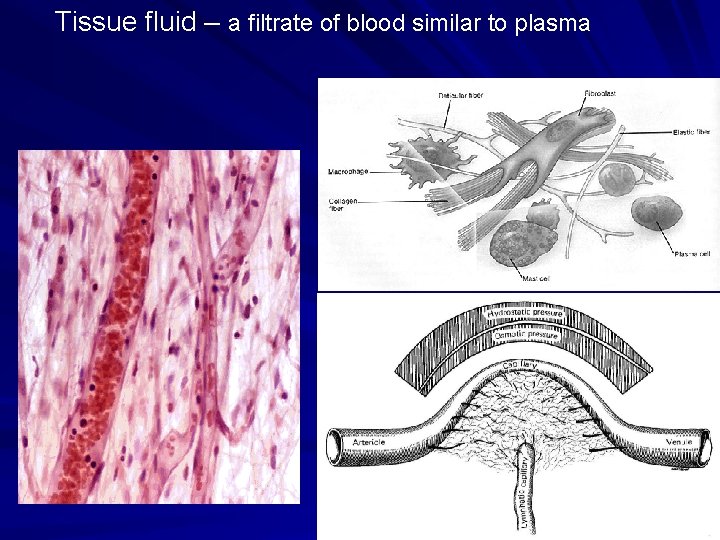
Tissue fluid – a filtrate of blood similar to plasma
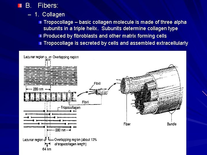
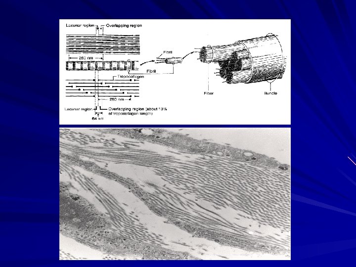
B. Fibers: – 1. Collagen Tropocollage – basic collagen molecule is made of three alpha subunits in a triple helix. Subunits determine collagen type Produced by fibroblasts and other matrix forming cells Tropocollage is secreted by cells and assembled extracellularly
Types of Collagen type I: found in dermis of skin, bone, tendon, dentin, fascia, sclera, organ capsules, fibrous cartilage; Collagen type II: very thin fibrins found in hyaline and elastic cartilage; Collagen type III: the major component of reticular fiber; Collagen type IV: does not form fibrils or fibers; found in the basal lamina underlying epithelial cells.
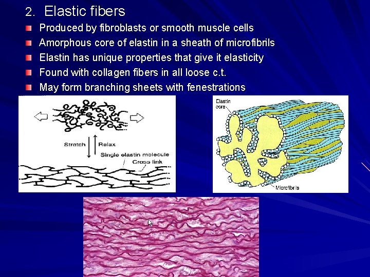
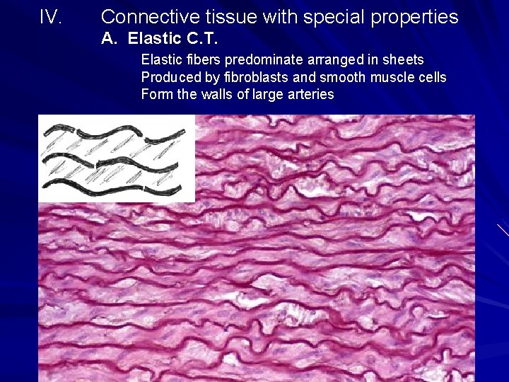
2. Elastic fibers Produced by fibroblasts or smooth muscle cells Amorphous core of elastin in a sheath of microfibrils Elastin has unique properties that give it elasticity Found with collagen fibers in all loose c. t. May form branching sheets with fenestrations
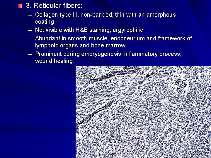
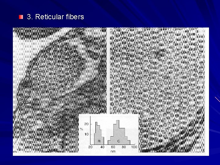
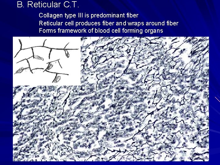
3. Reticular fibers: – Collagen type III, non-banded, thin with an amorphous coating – Not visible with H&E staining; argyrophilic – Abundant in smooth muscle, endoneurium and framework of lymphoid organs and bone marrow – Prominent during embryogenesis, inflammatory process, wound healing.
3. Reticular fibers
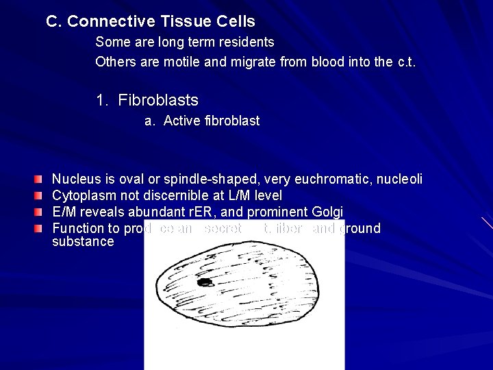
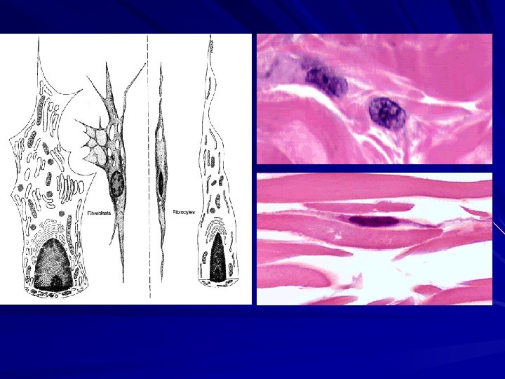
C. Connective Tissue Cells Some are long term residents Others are motile and migrate from blood into the c. t. 1. Fibroblasts a. Active fibroblast Nucleus is oval or spindle-shaped, very euchromatic, nucleoli Cytoplasm not discernible at L/M level E/M reveals abundant r. ER, and prominent Golgi Function to produce and secrete c. t. fibers and ground substance
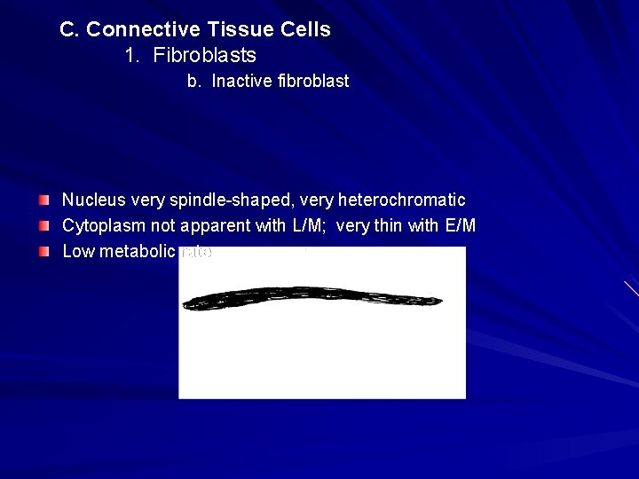
C. Connective Tissue Cells 1. Fibroblasts b. Inactive fibroblast Nucleus very spindle-shaped, very heterochromatic Cytoplasm not apparent with L/M; very thin with E/M Low metabolic rate
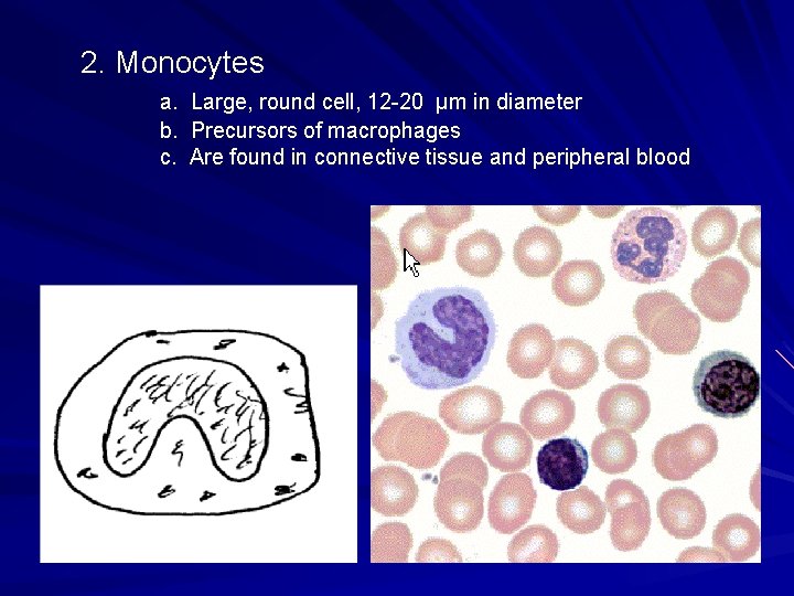
2. Monocytes a. b. c. Large, round cell, 12 -20 μm in diameter Precursors of macrophages Are found in connective tissue and peripheral blood
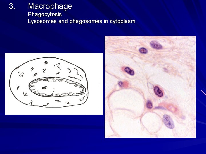
3. Macrophage Phagocytosis Lysosomes and phagosomes in cytoplasm
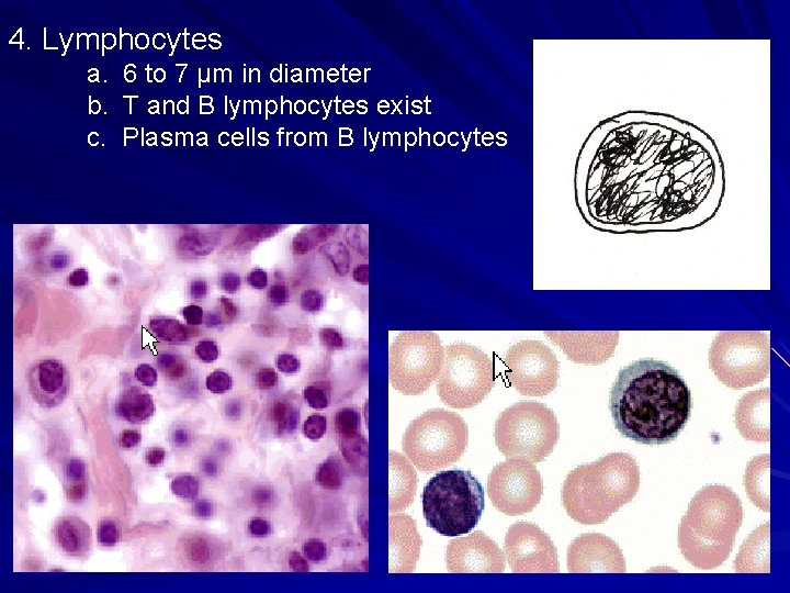
4. Lymphocytes a. b. c. 6 to 7 μm in diameter T and B lymphocytes exist Plasma cells from B lymphocytes
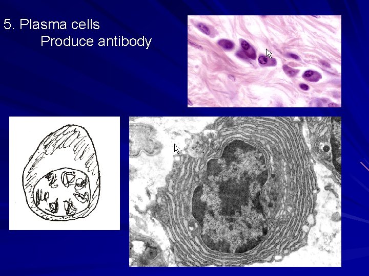
5. Plasma cells Produce antibody
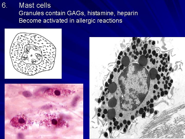
6. Mast cells Granules contain GAGs, histamine, heparin Become activated in allergic reactions
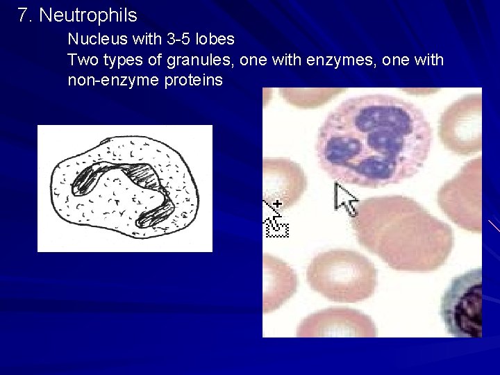
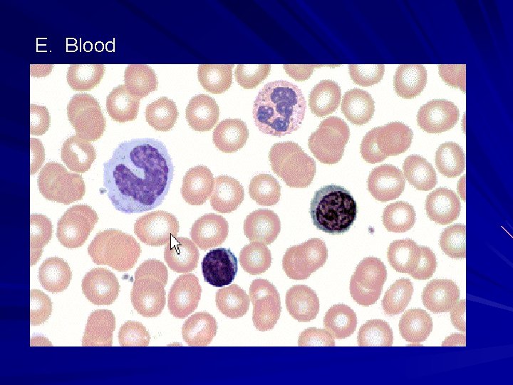
7. Neutrophils Nucleus with 3 -5 lobes Two types of granules, one with enzymes, one with non-enzyme proteins
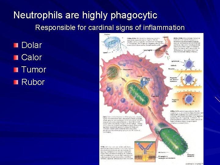
Neutrophils are highly phagocytic Responsible for cardinal signs of inflammation Dolar Calor Tumor Rubor
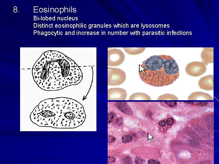
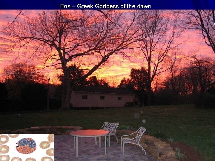
8. Eosinophils Bi-lobed nucleus Distinct eosinophilic granules which are lysosomes Phagocytic and increase in number with parasitic infections
Eos – Greek Goddess of the dawn
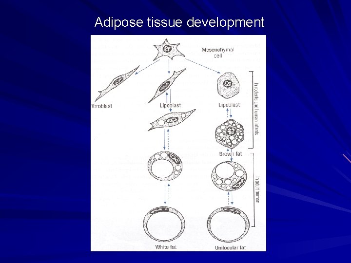
Adipose tissue development
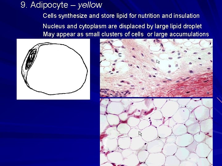
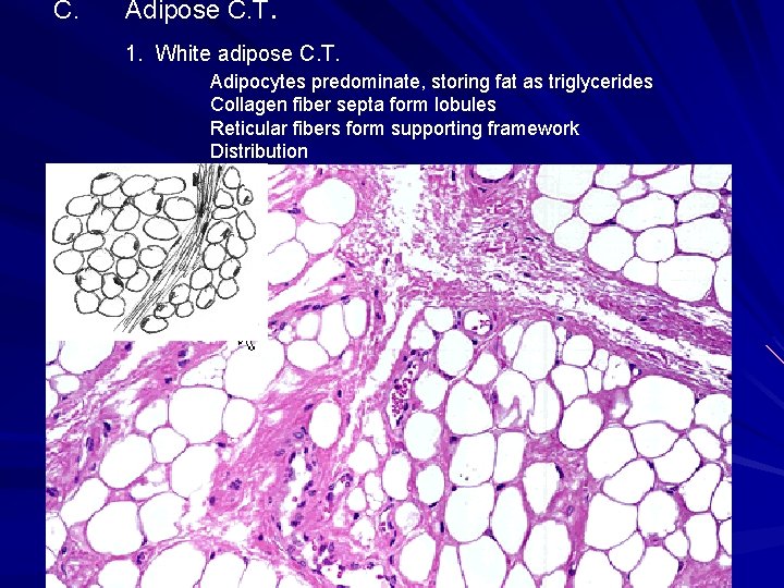
9. Adipocyte – yellow Cells synthesize and store lipid for nutrition and insulation Nucleus and cytoplasm are displaced by large lipid droplet May appear as small clusters of cells or large accumulations
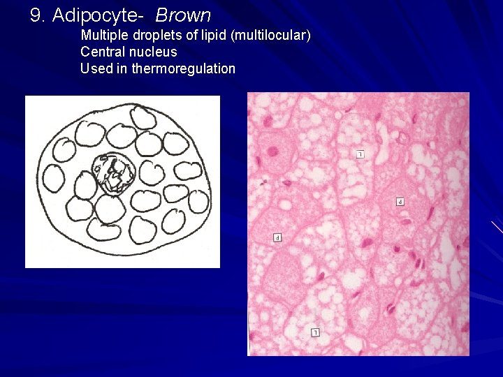
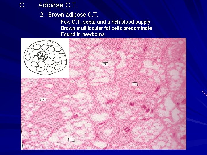
9. Adipocyte- Brown Multiple droplets of lipid (multilocular) Central nucleus Used in thermoregulation
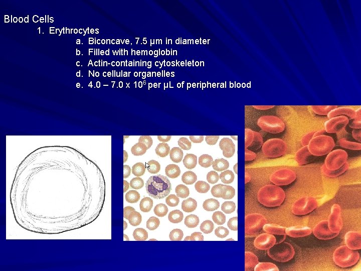
Blood Cells 1. Erythrocytes a. Biconcave, 7. 5 μm in diameter b. Filled with hemoglobin c. Actin-containing cytoskeleton d. No cellular organelles e. 4. 0 – 7. 0 x 106 per μL of peripheral blood
Leukocytes – 6, 000 to 10, 000 per μL of peripheral blood a. b. c. d. e. Neutrophils : 40 -75% of WBC’s Eosinophils: 5% of WBC’s Lymphocytes: 20 -50% of WBC’s Monocytes: 1 -5% of WBC’s Basophils: 0. 5% of WBC’s
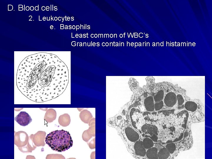
D. Blood cells 2. Leukocytes e. Basophils Least common of WBC’s Granules contain heparin and histamine
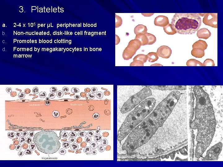
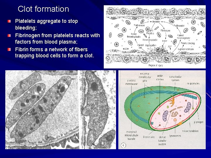
3. Platelets a. b. c. d. 2 -4 x 10⁵ per μL peripheral blood Non-nucleated, disk-like cell fragment Promotes blood clotting Formed by megakaryocytes in bone marrow
Clot formation Platelets aggregate to stop bleeding; Fibrinogen from platelets reacts with factors from blood plasma; Fibrin forms a network of fibers trapping blood cells to form a clot.
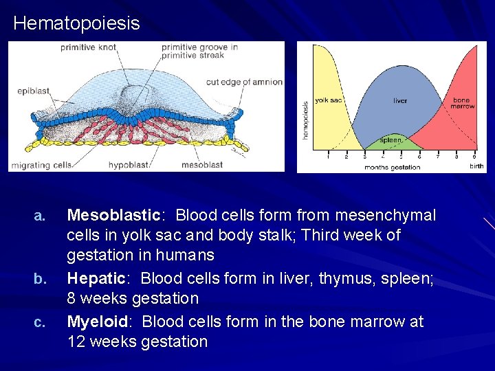
Hematopoiesis a. b. c. Mesoblastic: Blood cells form from mesenchymal cells in yolk sac and body stalk; Third week of gestation in humans Hepatic: Blood cells form in liver, thymus, spleen; 8 weeks gestation Myeloid: Blood cells form in the bone marrow at 12 weeks gestation
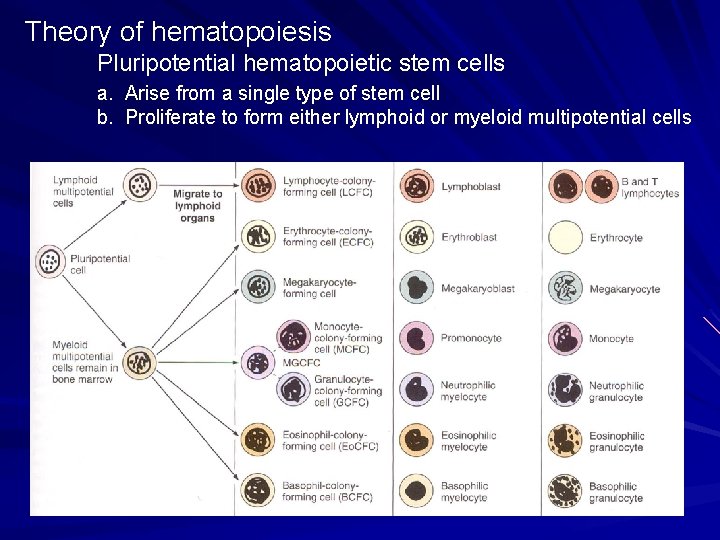
Theory of hematopoiesis Pluripotential hematopoietic stem cells a. Arise from a single type of stem cell b. Proliferate to form either lymphoid or myeloid multipotential cells
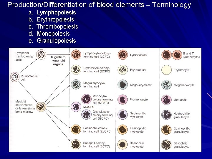
Production/Differentiation of blood elements – Terminology a. b. c. d. e. Lymphopoiesis Erythropoiesis Thrombopoiesis Monopoiesis Granulopoiesis
http: //www. path. uiowa. edu/virtualslidebox/ Table of Contents Supporting tissue Tendon #25 Dense regular CT Inactive fibroblast Adipocytes Integument Axilla-skin #78 Loose (areolar) CT Dense irrecular CT http: //java. vcu. edu/som-histology/
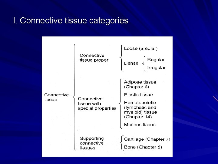
I. Connective tissue categories
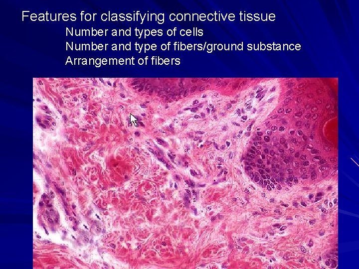
Features for classifying connective tissue Number and types of cells Number and type of fibers/ground substance Arrangement of fibers
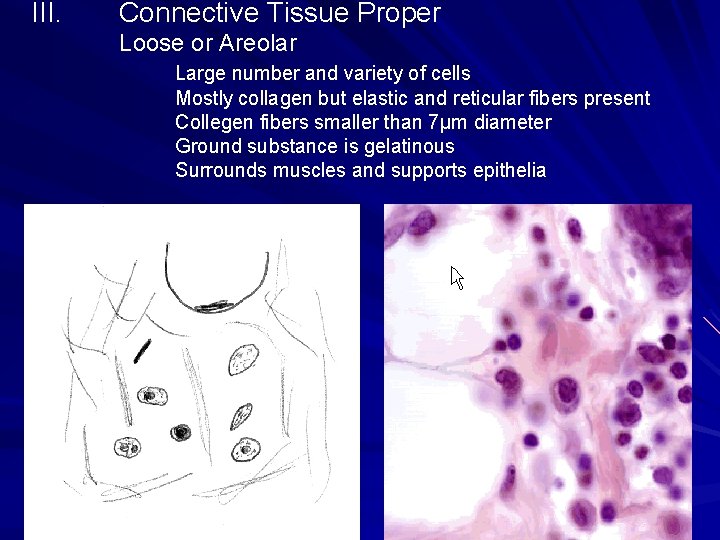
III. Connective Tissue Proper Loose or Areolar Large number and variety of cells Mostly collagen but elastic and reticular fibers present Collegen fibers smaller than 7μm diameter Ground substance is gelatinous Surrounds muscles and supports epithelia
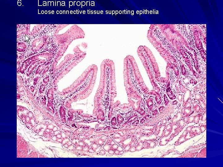
6. Lamina propria Loose connective tissue supporting epithelia
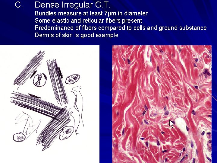
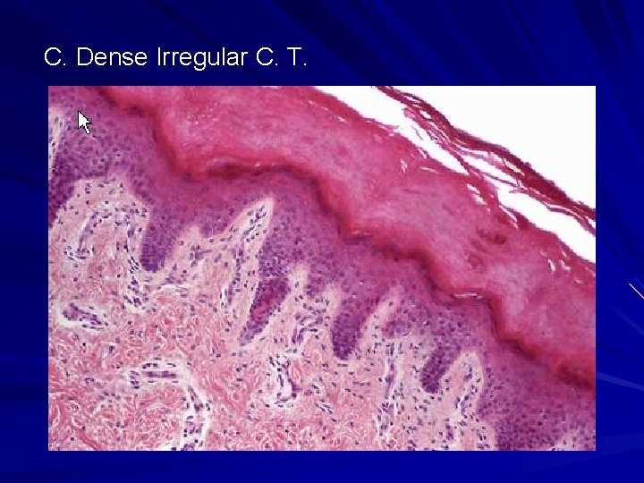
C. Dense Irregular C. T. Bundles measure at least 7μm in diameter Some elastic and reticular fibers present Predominance of fibers compared to cells and ground substance Dermis of skin is good example
C. Dense Irregular C. T.
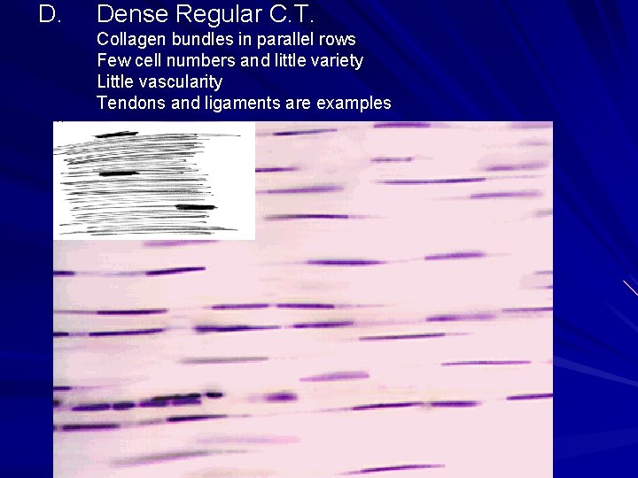
D. Dense Regular C. T. Collagen bundles in parallel rows Few cell numbers and little variety Little vascularity Tendons and ligaments are examples
IV. Connective tissue with special properties A. Elastic C. T. Elastic fibers predominate arranged in sheets Produced by fibroblasts and smooth muscle cells Form the walls of large arteries
B. Reticular C. T. Collagen type III is predominant fiber Reticular cell produces fiber and wraps around fiber Forms framework of blood cell forming organs
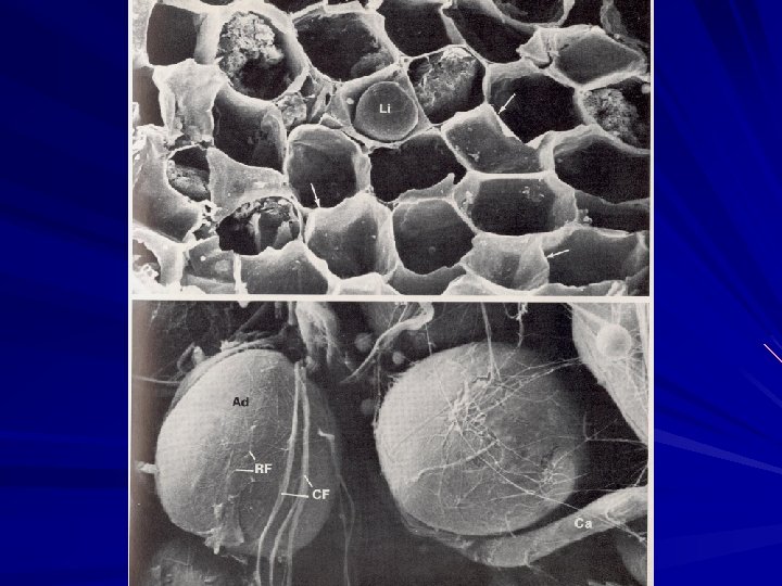
C. Adipose C. T. 1. White adipose C. T. Adipocytes predominate, storing fat as triglycerides Collagen fiber septa form lobules Reticular fibers form supporting framework Distribution
C. Adipose C. T. 2. Brown adipose C. T. Few C. T. septa and a rich blood supply Brown multilocular fat cells predominate Found in newborns
D. Mucous (Mucoid) C. T.
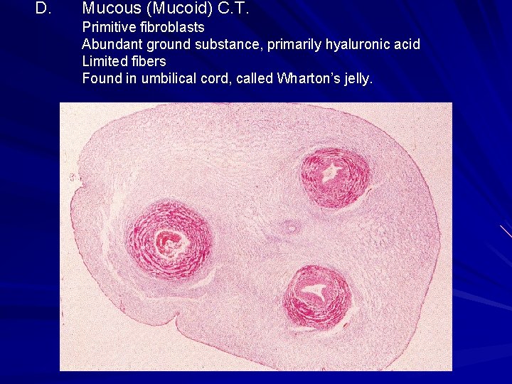
D. Mucous (Mucoid) C. T. Primitive fibroblasts Abundant ground substance, primarily hyaluronic acid Limited fibers Found in umbilical cord, called Wharton’s jelly.
E. Blood
http: //www. path. uiowa. edu/virtualslidebox/ Table of Contents Supporting tissue Tendon #25 Dense regular CT Inactive fibroblast Adipocytes Integument Axilla-skin #78 Loose (areolar) CT Dense irrecular CT
http: //www. path. uiowa. edu/virtualslidebox/ Table of Contents Supporting tissue Areolar connective tissue Fat (adipose tissue)
Digital Histology Site at VCU http: //java. vcu. edu/som-histology/