HISTOLOGY OF LOWER PART OF RESPIRATORY SYSTEM RESPIRATORY


 - Serous secretion – keeps lumen moist")

, blood vessels")


 • Type I alveolar cells: - simple squamous cells (flat")

: - Phagocytose the dust particles")


- Slides: 43
HISTOLOGY OF LOWER PART OF RESPIRATORY SYSTEM
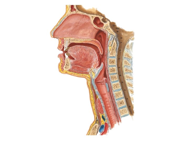
RESPIRATORY EPITHELIUM Most of the conducting portion is lined by pseudostratified ciliated columnar epithelium with rich population of goblet cells called respiratory epithelium.
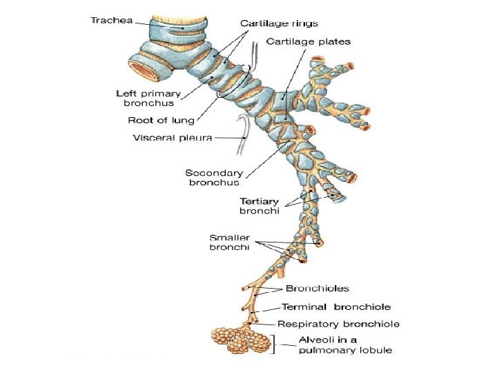
Bronchial tree
INTRAPULMONARY PART OF CONDUCTING SYSTEM Primary bronchus divides into , Secondary bronchus or lobar bronchus. Tertiary bronchus/ segmental bronchus Bronchioles Terminal bronchiole: last portion of conducting system
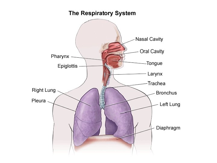
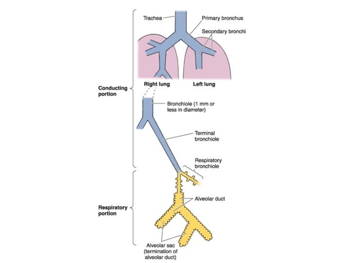
FUNCTIONAL COMPONENTS OF RESPIRATORY SYSTEM • CONDUCTING PORTION or cleaning system • RESPIRATORY PORTION or gaseous interchange system
TRACHEA
TRACHEA 1. Mucosa 2. Submucosa 3. Cartilaginous/smooth muscle layer 4. Adventitia
TRACHEA
TRACHEA
Mucosa • Common types of cells: - Pseudo-stratified ciliated columnar - Goblet cells - Basal cells (stem cells)
• Less common types of cells are – - Brush cells ( seen in association with nerve fibres, sensory function) - Argentaffin cells ( small granule cells): reflex regulating function
Lamina propria • Situated underneath the epithelium • Made up of loose connective tissue rich in elastic fibres
Submucosa • Contains seromucous glands (tracheal glands) - Serous secretion – keeps lumen moist - Mucous secretion- traps the small dust particles
Cartilaginous/smooth muscle layer • Hyaline cartilage with perichondrium forms anterior two thirds of tracheal circumference. • Posterior one third is completed by smooth muscles with fibroelastic tissue(trachealis muscle).
Adventitia • Connective tissue (elastic and collagen fibres), blood vessels
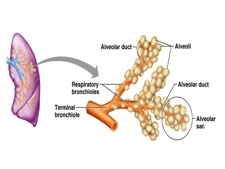
Lung • 2 parts – - Conducting part ( intrapulmonary bronchi, bronchioles and different orders of braching) - Respiratoty part ( alveolar ducts, alveolar sacs and alveoli)
LOBAR BRONCHUS OR SECONDARY BRONCHUS
BRONCHIOLE
Bronchus Bronchiole Lined by pseudostratified Lined by columnar epithelium ciliated epithelium Islands of hyaline cartilage are Cartilage is absent present More than 1 mm in diameter Less than 1 mm in diameter Seromucus glands are present Glands are absent Goblet cells are present Goblet cells are absent
RESPIRATORY EPITHELIUM Most of the conducting portion is lined by pseudostratified ciliated columnar epithelium with rich population of goblet cells called respiratory epithelium. Typical respiratory epithelium consists 8 types of cells that lines the tracheo-bronchial passages. They are, 1. Ciliated columnar cells. 2. Goblet cells.
3 Serous cells 4 Clara cells 5 Basal cells 6 Intermediate epitheliocytes 7 Brush cells 8 Kulchitsky (P cells or neuroendocrine or Argentaffin cells)
Different types of cells present in the intrapulmonary bronchial tree
CILIATED COLUMNAR CELL • Tall or low columnar cells • Beating of the cilia moves mucous forward • Each cell has 300 cilia
GOBLET CELL • These produce mucous which trap dust particles. • Present throughout except bronchiole.
SEROUS CELL • Apical – mucin vacuoles • Secretes Ig A antibody
• CLARA CELL Only in respiratory bronchiole • Similar to type II alveolar cell • Cuboidal non ciliated cells, has blunt projections • It contain electron dense secretory granules and many lysosomes. • Important source of surfactant
BRUSH CELL • These are sensory in function • Slender cells with microvilli. • They are in contact with afferent nerve fibers and so are consider as sensory receptor in function.
SMALL GRANULE Kulchitsky / CELL • Function neuroendocrine cell – Regulates bronchial secretion – Smooth muscle contraction – Ciliary action – Secrete serotonin, may stimulate the bronchial muscle. • Rounded shape, dense cored vesicles.
INTERMEDIATE CELL • These immature forms of other cells
BASAL CELL • These are stem cells. • These cells are mitotic stem cells for other type of epithelial cells. • Small rounded cells are in contact with basal lamina.
Respiratory part • Alveolar ducts • Alveolar sacs • Alveoli
Alveolar epithelial cells (pneumocytes) • Type I alveolar cells: - simple squamous cells (flat cells with scanty cytoplasm)
• Type II alveolar cells: - Rounded cells , protrudes from alveolar surface - Free surface bears microvilli - Produces surfactant
• Alveolar macrophages (Dust cells/ Heart failure cells): - Phagocytose the dust particles - Presence of these cells in sputum is of diagnostic importance. - If found with RBC’s , it is indicative of CCF.
Surfactant - complex mixture of lipid and protein - Forms a thin film over the alveolar surface - Derived from Clara cells and type II pneumocytes - Reduces the surface tension and makes the ventilation of alveoli much more efficient.
Identification points • TRACHEA 1. Lumen is lined by pseudostratified ciliated columnar epithelium with goblet cells. 2. Submucous layer has seromucous glands. 3. Anterior 2/3 rd has cartilagenous layer which has hayline cartilage with perichondrium. 4. Posterior 1/3 rdpart has smooth muscle which is called as trachealis.
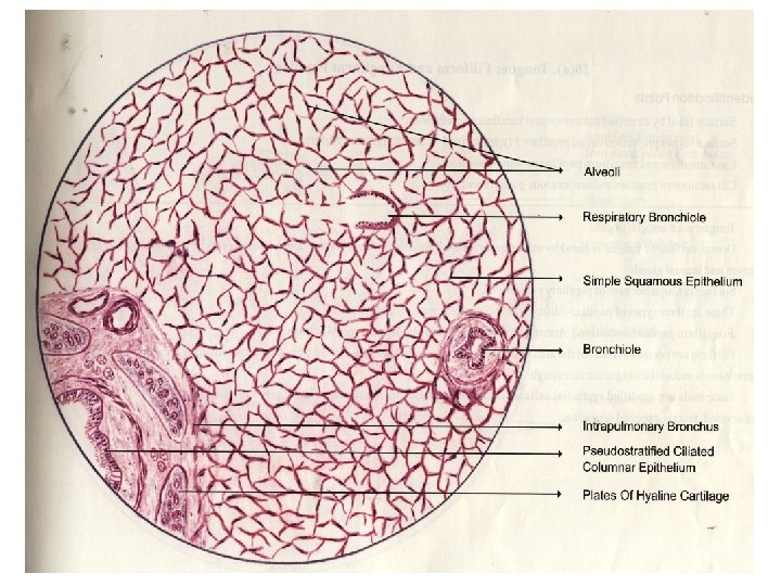
Identification points • LUNGS 1. Conducting part has cut section of bronchus and bronchiloes. 2. Respiratory part has alveoli lined by simple squamous epithelium. 3. Lumen of bronchus is lined by pseudostratified ciliated columnar epithelium with goblet cells. 4. Lumen of bronchiole is lined by simple columnar epithelium with cilia.