The ear The ear is divided into the


 is deficient superiorly which forms a notch. From the")


 cavity")


 cavity")


 wall : In its upper part, there is the opening of")


, Above and behind the promontory closed by")
 below the posterior end of the promontory closed")

 cavity: 1 -Air. 2 -Auditory ossicles. 3 -Muscles")


 to the")


 Insertion: into neck")


")


 in")


 At the geniculate ganglion: • Greater petrosal nerve. • Communicating branch with")

 IN the facial canal: 2 -Nerve to stapedius 3 -Chorda tympani.")

 Temporal bone, petrosal portion. b) Temporal bone, mastoid portion. c) Promontorium. d) Oval")
")

, the parotid, the")


- Slides: 126
The ear
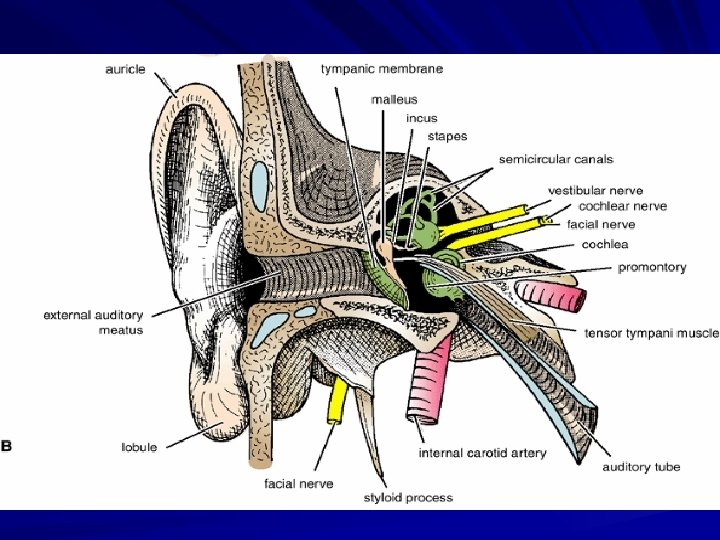
The ear is divided into the external ear , the middle ear and the inner ear
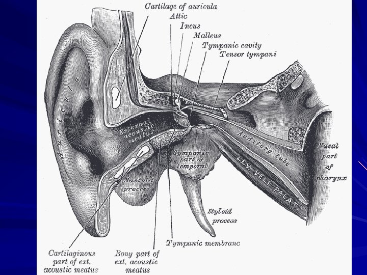
The external ear: It consists of the auricle, external auditory meatus and the tympanic membrane
1 -The auricle: it serves to collect air vibrations. it consists of a thin plate of elastic cartilage covered by skin. it has the following parts: 1 -Lobule 2 -The helix. 3 -The antihelix 4 -The tragus 5 - The antitragus 6 -The concha
antihelix antitragus
2 -The external auditory meatus: It is the passage between the concha of the auricle and the tympanic membrane. It is 1 inch long.
Its lateral 1/3 is cartilaginous directed upward and backward and its medial 2/3 are bony directed downwards and forwards.
One at the junction between the cartilaginous and osseus parts and the other lie about 5 mm from the tympanic membrane and is called the isthmus.
The meatus is lined by skin and its outer third is provided with hairs and ceruminous glands which secrete a yellowish-brown wax.
The hairs and the wax provide a sticky barrier that prevents the entrance of foreign bodies.
The sensory nerve supply of the lining skin is derived from the auriculotemporal nerve and the auricular branch of the vagus nerve.
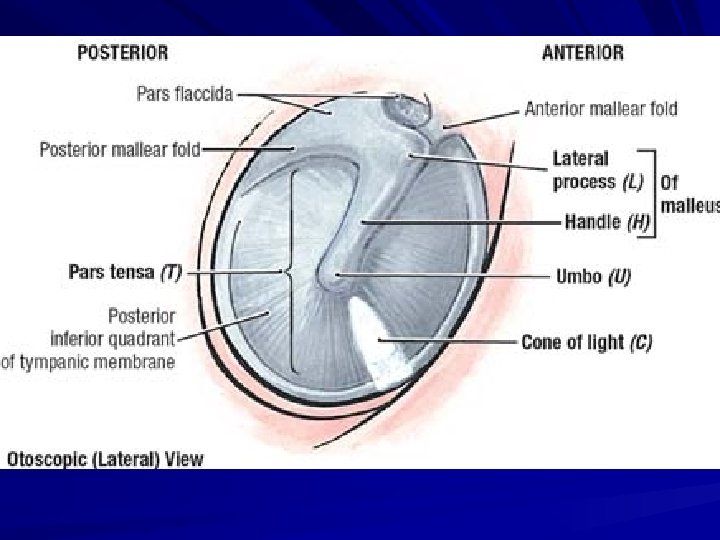
3 -The tympanic membrane: It is a thin membrane separates the external acoustic meatus from the middle ear cavity.
It is convex towards the middle ear and concave towards the outside.
The point of deepest concavity is called the umbo produced by the tip of the handle of the malleus.
The tympanic membrane is circular about 1 cm in diameter. The greater part of its circumference is fixed into a groove in the bone.
The groove or (tympanic sulcus) is deficient superiorly which forms a notch. From the sides of the notch, two bands, termed the anterior and posterior malleolar folds pass to the lateral process of the malleus.
The small triangular area on the tympanic membrane that is bounded by the folds is called the pars flaccida. The remainder of the membrane is called the pars tensa.
Tympanic membrane
The wall of the tympanic membrane is formed of three layers. The outer layer is formed by skin, the middle layer is formed of fibrous tissue which is deficient at the pars flaccida and the inner layer is formed of mucous membrane.
The outer surface of the tyrnpanic membrane is innervated by the auriculotemporal nerve and the auricular branch of the vagus. Its inner surface is supplied by the tympanic branch of the glossopharyngeal nerve.
Clinical notes: ►The tympanic membrane is extremely sensitive to pain. ► Myringotomy is an incision through the anteroinferior quadrant of the tympanic membrane and it provides drainage in individuals with chronic otitis media. Because the superior half of the tympanic membrane is much more vascular than the inferior half, incisions are made posteroinferiorly through the membrane. This incision also avoids injury to the chorda tympani nerve and auditory ossicles.
Middle ear (tympanic) cavity
The middle ear is an air-containing cavity in the petrous part of the temporal bone.
It is a narrow, oblique, slit-like cavity lined with mucous membrane. It is bounded laterally by the tympanic membrane and medially by the inner ear.
Posteriorly, it communicates with the mastoid antrum which connects it with the mastoid air cells. Anteriorly, it communicates with the nasopharynx through the auditory tube.
It is divided into: middle ear cavity proper and epitympanic recess
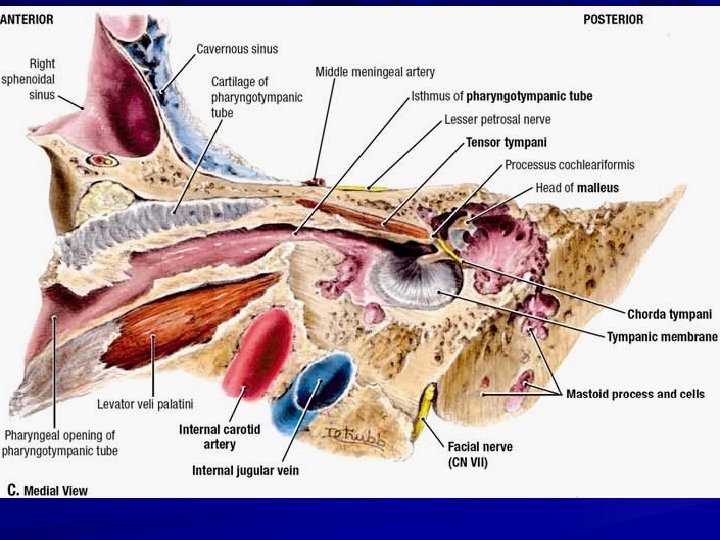
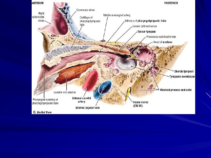
Boundaries of the middle ear (tympanic) cavity
The roof: is formed by the tegmen tympani which separates the tympanic cavity from the middle cranial fossa.
The floor is formed by a thin plate of bone, that separates the tympanic cavity from the superior bulb of the internal jugular vein.
The anterior (carotid) wall : In its upper part, there is the opening of the canal for the tensor tympani muscle. In its intermediate part, there is the opening of the auditory tube.
Its lower part is formed by a thin plate of bone which separates the tympanic cavity from the carotid canal.
The posterior wall : its upper part shows a large, irregular opening, the aditus to the mastoid antrum. Below this is a small, hollow, conical projection, the pyramid.
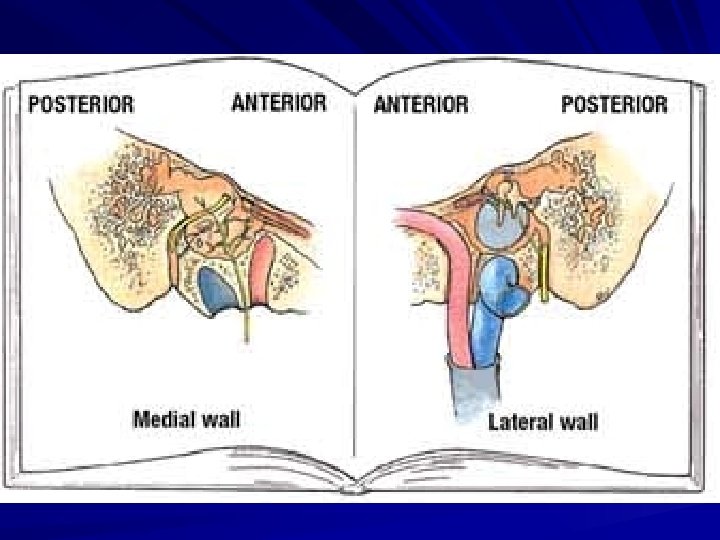
The lateral wall : is formed by the tympanic membrane. Above the tympanic membrane, the lateral wall is formed by the squamous part of the temporal bone.
The region of the cavity opposite the tympanic membrane is the tympanic cavity proper and the region above the level of the tympanic membrane is the epitympanic recess.
The medial wall is formed by the lateral wall of the inner ear. It shows: 1 -The promontory, is a rounded projection results from underlying first turn of cochlea.
2 - The oval window (fenestra vestibuli), Above and behind the promontory closed by the base of the stapes.
3 -The round window (fenestra cochleae) below the posterior end of the promontory closed by the secondary tympanic membrane.
The medial side of this window faces the perilymph of the scala tympani. The facial canal runs horizontally backward above the promontory and the oval window. Then, it curves downwards to descend in the posterior wall.
Contents of the middle ear (tympanic) cavity: 1 -Air. 2 -Auditory ossicles. 3 -Muscles of the middle ear. 4 -The chorda tympani.
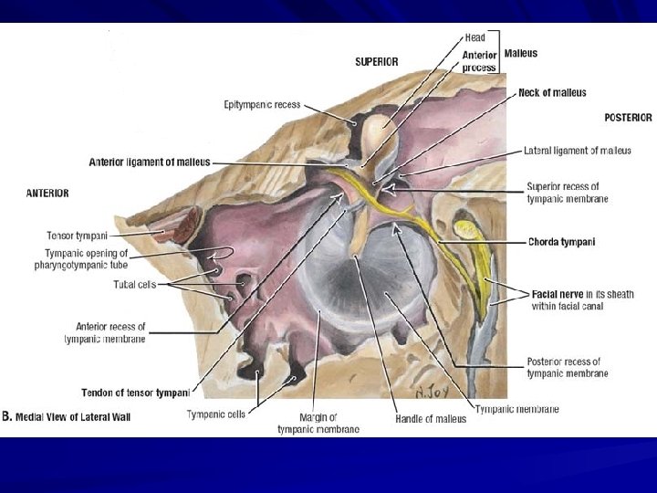
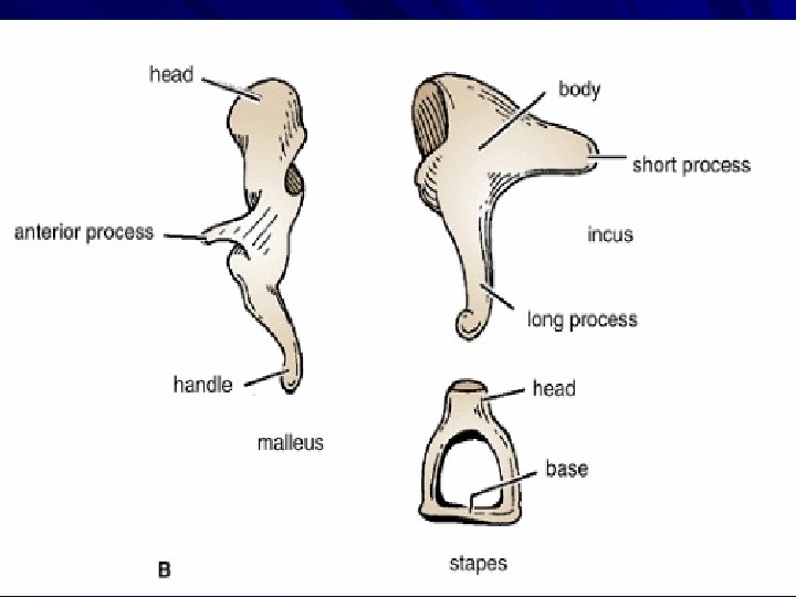
Auditory ossicles: they are the malleus, incus, and stapes. arranged from the lateral to the medial side.
The malleus is the largest ossicle and possesses a head, a neck, a long process or handle, an anterior process, and a lateral process.
The incus possesses a large body and two processes. The stapes has a head, a neck, two limbs, and a base.
The ossicles serve to transmit the vibrations of the tympanic membrane (eardrum) to the perilymph of the internal ear.
1 - Tensor tympani: Origin: from the wall of auditory tube and its own canal Insertion: into handle of malleus Nerve supply: Mandibular nerve
Action: It pulls the handle of malleus; thus increasing the inward convexity of the tympanic membrane and rendering it more tense. So, it dampens down vibrations of tympanic membrane. As a result of this, it hinders the movements of the ear ossicles to protect the inner car during exposure to loud sounds.
2 -Stapedius: Origin: from pyramid (on posterior wall of middle ear) Insertion: into neck of stapes. Nerve supply : Facial nerve.
Action: It pulls on the neck of the stapes during loud sounds, thus providing a damping effect on the sound vibrations reaching to the internal ear i. e Dampens down vibrations of stapes
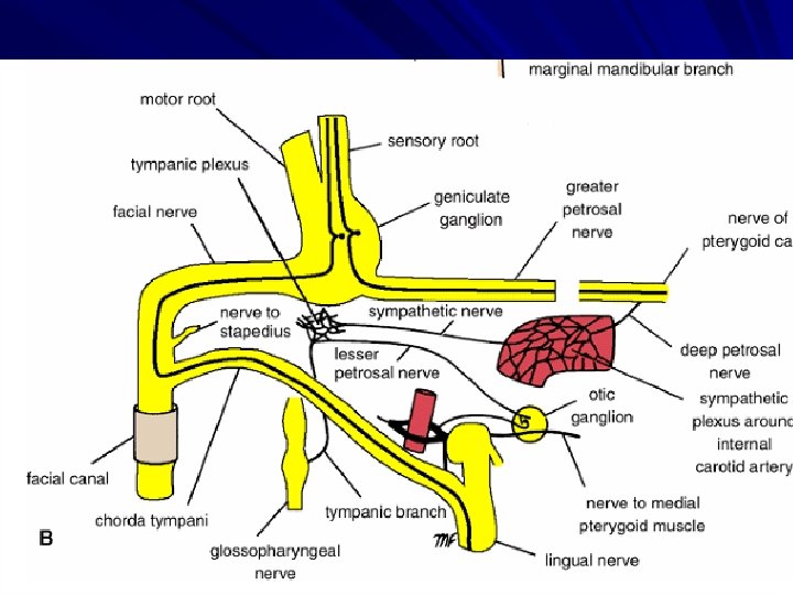
4 -The chorda tympani arises from the facial nerve just above the stylomastoid foramen. It enters the middle ear close to the posterior border of the tympanic membrane.
It then runs forward over the tympanic membrane and crosses the root of the handle of the malleus.
It lies in the interval between the mucous membrane and the fibrous layers of the tympanic membrane.
The nerve leaves the middle ear through the petrotympanic fissure and enters the infratemporal fossa, where it joins the lingual nerve.
Auditory tube (Eustachian tube, pharyngotympanic tube)
The auditory tube connects the anterior wall of the tympanic cavity to the nasaopharynx.
It is about 36 mm long extending from the anterior wall of the tympanic cavity downward, forward and medially to the nasopharynx.
Its lateral third is bony, and its medial two thirds are cartilaginous. The narrowest part of the tube (isthmus) is at the junction of the bony with the cartilaginous part.
Its function is to equalize air pressure on the tympanic membrane. Note: It is a pathway for the spread of infection from the nasopharynx to the middle ear.
The mastoid antrum It is an air sinus which lies behind the tympanic cavity in the petrous part of the temporal bone. It is rounded in shape and may be large up to 1 cm in diameter.
It is lined with mucous membrane. It communicates anteriorly with the epitympanic recess by the aditus and posteroinferiorly with the mastoid air cells.
Relations of the mastoid antrum: These are important in understanding the spread of infection.
Anterior wall is related to middle ear and contains aditus to the mastoid antrum.
Posterior wall separates the antrum from the sigmoid venous sinus and the cerebellum.
Lateral wall is thick and forms the floor of the suprameatal triangle
Inferior wall is perforated with holes, through which the antrum communicates with the mastoid air cells.
Medial wall is related to the posterior semicircular canal
Superior wall is the thin plate of bone, the tegmen tympani, which is related to the meninges of the middle cranial fossa and the temporal lobe.
Mastoid Air Cells The mastoid air cells are a series of communicating cavities within the mastoid process.
They are continuous above with the antrum. They are lined with mucous membrane. Their degree of development shows considerable individual variations.
Clinical note: Inadequate treatment of otitis media may result in the spread of the infection into the mastoid antrum. and mastoid air cells (acute mastoiditis). Acute mastoiditis may be followed by the further spread of infection to the menings, temporal lobe of the brain, inner ear and sigmoid venous sinus.
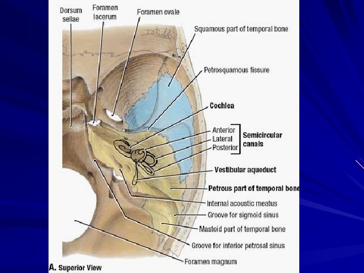
The Internal Ear, or Labyrinth
The labyrinth is situated in the petrous part of the temporal bone, medial to the middle ear It consists of:
1 -Bony labyrinth: comprising a series of cavities within the bone.
2 -Membranous labyrinth: comprising a series of membranous sacs and ducts contained within the bony labyrinth.
1 -The bony labyrinth consists of: 1 - vestibule 2 - semicircular canals cochlea 3 -
These cavities are lined by endosteum and contain a clear fluid (the perilymph) in which is suspended the membranous labyrinth.
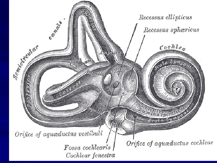
Bony labyrinth
Membranous labyrinth
The bony labyrinth 1 -The vestibule, the central part of the bony labyrinth, lies posterior to the cochlea and anterior to the semicircular canals.
In its lateral wall are the fenestra vestibuli which is closed by the base of the stapes and the fenestra cochleae which is closed by the secondary tympanic membrane.
2 -The three semicircular canals: superior, posterior, and lateral, lie in three different planes perpendicular to each other. open into the posterior part of the vestibule. Each canal has a swelling at one end called the ampulla.
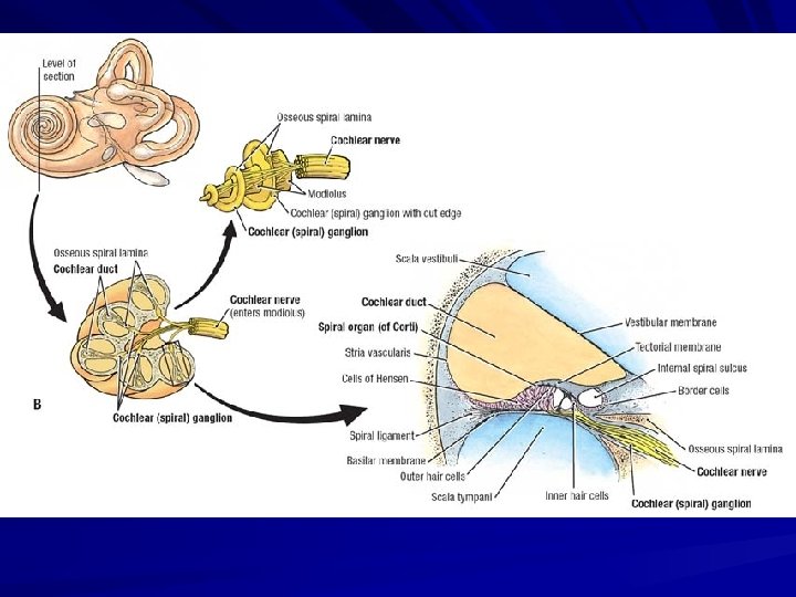
3 -The cochlea: resembles a snail shell. It opens into the anterior part of the vestibule. The whole structure is cone-shaped with the apex faces anterolaterally and the base posteromedially.
The first basal turn of the cochlea is responsible for the promontory seen on the medial wall of the tympanic cavity.
Basically, it consists of a central pillar, the modiolus, around which a hollow bony tube makes two and one half spiral turns.
2 -The membranous Labyrinth is lodged within the bony labyrinth. It is filled with endolymph and surrounded by perilymph.
It consists of: 1 - Utricle and saccule, which are lodged in the bony vestibule. 2 - Three semicircular ducts, which lie within the bony semicircular canals.
3 -Duct of the cochlea, which lies within the bony cochlea. It is triangular in cross section and contains the sensory organ of hearing (Organ of Corti) All these structures freely communicate with one another.
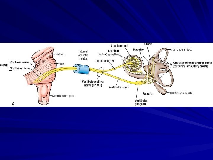
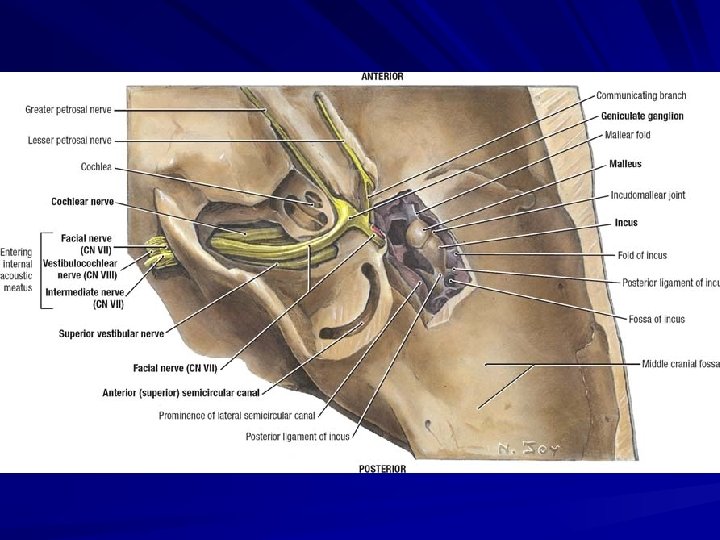
Vestibulocochlear Nerve It is the eighth cranial nerve. It emerges on the lower border of pons lateral to facial nerve. On reaching the bottom of the internal acoustic meatus the nerve divides into vestibular and cochlear portions.
1 -The vestibular nerve is expanded to form the vestibular ganglion. Its branches supply the utricle, the saccule, and the ampullae of the semicircular ducts.
2 -The cochlear nerve divides into numerous branches, which enter foramina at the base of the modiolus. The peripheral branches of this nerve pass from the ganglion to spiral organ of Corti.
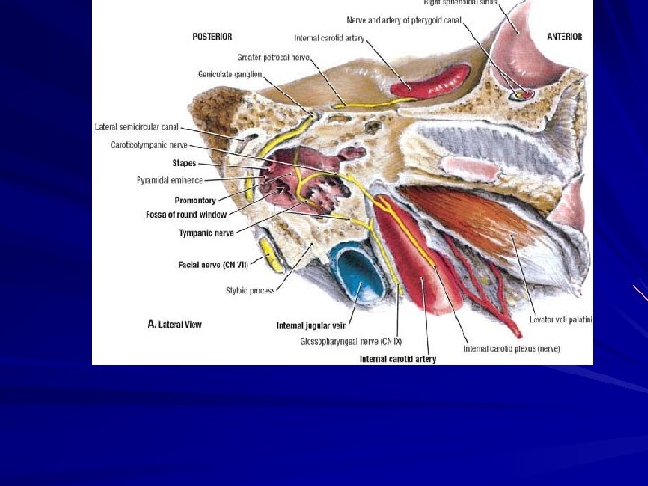
Facial nerve The facial nerve has a motor root and a sensory root (nervus intermedius). The nerve emerges on the lower border of the pons medial to vestibulocochlear nerve.
The roots pass laterally in the posterior cranial fossa with the vestibulocochlear nerve and enter the internal acoustic meatus. On reaching the bottom of the internal acoustic meatus, the facial nerve enters the facial canal
The nerve runs laterally above the vestibule of the internal ear until it reaches the medial wall of the middle ear. Here, the nerve expands to form the sensory geniculate ganglion.
The nerve then bends sharply backward above the promontory.
On arriving at the posterior wall of the middle ear, it curves downward on the medial side of the aditus of the mastoid antrum.
It descends in the posterior wall of the middle ear, behind the pyramid, and finally emerges through the stylomastoid foramen into the neck
Branches: 1) At the geniculate ganglion: • Greater petrosal nerve. • Communicating branch with the lesser petrosal. • External petrosal nerve which join the sympathetic plexus around the middle meningeal artery.
Branches: 1 -Greater petrosal nerve arises at the geniculate ganglion. It carries secretomotor fibers to the lacrimal glands of nose and palate. It also contains taste fibers from palate.
2) IN the facial canal: 2 -Nerve to stapedius 3 -Chorda tympani.
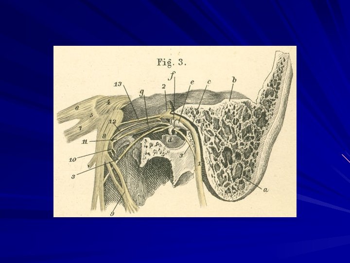
facial nerve. genu of facial nerve. greater superficial petrosal nerve. lesser superficial petrosal nerve. glossopharyngeal nerve. tympanic nerve (s. Jacobsonii) with branches to the round and oval windows, to the tubes, and to the internal carotid plexus. 7. lesser deep petrosal nerve. 8. vagus nerve with auricular ramus. 1. 2. 3. 4. 5. 6.
a) Temporal bone, petrosal portion. b) Temporal bone, mastoid portion. c) Promontorium. d) Oval window. e) Round window. f) Auditory tube (Eustachian tube). g) m. Tensor tympani. h) m. Stapedius. i) Internal carotid artery. k) Internal jugular vein.
1. 2. 3. 4. 5. 6. 7. 8. 9. 10. 11. 12. 13. a) Mastoid process. b) Petrous portion of temporal bone. c) Facial canal ( s. Fallopian canal). d) Tympanic cavity. e) Incus. f) Malleus. g) m. Tensor tympani. facial nerve. genu of the facial nerve. chorda tympani. trigeminal nerve. semilunar ganglion. trigeminal nerve, ramus I (ophthalmic division. ( trigeminal nerve, ramus II (maxillary division. ( trigeminal nerve, ramus III (mandibular division. ( anterior auricular nerve (with its two branches. ( otic ganglion. nerve for m. tensor tympani , lesser superficial petrosal nerve. greater superficial petrosal nerve.
Lymphatic drainage of head and neck The lymph nodes in the head and neck are made up of a number of regional groups and a terminal group. The regional groups include both the horizontal series of superficial nodes which surrounds the junction of head and neck and the deep groups of lymph nodes. The terminal group of nodes receives all the lymph vessels of the head and neck, either directly or indirectly (via one of the regional groups). It is called the deep cervical group.
The superficial groups of lymph nodes are the occipital, retroauricular (mastoid), the parotid, the buccal, the submandibular, the submental, the anterior cervical and the superficial cervical. • The deep groups of lymph nodes are the retro-pharyngeal, lingual, laryngeal and the tracheal.
The deep cervical lymph nodes: They form a chain along the course of the intemal jugular vein, from the skull to the root of the neck. The majority of them lie on the anterolateral aspect of the internal jugular vein deep to the stemomastoid muscle.
They receive lymph from the superficial and deep lymph nodes. The inferior belly of omohyold subdivides them into an upper and a lower group
Upper deep cervical lymph nodes: They are arranged along the upper part of the internal jugular vein. Of this group, an Important lymph node lies in the angle between the posterior belly of digastric and the internal jugular vein, This is called jugulo-digastric lymph node.
Lower deep cervical lymph nodes: They are arranged along the lower part of the internal jugular vein. Of this group an important lymph node lies along the intermediate tendon of omohyoid called the jugulo-omohoid lymph node.
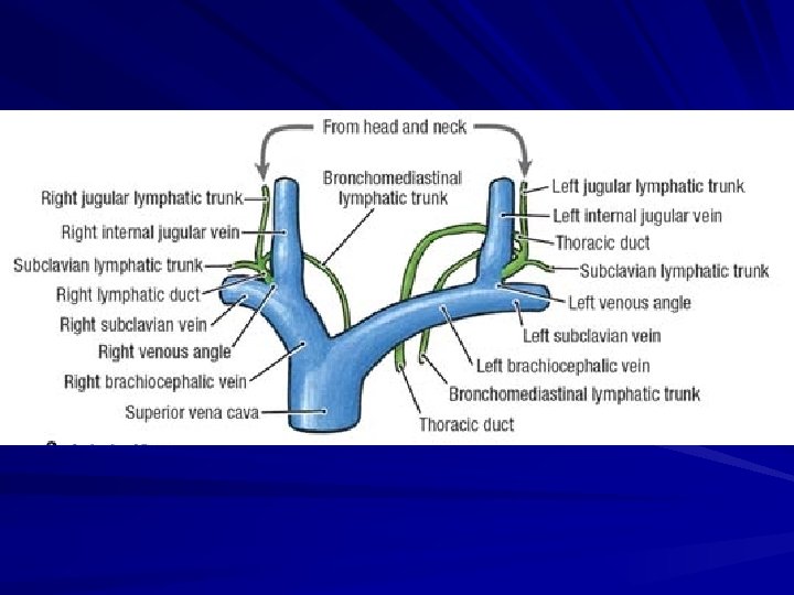
Efferent lymph vessels of the deep cervical lymph nodes collect to form the jugular lymph trunk. This trunk drains into the thoracic duct on the left side or the right lymphatic duct on the right side. Alternatively, it may open into the angle between the internal jugular vein and the subclavian vein.