EAR Three parts of the Ear 1 External


 and external acoustic meatus Auricle: made up of")


 Small, air-containing cavity in petrous part of temporal bone Contains")
 Walls (cont. ): • Anterior wall separates the tympanic cavity")
 Walls (cont. ): Posterior wall (cont. ): • pyramidal eminence")
 Medial wall (cont. ): Oval (vestibular) window which is closed")


: • articulates with head of stapes • articulates")


 Tube Canal that communicates nasopharynx with tympanic cavity & allows for equalization")

 2. cochlea (anterior to")
: Contains clear fluid (perilymph) that circulates through the")


: 1. utricle and saccule contain specialized receptors (maculae)")

- Slides: 41
EAR
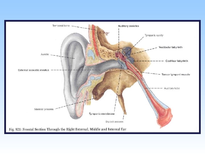
Three parts of the Ear 1. External 2. Internal 3. Middle
Objectives 1. Describe the main anatomical features of the auricle, the external acoustic meatus and the tympanic membrane 2. Describe the walls and anatomical relationships of the middle ear 3. Describe the anatomical features of the auditory ossicles 4. Describe the location, action and nerve supply of the tensor tympani and stapedius muscles 5. Describe the openings, structure and function of the auditory tube 6. Name the parts of the bony and membranous labyrinths and describe their arrangement 7. Name the receptors found in the membranous labyrinth and indicate their functions
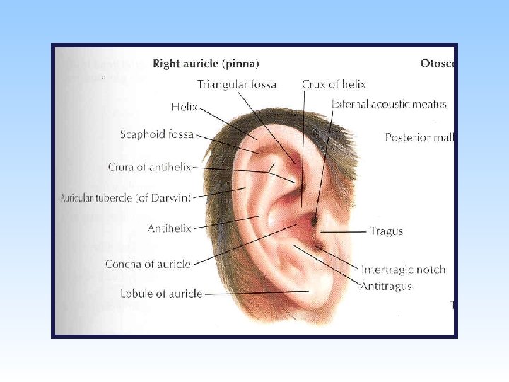
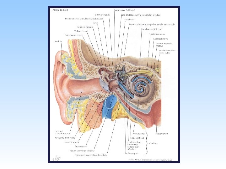
External Ear Consists of auricle (pinna) and external acoustic meatus Auricle: made up of elastic cartilage covered with skin. • helix: rim of auricle (resembles beginning of spiral) • antihelix: curved eminence anterior to helix • concha: most depressed area of auricle, anterior to antihelix • tragus: small prominence that overhangs entrance to external acoustic meatus • antitragus: small tubercle posterior to tragus • lobule non-cartilaginous part of auricle (consists of loose connective tissue and fat covered with skin)
External Ear • external acoustic meatus: • curved, blind-ended canal that conducts sound waves from auricle to tympanic membrane • can be straightened for examination by pulling auricle upward and backward • lateral part has cartilaginous walls (cartilage is continuous with that of auricle) • medial part has bony walls (squamous and tympanic parts of temporal bone) • lined by skin (continuous with skin of auricle)
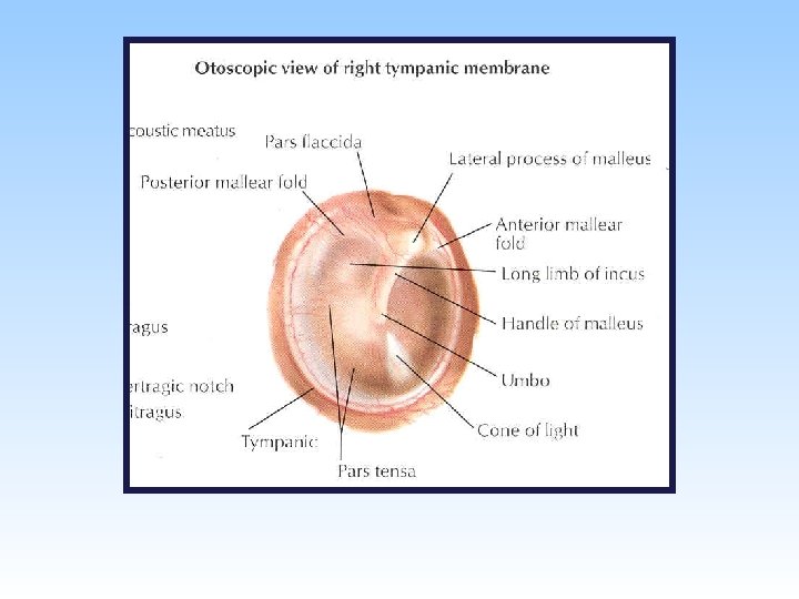
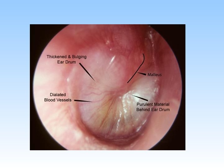
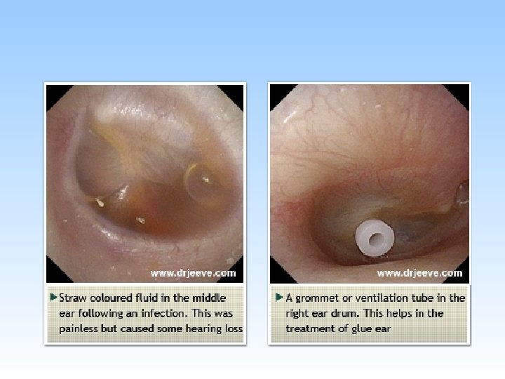
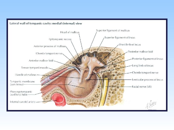
Tympanic Membrane Thin membrane located at medial end of external acoustic meatus & forms boundary between external ear and middle ear. Concave laterally (umbo most indrawn part of membrane) Handle and lateral process of malleus are firmly attached to its inner surface (can be seen on otoscopic examination) When membrane is illuminated, its concavity produces a “cone of light” that radiates from the umbo. Covered with thin skin externally and mucous membrane of tympanic cavity internally. Between them there is a layer of fibrous tissue (tense part) that is deficient superiorly (flaccid part).
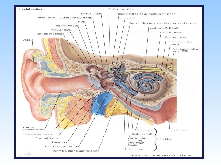
Middle Ear (Tympanic Cavity) Small, air-containing cavity in petrous part of temporal bone Contains auditory ossicles (malleus, incus and stapes), which transmit vibrations from tympanic membrane to internal ear Walls: • roof is a thin bony plate that separates tympanic cavity from middle cranial fossa. • floor is a thin bony plate that separates tympanic cavity from the internal jugular vein.
Middle Ear (Tympanic Cavity) Walls (cont. ): • Anterior wall separates the tympanic cavity from internal carotid artery. Two openings leads into auditory tube & a canal for tensor tympani muscle. • Posterior wall: It has an irregular opening to mastoid antrum which communicates tympanic cavity with mastoid antrum. Mastoid antrum communicates inferiorly and posteriorly with mastoid air cells
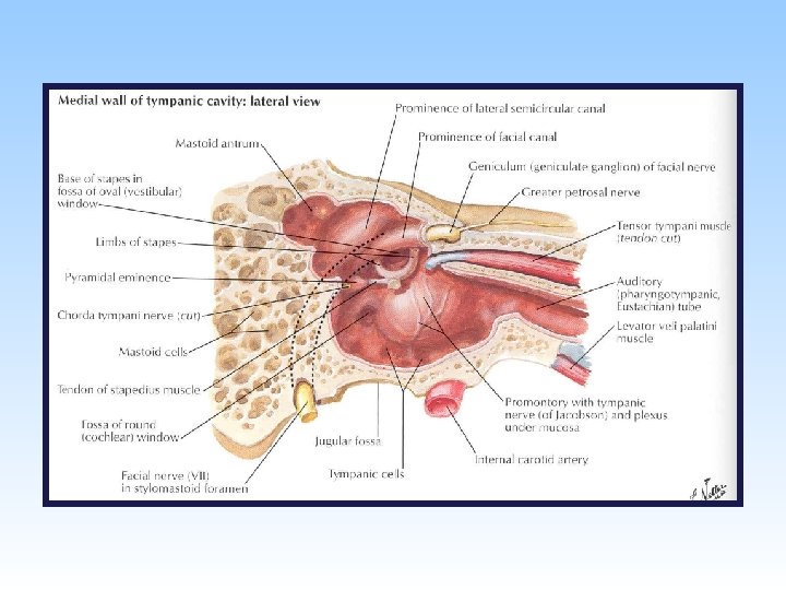
Middle Ear (Tympanic Cavity) Walls (cont. ): Posterior wall (cont. ): • pyramidal eminence houses the stapedius muscle. The tendon of stapedius emerges through small opening at tip of pyramidal eminence and inserts into stapes. Lateral wall: formed mostly by tympanic membrane Medial wall: • separates middle ear from internal ear • promontory rounded elevation produced by underlying 1 st turn of cochlea
Middle Ear (Tympanic Cavity) Medial wall (cont. ): Oval (vestibular) window which is closed by base of stapes. Round (cochlear) window closed by secondary tympanic membrane Prominence of facial canal
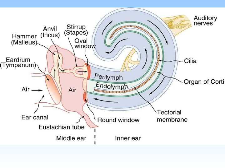
Three Auditory Ossicles 1. Malleus: • articulates with body of incus • receives insertion of tensor tympani muscle
Auditory Ossicles 2. Incus (cont. ): • articulates with head of stapes • articulates with head of malleus 3. Stapes: • head articulates with the incus • neck receives insertion of stapedius muscle • base attached to margins of oval window
head body short limb lateral process long limb anterior process handle head neck posterior limb anterior limb base
Muscles Associated with the Ossicles Movements of ossicles are modified and dampened by protective action of 2 small muscles: tensor tympani and stapedius Tensor tympani: • inserts into handle of malleus • innervated by the mandibular nerve • action: draws handle of malleus and tympanic membrane medially to dampen the vibrations of tympanic membrane
Muscles Associated with the Ossicles Stapedius: • located within pyramidal eminence of posterior wall of tympanic cavity & inserts into stapes • innervated by facial nerve (nerve to stapedius branch) • tilts stapes and dampens its vibrations
Eustachian (Pharyngotympanic) Tube Canal that communicates nasopharynx with tympanic cavity & allows for equalization of pressure on both sides of tympanic membrane. • begins in anterior wall of tympanic cavity & ends in lateral wall of nasopharynx • posterior ⅓ (close to tympanic cavity) has bony walls • anterior ⅔ (close to pharynx) has cartilaginous walls • contraction of tensor veli palatini (swallowing, yawning) helps to open lumen of auditory tube
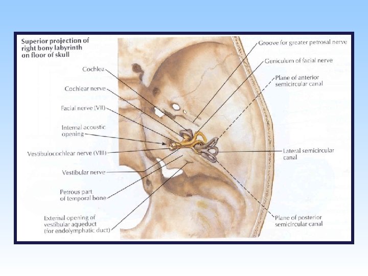
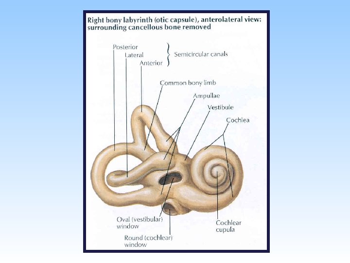
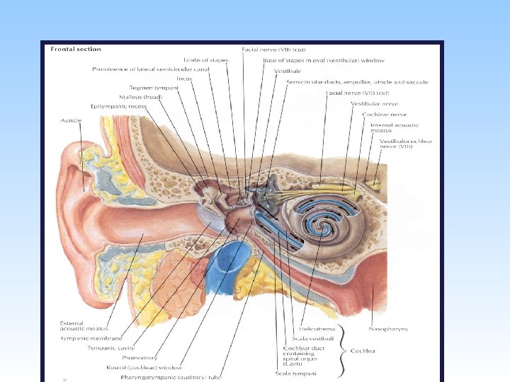
Internal Ear Parts: Bony labyrinth: series of cavities within petrous part of temporal bone Membranous labyrinth: series of membranous sacs and ducts contained within bony labyrinth
Bony labyrinth within the temporal bone. 1. vestibule (central part) 2. cochlea (anterior to vestibule) 3. semicircular canals (posterior to vestibule)
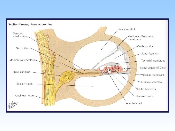
Internal Ear Bony labyrinth (cont. ): Contains clear fluid (perilymph) that circulates through the sacula vestibula and sacula tempani. Three semicircular canals (anterior, posterior and lateral) are perpendicular to each other and open into posterior part of vestibule. Each canal has a swelling at one end called the ampulla. Cochlea resembles a snail shell & consists of central pillar (modiolus) around which a bony tube makes approximately 2 ½ turns. It contains circulating endolymph.
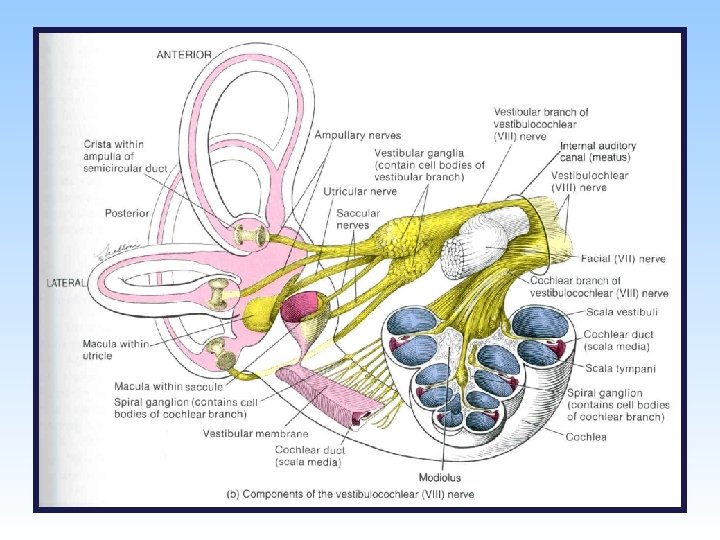
Internal Ear Membranous labyrinth: • The channels created by the bony labyrinth and are filled with perilymph • Central canal is filled with endolymph • Main channels: • utricle and saccule (lie within vestibule of bony labyrinth) • 3 semicircular ducts (lie within 3 semicircular canals) • cochlear duct (lies within bony cochlea)
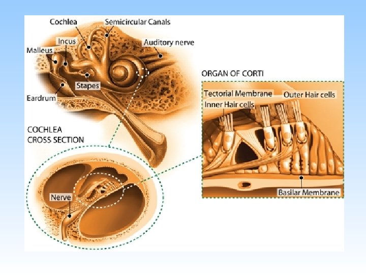
Internal Ear Membranous labyrinth (cont. ): 1. utricle and saccule contain specialized receptors (maculae) sensitive to position of head with respect to gravity and linear acceleration 2. ampulla of each semicircular duct contains a specialized receptor (crista ampullaris) sensitive to angular acceleration 3. cochlear duct contains a specialized hearing receptor called the spiral organ/organ of Corti. 4. maculae and cristae ampullares are innervated by vestibular part of CN VIII 5. spiral organ is innervated by cochlear part of CN VIII
Putting it all together • https: //www. youtube. com/watch? v=f. IAj. Jog J 0 cs