Anatomy of the Ear Parts of the Ear


- Slides: 24
Anatomy of the Ear Parts of the Ear
Is this your ear?
NOPE – This is the Ear!
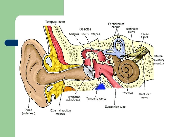
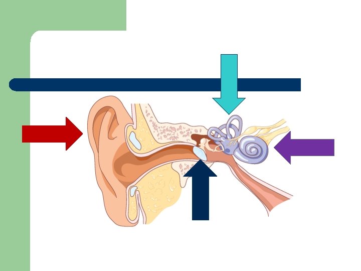
There are three sections of the ear! l OUTER l MIDDLE l INNER
Outer Ear l l Pinna -also known as the Auricle Ear Canal or External Auditory Canal
Middle Ear l l l Tympanic Membrane - Ear Drum Eustachian Tube Three Bones known as the Ossicular Chain – – – Malleus Incus Stapes
Inner Ear l l Semicircular canals Cochlea l l l Organ of Corti 3 Chambers 2 Membranes
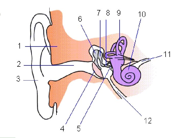
Review
Anatomy of the Ear Functions of the Ear
Outer Ear l Pinna -also known as the Auricle. l l Made entirely of cartilage Varies in size and shape Gathers sound waves from the environment Ear Canal l l Lined with hair follicles to keep foreign objects out Lined with skin-supporting glands Sebaceous- secretes an oily, fatty substance is cerumen (also known as ear wax) – Cerumen exits ear naturally –
Middle Ear l Tympanic Membrane - Ear Drum l l Must be protected, extremely thin Keeps constant temperature and humidity Vibrates when sound waves hit it Eustachian Tube – Equalizer – 3 Primary Functions l l l Helps ventilate middle ear Equalize pressure Maintain proper drainage
Middle Ear - The Ossicular Chain l l l Malleus Incus Stapes Purpose: helps transmit sound waves from the tympanic membrane to the inner ear
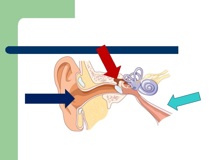
Look Again.
Inner Ear l Cochlea – – Twisting bony shell Diagrams show the inside 3 to 5 rows of thousands of tiny hairs Three Scala (or chambers) l Scala Vestibuli - oval window l Scala Media (cochlear duct) l Scala Tympani - round window
Inside the Cochlea l “Organ of Corti – Also Known As: Organ of Hearing” l l l full length of Scala Media resides on basilar membrane lined with nerve endings come together to make 8 th nerve (from here it gets really complicated so we will stop)
Inside the Cochlea Each chamber had different fluids that cannot mix – if these fluids cannot mix - will kill all nerves. l These fluids separated by membranes – Reissner’s Membrane – Basilar Membrane l
Semicircular canals l l The canals sit at right angles to each other, are all attached. They detect horizontal and vertical movement as well as acceleration.
How the Ear Work! l https: //www. youtube. com/watch? v=qgdqpo. Pb 1 Q