EAR Three parts of the Ear 1 External


 Tube Canal that communicates nasopharynx with tympanic cavity & allows for equalization")

 2. cochlea (anterior to")


: • greatest bulk is contributed by ciliary muscle (muscle")
 • contains 2 smooth")
: • optic part posterior part of retina which covers")
: • at posterior pole of eyeball, retina has")


 to inferior border")


 • separated posteriorly from vertebral column")


 is a collection of lymphoid tissue at entrance of")

 • extends from superior border of epiglottis to inferior border of cricoid")


 • facial")


 Part of Maxillary Artery • maxillary artery leaves infratemporal fossa and")


 surface of body of maxilla • posterior:")


: 2. Inferior alveolar nerve: • contains")
: 3. Lingual nerve: • descends medial")


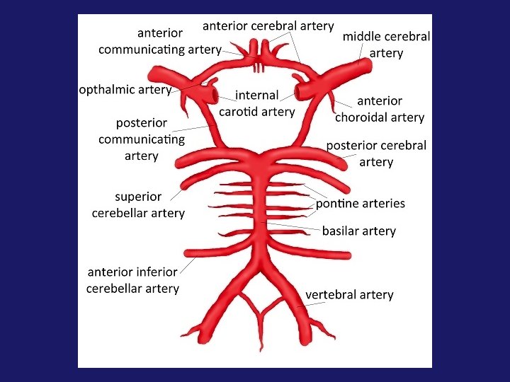
 • formed by large cerebral arteries and their interconnections")
- Slides: 106
EAR
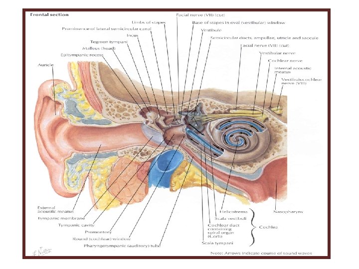
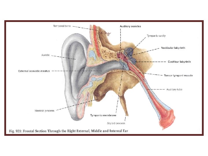
Three parts of the Ear 1. External 2. Internal 3. Middle
External Ear • external acoustic meatus: • curved, blind-ended canal that conducts sound waves from auricle to tympanic membrane • can be straightened for examination by pulling auricle upward and backward • lateral part has cartilaginous walls (cartilage is continuous with that of auricle) • medial part has bony walls (squamous and tympanic parts of temporal bone) • lined by skin (continuous with skin of auricle)
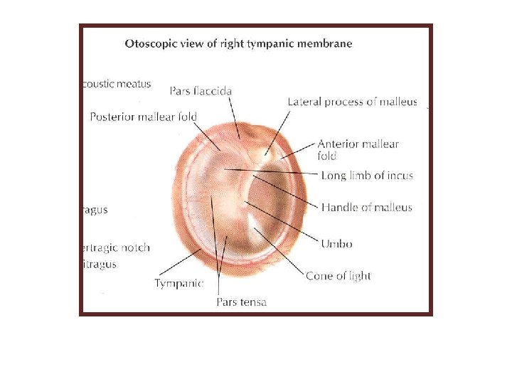
Tympanic Membrane Thin membrane located at medial end of external acoustic meatus & forms boundary between external ear and middle ear. Concave laterally (umbo most indrawn part of membrane) Handle and lateral process of malleus are firmly attached to its inner surface (can be seen on otoscopic examination) When membrane is illuminated, its concavity produces a “cone of light” that radiates from the umbo. Covered with thin skin externally and mucous membrane of tympanic cavity internally. Between them there is a layer of fibrous tissue (tense part) that is deficient superiorly (flaccid part).
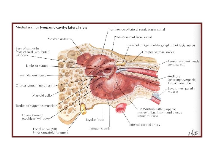
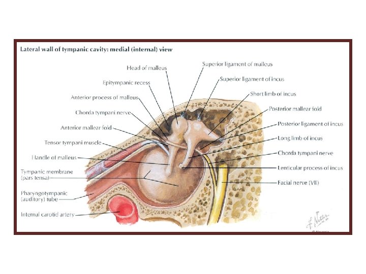
Three ear ossicles in the middle ear.
Muscles Associated with the Ossicles Movements of ossicles are modified and dampened by protective action of 2 small muscles: tensor tympani and stapedius Tensor tympani: • inserts into handle of malleus • innervated by the mandibular nerve • action: draws handle of malleus and tympanic membrane medially to dampen the vibrations of tympanic membrane
Eustachian (Pharyngotympanic) Tube Canal that communicates nasopharynx with tympanic cavity & allows for equalization of pressure on both sides of tympanic membrane. • begins in anterior wall of tympanic cavity & ends in lateral wall of nasopharynx • posterior ⅓ (close to tympanic cavity) has bony walls • anterior ⅔ (close to pharynx) has cartilaginous walls • contraction of tensor veli palatini (swallowing, yawning) helps to open lumen of auditory tube
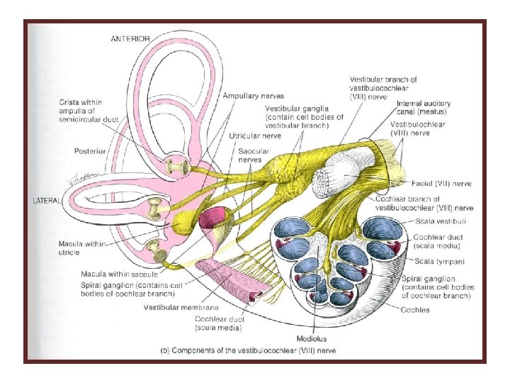
Internal Ear Parts: Bony labyrinth: series of cavities within petrous part of temporal bone Membranous labyrinth: series of membranous sacs and ducts contained within bony labyrinth
Bony labyrinth within the temporal bone. 1. vestibule (central part) 2. cochlea (anterior to vestibule) 3. semicircular canals (posterior to vestibule)
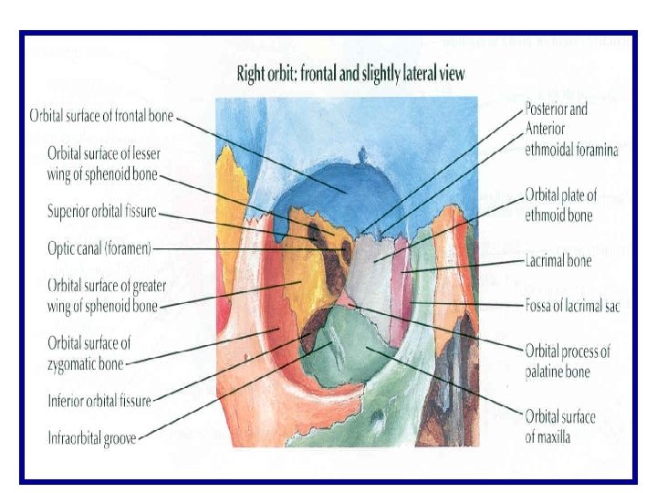
ORBIT and EYE
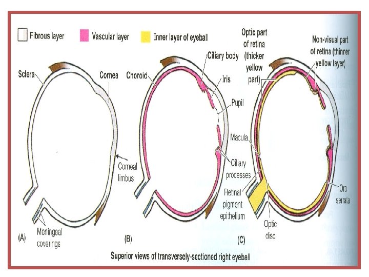
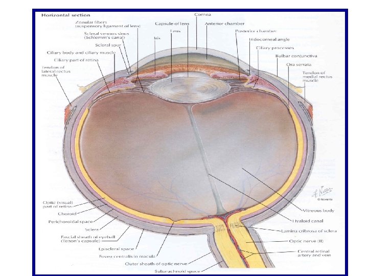
Eyeball • peripheral organ for special sense of sight • shaped like a hollow sphere • consists of 3 concentric layers (coats/tunics): fibrous layer, vascular layer and inner layer (retina) • fibrous layer: • has 2 parts: sclera and cornea • sclera (“white of the eye”): firm, smooth, opaque fibrous cup which forms posterior 5/6 of fibrous layer continuous in front with cornea at corneoscleral junction • cornea: transparent, anterior 1/6 of fibrous layer represents a segment of a smaller sphere (compared to sclera) bulges forward from sclera has no blood vessels and receives rich sensory nerve supply from ophthalmic nerve
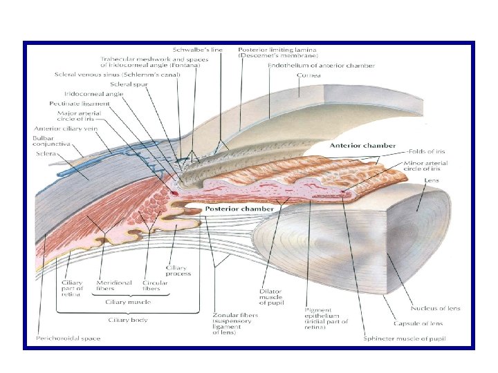
Eyeball • vascular layer: • has 3 parts: choroid, ciliary body and iris (in posterior-to-anterior direction) • choroid: thin, vascular membrane immediately deep to sclera consists of a dense capillary plexus and small arteries and veins held together by connective tissue with abundant melanocytes • ciliary body: • raised area in anterior part of vascular layer • wedge-shaped in cross-section inwardly projecting angle of wedge is directed toward lens and connected to it by fibers of suspensory ligament of lens (zonular fibers)
Eyeball Ciliary body (cont. ): • greatest bulk is contributed by ciliary muscle (muscle of accommodation reflex helping to maintain a clear visual image as gaze is shifted from a distant to a near point or vice versa) • when shifting gaze from a distant to a near point, ciliary muscle contracts and its contraction reduces tension of suspensory ligament of lens reduces tension exerted by ligament on lens under less tension, because of its natural elasticity, lens increases its curvature (becomes thicker in anteroposterior direction) increases refractive power of lens for vision of close objects (opposite happens when shifting gaze from a near point to a distant one) • ciliary processes: internal projections of ciliary body its covering epithelium secretes aqueous humor (watery fluid filling space between cornea and lens)
Eyeball • iris: • pigmented diaphragm with central aperture (pupil) • contains 2 smooth muscles which control pupillary size: • sphincter (constrictor) pupillae muscle formed by circular fibers surrounding margin of pupil decreases pupillary size • dilator pupillae muscle formed by fibers that radiate from pupillary margin toward outer circumference of iris increases pupillary size • ciliary and sphincter pupillae muscles receive parasympathetic innervation via oculomotor nerve • dilator pupillae muscle receives sympathetic innervation from superior cervical ganglion
Eyeball • inner layer (retina): • optic part posterior part of retina which covers inner aspect of choroid contains photoreceptors (rods and cones), 1 st and 2 nd order neurons of visual pathway, interneurons and supporting cells ends anteriorly at a jagged border (ora serrata), a short distance behind ciliary body • nonvisual (ciliary and iridial) part thin, anterior part of retina which lines internal aspect of ciliary body and posterior surface of iris has no nervous elements
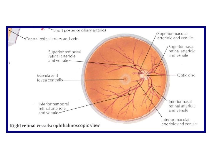
Eyeball • inner layer (cont. ): • at posterior pole of eyeball, retina has oval yellowish area (macula lutea) with a central depression (fovea centralis) • fovea centralis is the part of retina that provides maximal visual acuity contains only cones • as we move peripherally from fovea centralis, there are less cones and more rods • optic disc: located medial to macula lutea area where axons of ganglion cells of all retina converge to form optic nerve contains no photoreceptors (blind spot of retina) branches of central artery and vein of retina spread out from center of optic disc over inner surface of retina
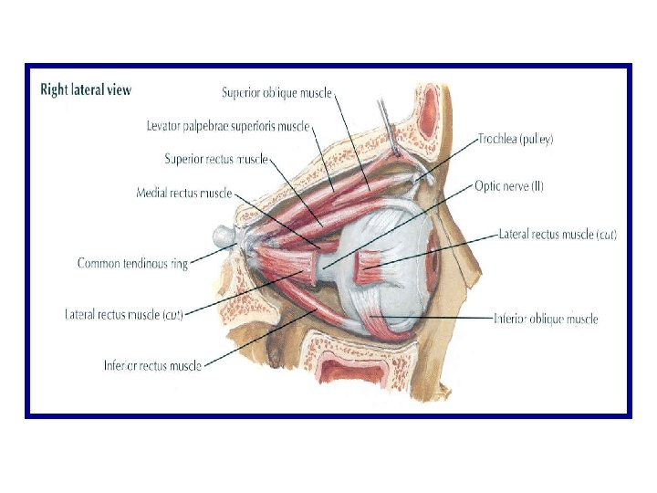
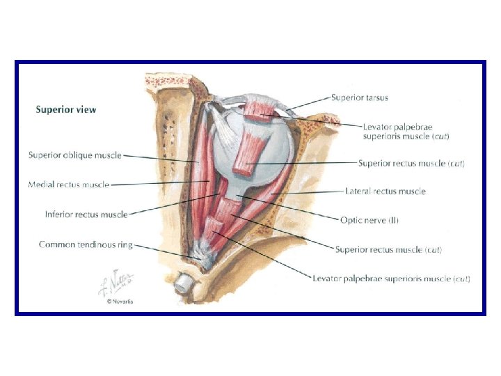
Clinical Testing of the Extraocular Muscles that Move Eyeball • eye abduction lateral rectus (abducens nerve) • eye adduction medial rectus (oculomotor nerve) • elevation of abducted eye superior rectus (oculomotor nerve) • depression of abducted eye inferior rectus (oculomotor nerve) • elevation of adducted eye inferior oblique (oculomotor nerve) • depression of adducted eye superior oblique (trochlear nerve)
LARYNX
Larynx Located in anterior neck, at level of bodies of C 3 -C 6 vertebrae Functions: • part of air passages that links pharynx above to trachea below • sphincter action prevents passage of solids and liquids from pharynx into air passages below (aspiration) • organ of phonation controls the flow of air from lungs to produce sounds, determine pitch & volume. • skeleton: • 3 large unpaired cartilages: thyroid, cricoid and epiglottis • 3 smaller paired cartilages: arytenoid, corniculate and cuneiform
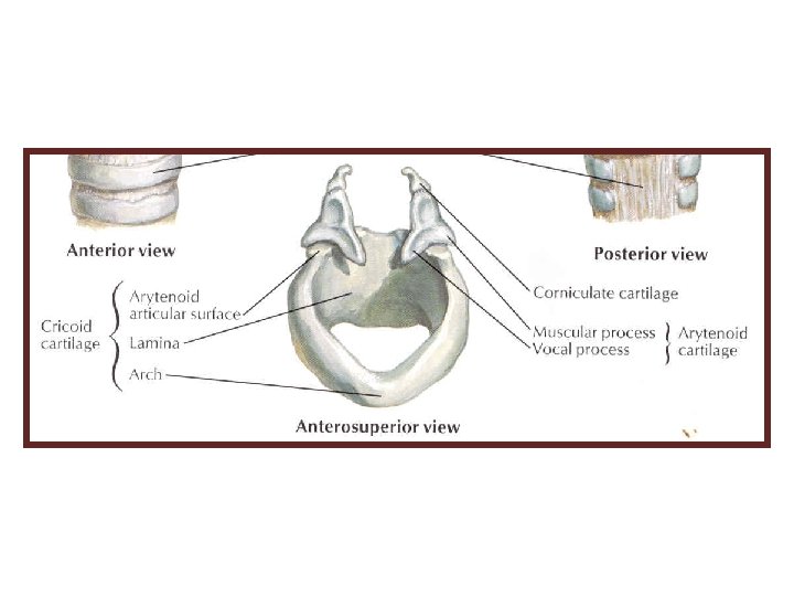
Skeleton of Larynx 3 large unpaired cartilages: 1. thyroid 2. cricoid 3. epiglottis
Epiglottis • leaf-shaped plate of elastic cartilage that stands almost vertically posterior to root of tongue and hyoid bone • superior end is broad and free
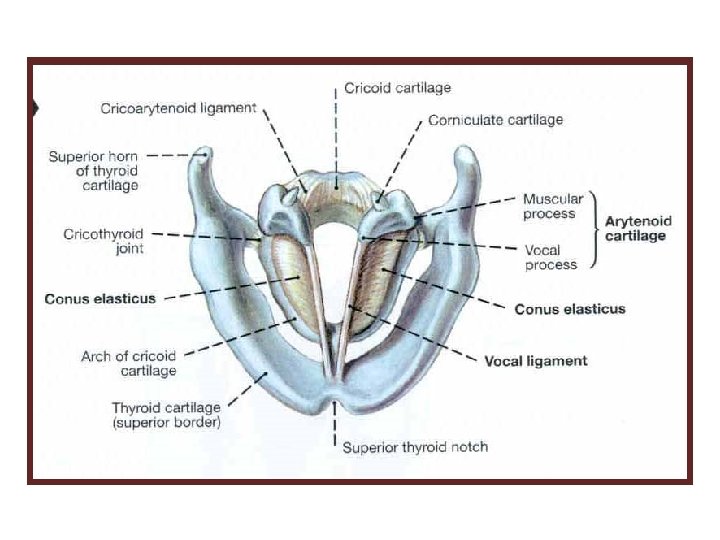
Larynx Skeleton Paired cartilages: 1. Arytenoid 2. Corniculate
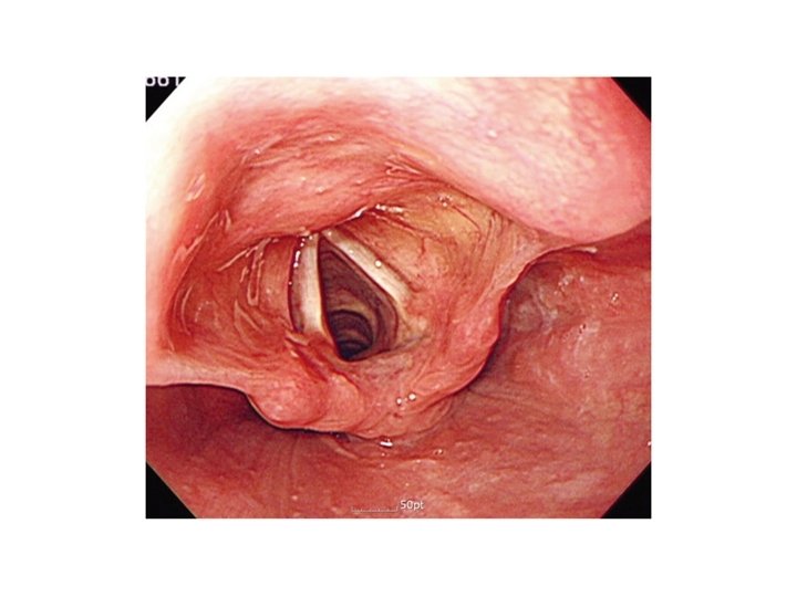
Interior of larynx • • laryngeal inlet: • communication between laryngopharynx and larynx • obliquely The inlet passes from epiglottis to arytenoid cartilages laryngeal cavity is roughly shaped like an hour glass
Interior of the Larynx • on each side 2 mucosal folds project into laryngeal lumen: vestibular fold (superior) and vocal fold (inferior) • vestibular fold is also known as false vocal cord • vocal fold is also known as true vocal cord
Interior of Larynx • Vocal fold is a prominent, wedge-shaped mucosal fold that contains vocal ligament close to its free margin • Rima glottidis is the space between right and left vocal folds. The width and shape of rima glottidis vary with movements of vocal folds and arytenoid cartilages.
Interior of Larynx • glottis is a part of larynx most directly concerned with production of sounds & consists of two vocal folds and space between them (rima glottidis) https: //www. youtube. com/watch? v=i. Yp. Dwhp. ILk. Q https: //www. youtube. com/watch? v=pc. TPOm. NSt. PI
Infraglottic larynx • infraglottic part of extends from vocal folds (superiorly) to inferior border of cricoid cartilage (inferiorly)
Innervation of the larynx Motor nerve supply: • external laryngeal nerve (branch of superior laryngeal nerve) supplies cricothyroid muscle • all other muscles are supplied by recurrent laryngeal nerve
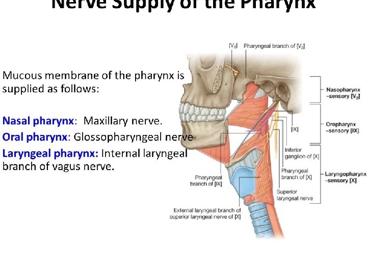
Innervation of the layrnx Sensory nerve supply: • internal laryngeal nerve (branch of superior laryngeal nerve) pierces thyrohyoid membrane supplies laryngeal mucosa from inlet to vocal folds • recurrent laryngeal nerve supplies laryngeal mucosa below vocal folds
Vasculature of the larynx Superior laryngeal artery branch of superior thyroid artery that accompanies internal laryngeal nerve Inferior laryngeal artery branch of inferior thyroid artery that accompanies recurrent laryngeal nerve
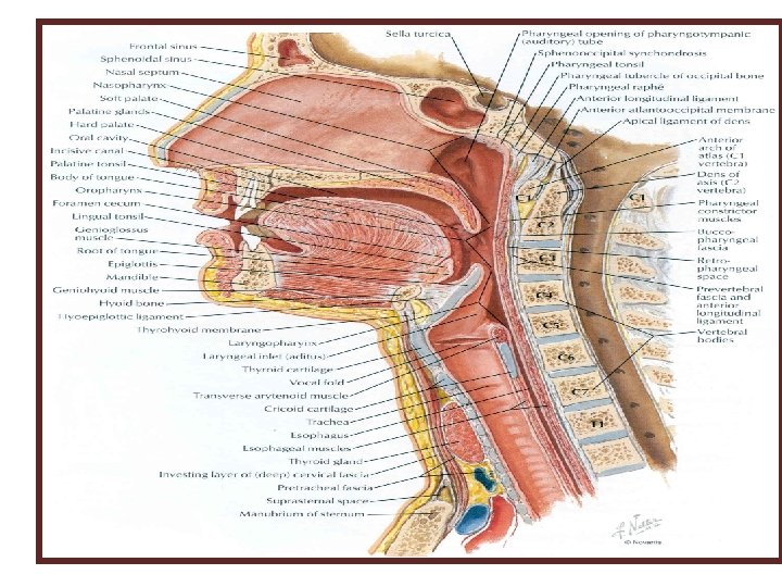
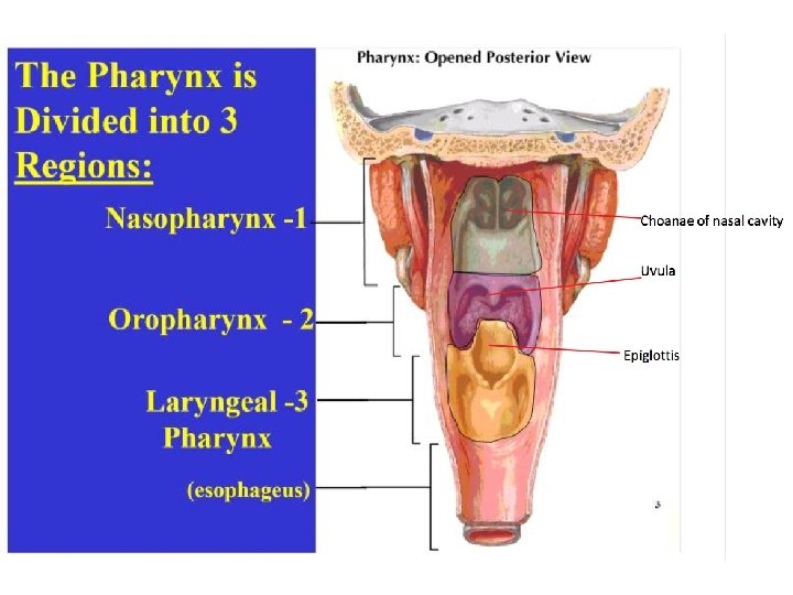
PHARYNX
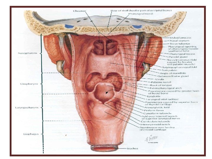
Pharynx • Fibromuscular, funnelshaped organ that has a common chamber for the digestive and respiratory systems. • Extends from base of skull to inferior border of cricoid cartilage (lower border of body of C 6), where it becomes continuous with esophagus. • It has lateral and posterior walls but the anterior wall is incomplete due to its’ communicates anteriorly with nasal cavity, oral cavity and larynx.
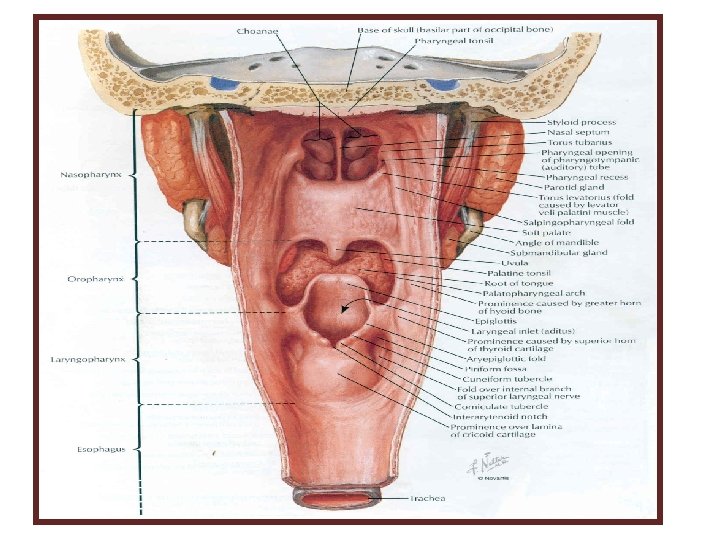
Parts: 1. Nasopharynx 2. Oropharynx 3. Laryngopharynx (hypopharynx) • separated posteriorly from vertebral column and anterior vertebral muscles and prevertebral fascia by loose connective tissue of retropharyngeal space
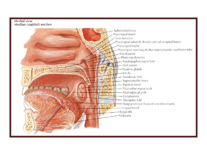
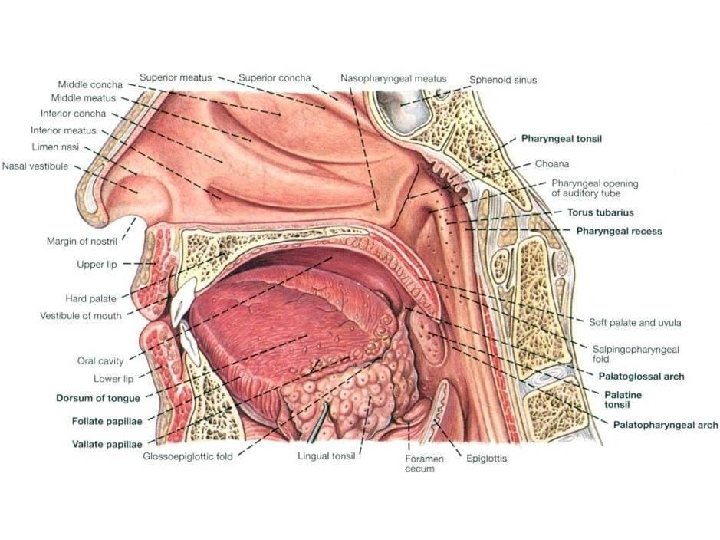
Nasopharynx • It is entirely respiratory in function. • It extends from base of skull to soft palate. • It communicates anteriorly with nasal cavity via choanae (posterior nasal apertures). • Pharyngeal tonsil: collection of lymphoid tissue located in roof of nasopharynx (known as adenoids) • Pharyngeal opening of auditory tube is located in lateral wall of nasopharynx. • The medial end of the auditory tube cartilage elevates the mucosa (torus tubarius).
The salpingopharyngeal fold is a mucosal fold that extends downward from posterior limb of torus tubarius & covers salpingopharyngeus muscle Tubal tonsil is a collection of lymphoid tissue around pharyngeal opening of auditory tube.
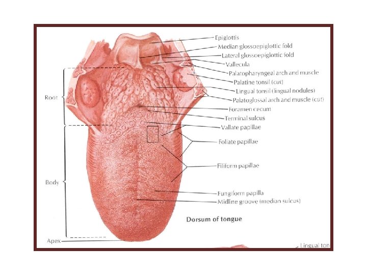
Oropharynx • Extends from soft palate to superior border of epiglottis. • Communicates anteriorly with oral cavity via oropharyngeal isthmus. • Palatine tonsil is located in its lateral wall between palatoglossal and palatopharyngeal arches (in tonsillar fossa/sinus). • Anterior surface of epiglottis is connected to root of tongue by 3 glossoepiglottic folds (1 median and 2 lateral). • Epiglottic valleculae are 2 small depressions located between epiglottis and root of tongue; one on either side of median glossoepiglottic fold.
Pharyngeal lymphoid ring (Waldeyer’s ring) is a collection of lymphoid tissue at entrance of pharynx from nasal and oral cavities & includes the: 1. pharyngeal 2. tubal 3. lingual 4. palatine tonsils
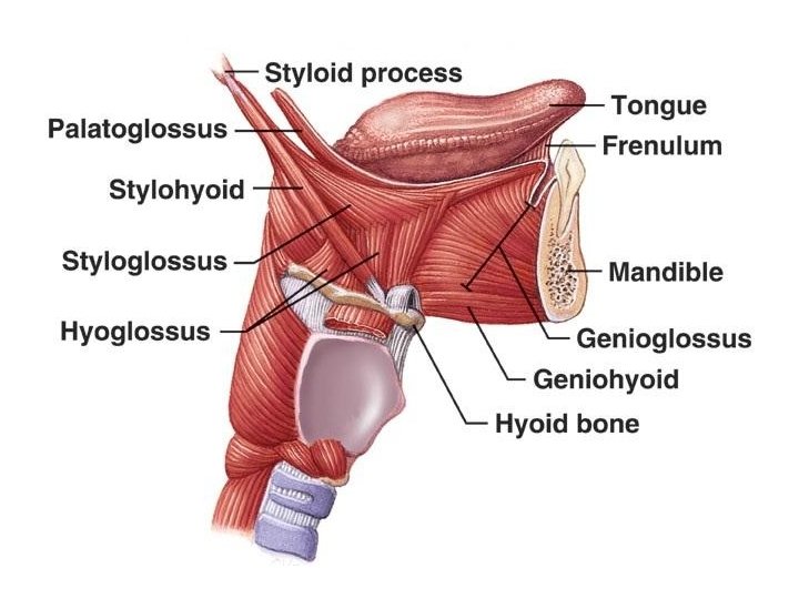
External & Internal Tongue Muscles
Laryngopharynx (Hypopharynx) • extends from superior border of epiglottis to inferior border of cricoid cartilage, where pharynx becomes continuous with esophagus • communicates with laryngeal cavity via laryngeal inlet • piriform fossa (recess): depression located inferolateral to laryngeal inlet and medial to lamina of thyroid cartilage internal laryngeal nerve is located under mucosa of piriform fossa
3 Pharyngeal Constrictor Muscles 1. Superior 2. Middle 3. Inferior
Gaps Between the Constrictor Muscles Overlapping of constrictor muscles leaves 4 gaps in musculature for structures to enter or leave pharynx. Structures passing through gap between superior constrictor and base of skull: 1. pharyngotympanic (auditory) tube 2. levator veli palatini muscle 3. ascending palatine artery (branch of facial artery)
Motor nerve supply of the pharynx: All muscles are supplied by vagus nerve via its pharyngeal branch, except stylopharyngeus (supplied by glossopharyngeal nerve). The inferior constrictor receive some innervation from external laryngeal nerve.
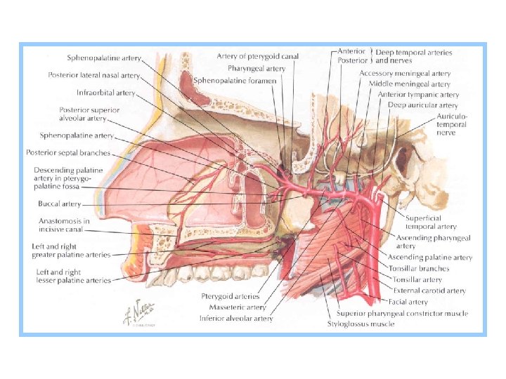
Blood Supply of the Pharynx Arteries: • ascending pharyngeal artery (pharyngeal branches) • facial artery (ascending palatine and tonsillar branches) • maxillary artery (descending palatine and pharyngeal branches) • lingual artery (dorsal lingual branches) • superior thyroid artery (muscular branches)
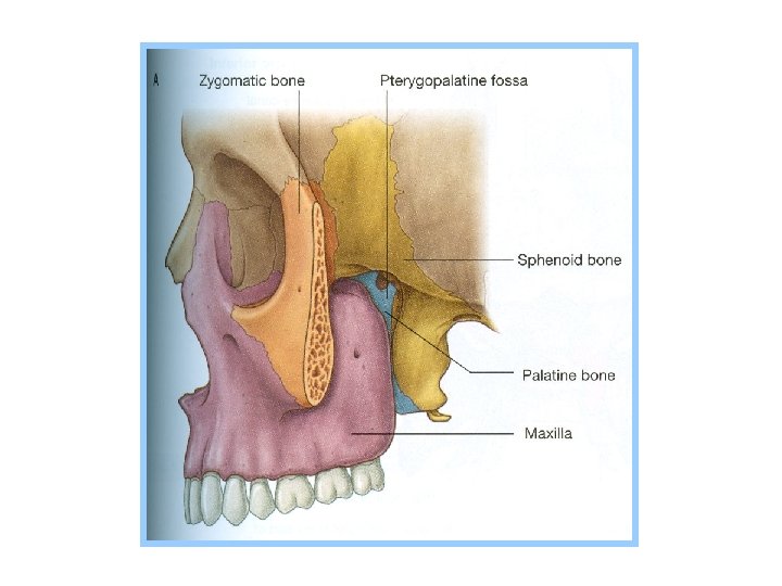
Pterygopalatine Fossa
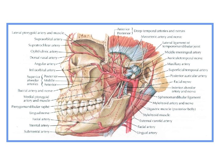
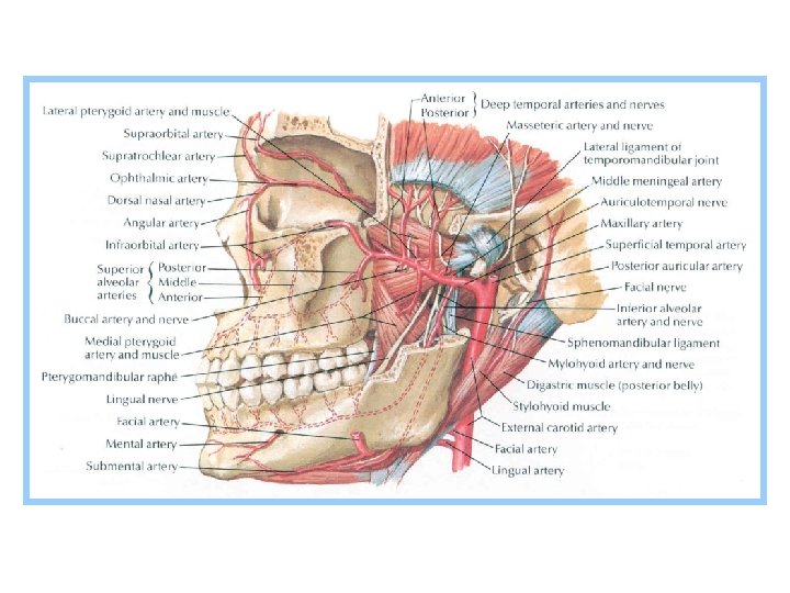
Maxillary Artery Larger terminal branch of external carotid artery. Passes anteriorly, deep to neck of mandible, runs through infratemporal fossa and terminates in pterygopalatine fossa. Parts: 1. mandibular (1 st) 2. pterygoid (2 nd) 3. pterygopalatine (3 rd)
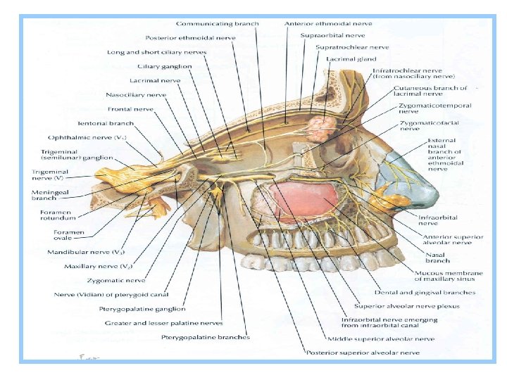
Pterygopalatine (3 rd) Part of Maxillary Artery • maxillary artery leaves infratemporal fossa and enters pterygopalatine fossa via pterygomaxillary fissure • its branches follow branches of maxillary nerve and pterygopalatine ganglion • branches: • posterior superior alveolar artery: accompanies posterior superior alveolar nerve descends on posterior surface of maxilla divides into branches which pass through alveolar foramina of maxilla supplies maxillary molar and premolar teeth and mucosa of maxillary sinus
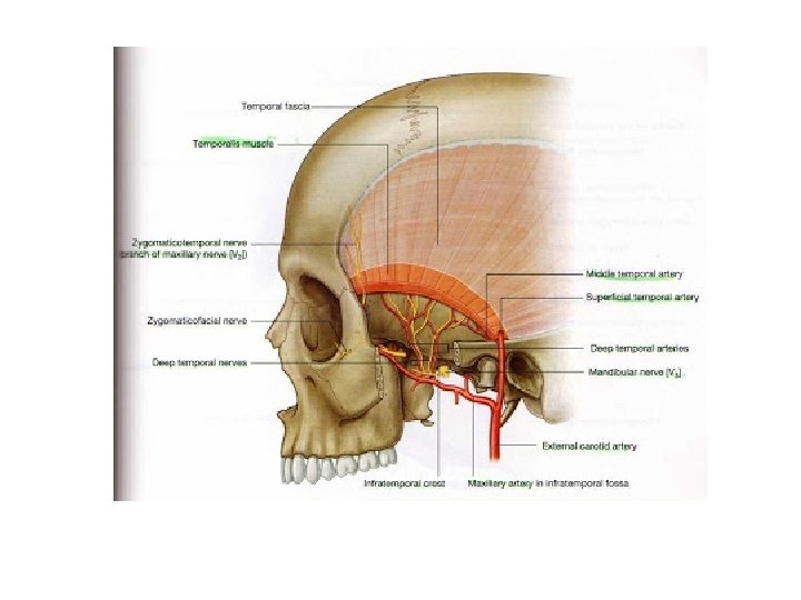
TEMPORAL AND INFRATEMPORAL FOSSAE
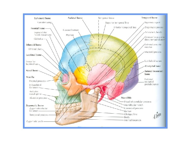
Temporal Fossa Contents: • temporalis muscle and its fascia • deep temporal nerves and vessels • superficial temporal vessels • auriculotemporal nerve • zygomaticotemporal nerve and vessels
Infratemporal Fossae
Infratemporal Fossa Boundaries: • anterior: infratemporal (posterior) surface of body of maxilla • posterior: tympanic part and styloid process of temporal bone • medial: lateral pterygoid plate • lateral: ramus of mandible • superior: infratemporal surface of greater wing of sphenoid • has no inferior bony boundary
Contents: • lower part of temporalis muscle • medial and lateral pterygoid muscles • mandibular nerve and its branches • chorda tympani • otic ganglion • maxillary artery and its branches • pterygoid venous plexus and maxillary vein
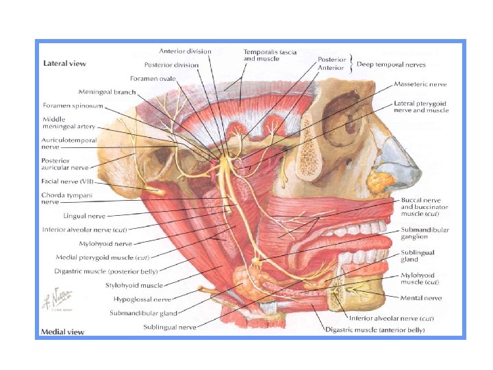
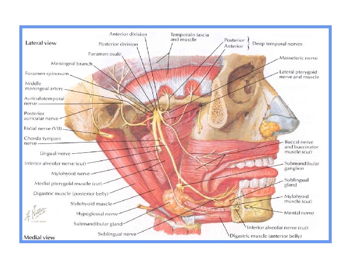
Mandibular Nerve Branches from anterior division: 1. masseteric nerve: passes through mandibular notch to reach masseter muscle, which it supplies. It also contains a few sensory fibers for anterior aspect of temporomandibular joint. 2. lateral pterygoid nerve: supplies lateral pterygoid muscle 3. deep temporal nerves (anterior and posterior): ascend deep to temporalis muscle to supply it 4. buccal nerve: passes between 2 heads of lateral pterygoid and runs anteriorly and inferiorly emerges onto cheek from under ramus of mandible gives sensory innervation to skin and mucosa of cheek
Mandibular Nerve Branches from posterior division: 1. Auriculotemporal nerve: • originates by 2 roots that encircle middle meningeal artery and join posterior to it • passes posteriorly, medial to lateral pterygoid and neck of mandible passes posterior to temporomandibular joint ascends superficial to zygomatic process of temporal bone toward temporal region (with superficial temporal vessels) • gives sensory innervation to auricle, external acoustic meatus, tympanic membrane, temporomandibular joint and scalp • gives off parotid branches contain postganglionic parasympathetic fibers from otic ganglion
Mandibular Nerve Branches from posterior division (cont. ): 2. Inferior alveolar nerve: • contains motor and sensory fibers • passes inferiorly, medial to lateral pterygoid, to enter mandibular canal via mandibular foramen & runs within mandibular canal and supplies sensory innervation to all mandibular teeth of corresponding side • gives off mental nerve, which emerges onto face via mental foramen & supplies sensory innervation to lower lip and chin • before entering mandibular canal it gives off mylohyoid nerve & supplies mylohyoid anterior belly of digastric muscles
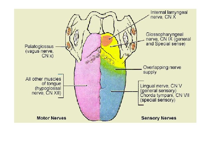
Mandibular Nerve Branches from posterior division (cont. ): 3. Lingual nerve: • descends medial to lateral pterygoid, anterior to inferior alveolar nerve • gives general sensory innervation (touch, pain, temperature) to mucosa of anterior ⅔ of tongue and mucosa of floor of mouth • high in infratemporal fossa is joined by chorda tympani (intrapetrosal branch of facial nerve)
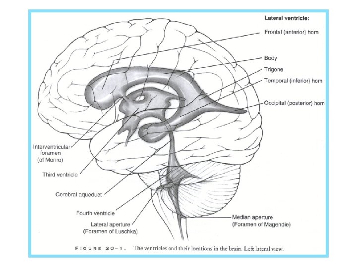
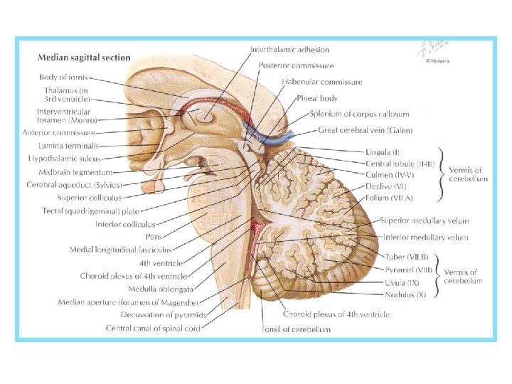
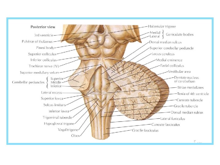
Brain
Parts of the Brain • brainstem: • medulla oblongata • pons • midbrain • cerebellum • diencephalon: • thalamus • hypothalamus • telencephalon (cerebral hemispheres)
thalamus hypothalamus
cerebellar nuclei cerebellar cortex subcortical white matter
hypothalamus
left thalamus right thalamus
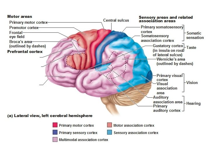
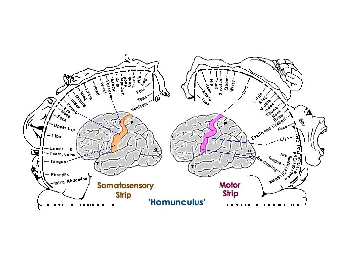
central sulcus lateral sulcus insula
Arterial Supply of the Brain • it is provided by 2 pairs of arteries: vertebral and internal carotid • vertebral artery: • enters cranial cavity via foramen magnum • right and left vertebral arteries join each other at pontomedullary junction to form basilar artery • basilar artery runs along midline of anterior surface of pons and ends at upper end of pons by dividing into 2 posterior cerebral arteries • vertebral and basilar arteries give branches that supply spinal cord, brainstem, cerebellum and posterior parts of cerebral hemispheres
Cerebral Arterial Circle (of Willis) • formed by large cerebral arteries and their interconnections on ventral surface of the brain • components: • anterior communicating a. • anterior cerebral a. • internal carotid a. • posterior communicating a. • posterior cerebral a. • serves as a potential vascular shunt, assisting in development of collateral circulation if one of the proximal vessels is occluded