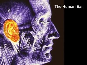
The Ear Three parts of the ear Outer


- Slides: 25

The Ear

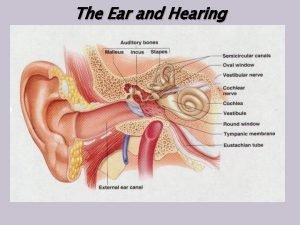
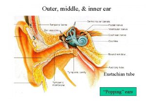
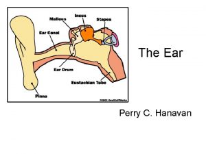
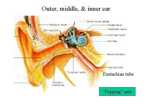
Three parts of the ear • Outer ear – The Pinna – what we see on the sides of our head – – The pinna gather sound into our ears • The Ear canal leads to the: • Middle Ear – The section from the ear drum to the oval window – Contains three small bones, the malleus, incus, and stapes (hammer, anvil, and stirrup), ILLUSTRATION 1

The Inner Ear • The inner ear is composed of the cochlea, from the Greek word for snail, and the semicircular canals • The cochlea is filled with fluid, and the pushing and pulling of the stapes on the oval window moves the fluid in this coiled tube ILLUSTRATION 1

External ear • The folds in the ear channel sounds into our inner ear. Since each person has slightly different folds and channels, we each hear slightly differently. ILLUSTRATION 1

Sound Amplification The Pinna and ear canal amplify sound coming into the ear 20 times Bonus slide –no illustration

Middle Ear • carries vibrations from the eardrum to the inner ear. • The stapes, pulls or pushes on the membranous oval window The oval window is a closed membrane, but acts as the entrance to the inner ear for sound energy. ILLUSTRATION 2

Inner Ear – Cochlea stretched out • The vibrations travel into the fluid of the upper tube of the cochlea and around the tip of the organ into the fluid of the lower tube. • the round window releases the pressure of this motion

Inner Ear • Mechanical to electrical impulse • Fluid -The motion of the stapes sets the fluid in the top moving • Hair Receptors

Hearing – Cochlear Duct • Hair Receptors - Displacement

Geography of the Cochlea 20, 000 Hz 1500 hz 500 Hz 20 Hz • Where different sound frequencies will trigger these hair cells has been mapped

20, 000 Hz 1500 Hz 20 Hz

Cochlear Duct • Vestibular duct and Tympanic duct – Fluid and bone

Equilibrium • Hair Cells • Vestibular complex – 3 basic directional movements • When you tilt your head the fluid in the cochlear duct pushes the hair receptors in different directions

Hearing Process • Step 1 –sound waves arrive at the tympanic Membrane • Directionality

Sound Location • We can distinguish the slightest variation in amplitude (volume) and therefore determine direction of sounds

Step 2 • Movement of the tympanic membrane causes displacement of the auditory ossicles

Step 3 • Movement of the stapes establishes pressure waves in the perilymph

Step 4 • Pressure waves distort the basilar membrane

Step 5 • Transformation of mechanical impulse

Step 6 • Information to the Nervous system

Organ of Corti

Nerves

Signals • Two types of signalling: • Dendrites • Axons

Chemical messengers

Space and joining • Synapse • Synaptic Cleft
 Evs ppt for class 4
 Animal ear like leaves
 External parts of computer
 Names of parts of ear
 Auris ear
 Hát kết hợp bộ gõ cơ thể
 Slidetodoc
 Bổ thể
 Tỉ lệ cơ thể trẻ em
 Gấu đi như thế nào
 Chụp phim tư thế worms-breton
 Chúa yêu trần thế
 Các môn thể thao bắt đầu bằng tiếng nhảy
 Thế nào là hệ số cao nhất
 Các châu lục và đại dương trên thế giới
 Công thức tính thế năng
 Trời xanh đây là của chúng ta thể thơ
 Cách giải mật thư tọa độ
 Phép trừ bù
 Phản ứng thế ankan
 Các châu lục và đại dương trên thế giới
 Thể thơ truyền thống
 Quá trình desamine hóa có thể tạo ra
 Một số thể thơ truyền thống
 Cái miệng xinh xinh thế chỉ nói điều hay thôi
 Vẽ hình chiếu vuông góc của vật thể sau
