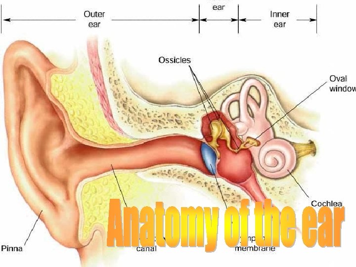
3 Parts External ear Pinna auricle External auditory


 ® Mastoid air")


 Mesotympanum Hypotympanum")


- Slides: 34
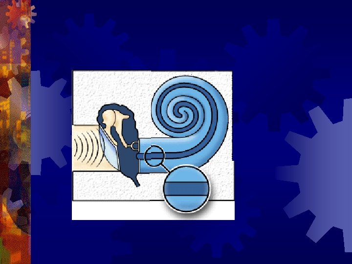
3 Parts: ® External ear ® Pinna/ auricle ® External auditory canal/ meatus ® Middle ear cleft ® Eustachian tube ® Tympanic cavity (middle ear) ® Mastoid air cell system ® Inner ear (labyrinth) ® Cochlea ® Vestibular apparatus
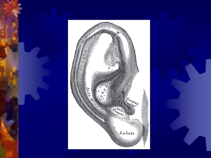
External ear- Pinna ® Skin draping the auricular cartilage ® Lateral and medial surface ® Ridges and depressions ® Skin-perichondrium: tightly adhered on the lateral surface ® Inflammatory conditions are extremely painful ® Perichondritis --‘Cauliflower ear’ ® Congenital anomalies may indicate more anomalies inside
External auditory canal ® 24 mm long ® 2 parts Lateral 1/3 Cartilaginous (8 mm) ® Medial 2/3 Bony (16 mm) ® ® Tortuous- straighten to examine ® Skin lined- Differences ® TM- oblique ® Anterior and inferior recesses ® Fissures/ foramina ® Pathological conditions
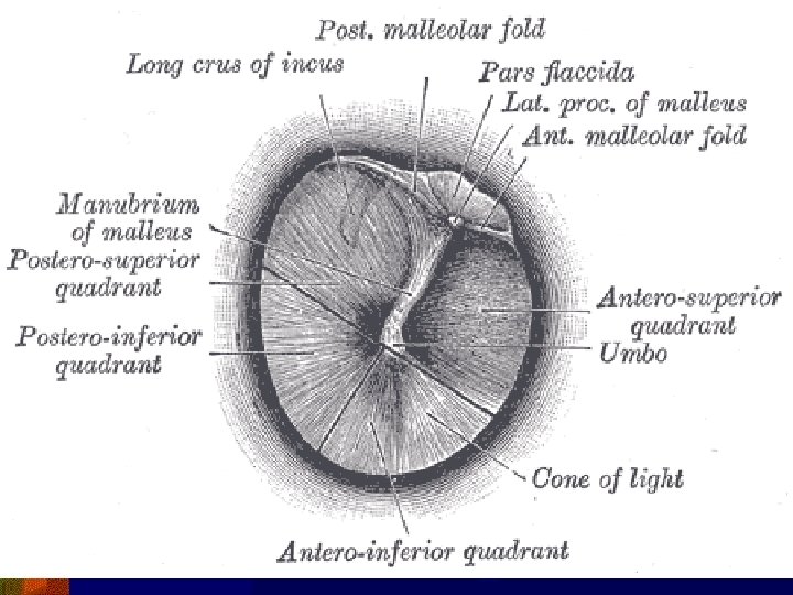
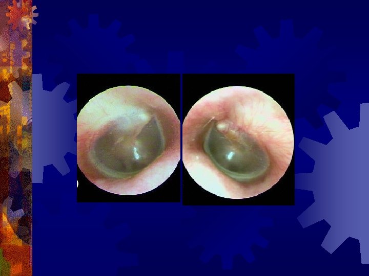
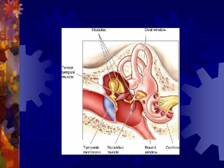
Tympanic membrane ® Pearly white translucent membrane‘Lusture’ ® Obliquely placed ® 2 parts: Pars tensa and pars flaccida ® 3 layers of pars tensa: ® Skin-fibrous-mucosa ® Annulus ® Notch of Rivinus
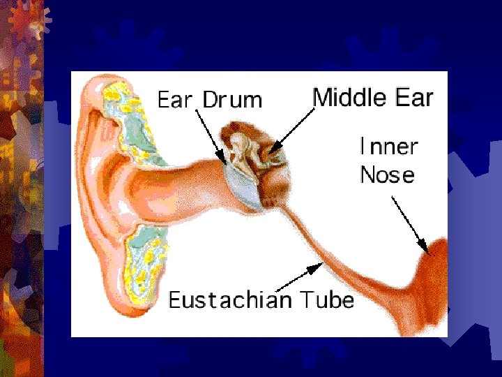
Middle ear cleft ® Eustachian tube ® Tympanic cavity (middle ear) ® Mastoid air cell system
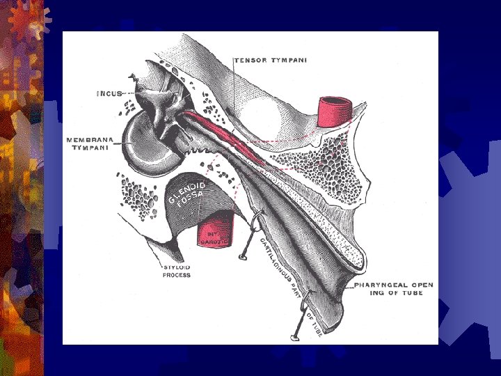
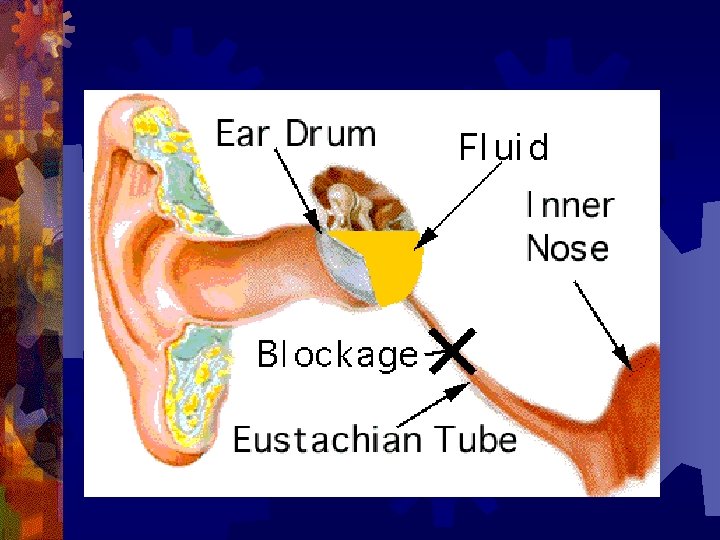
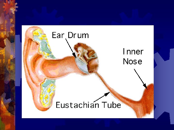
Eustachian tube ® 36 mm long ® 2 parts ® Medial 2/3 Cartilagenous- 24 mm ® Lateral 1/3 Bony- 12 mm ® Child v/s Adult ® Closed at rest ® Respiratory epithelium
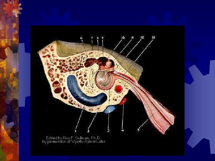
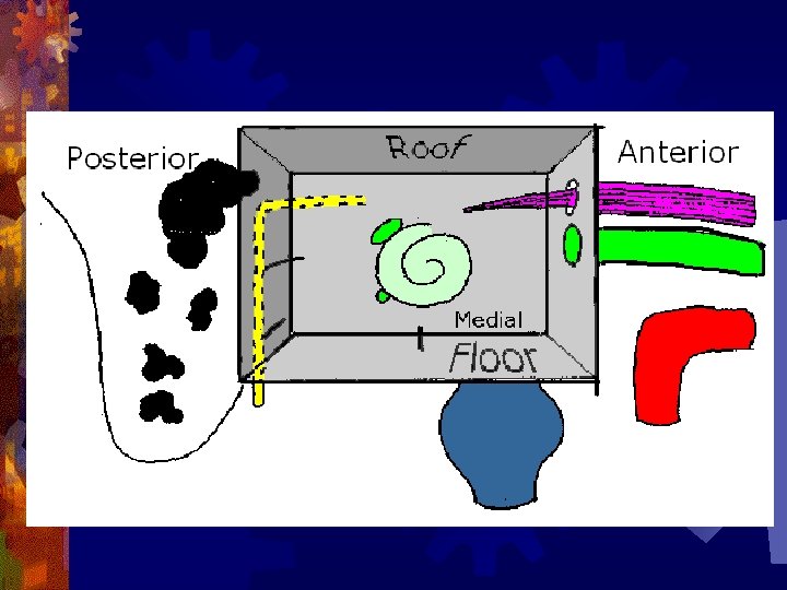
Tympanic cavity: 6 walls ® Lateral ® Medial ® Roof ® Floor ® Anterior ® Posterior
Epitympanum (attic) Mesotympanum Hypotympanum
Lateral wall ® Membranous ® Tympanic membrane ® Bony Lateral attic wall ® Lateral wall of hypotympanum ®
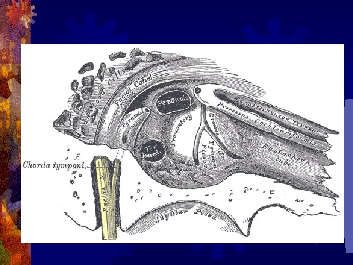
Medial wall ® Promontry ® Oval window ® Round window ® Processus cochleariformis ® Horizontal part of facial canal forms a bulge
Anterior wall ® Canal for tensor tympani ® Eustachian tube opening ® Corda tympanic nerve exits ® Internal carotid artery is related in its inferior part
Posterior wall ® Aditus ad antrum ® Bulge produced by lateral semicircular canal ® Pyramid ® Bulge produced by vertical part of facial N ® Sinus tympani ® Facial recess
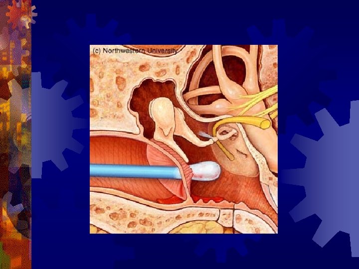
Contents of middle ear ® 3 ossicles: Malleus, incus and stapes ® 2 muscles: Tensor tympani and stapedius ® 2 nerves: Corda tympani and tympanic plexus (IX) ® Mucosal folds and ligaments ® Vessels
Malleus
Incus
Stapes
® Blood supply ® Anterior, posterior, superior and inferior tympanic vessels and carotico-tympanic arteries ® Nerve supply: ® Tympanic plexus (IX cranial nerve) ‘Jacobson’s nerve’