Skeletal System The skeletal system is composed of


 Coccyx (4) Lumbar Vertebrae (5) Cervical Vertebrae (7) Thoracic Vertebrae")


 and the Sternum • The ribs")


 Sternal End Acromial End Conoid Tubercle")


 is the process of bone tissue")

 of the epiphyseal")


![Decreased Blood [Ca 2+] Increased PTH release by parathyroid gland Binds to osteoblast causing](https://slidetodoc.com/presentation_image_h2/a9f1ea194a0f4f8c5df3a6ce2d7dee75/image-74.jpg "Decreased Blood [Ca 2+] Increased PTH release by parathyroid gland Binds to osteoblast causing")


- Slides: 77
Skeletal System
• The skeletal system is composed of 206 bones. • The skeletal system provides 1. ) protection 2. )support 3. ) production of red blood cells 4. ) movement 5. ) Triglyceride Storage (yellow fat) 6. ) Storing Minerals • • Bones are attached to other bones by ligaments.
Axial Skeleton Skull, Vertebral Column, Ribs, & Sternum • 80 bones
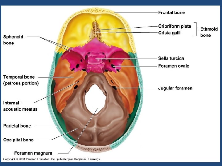
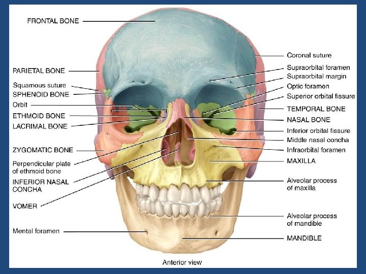
Skull • Cranium • Occipital • Temporal • Frontal • Parietal Sphenoid 29 bones 8 1 2 2 Butterfly shaped or winged shaped
• Face • Mandible • Maxillae • Zygomatic Lacrimal Ethmoid 14 1 2 2
Vertebral column 26 bones • • • Vertebral column 26 bones 24 vertebrae & 1 sacrum & 1 coccyx 7 Cervical Vertebrae Atlas 1 st cervical vertebrae (nod yes) Axis 2 nd cervical vertebrae ( no )
Vertebral Column Sacrum (5) Coccyx (4) Lumbar Vertebrae (5) Cervical Vertebrae (7) Thoracic Vertebrae (12)
Cervical Vertebrae C 1 & C 2 Axis Dens Atlas Body Transverse Foramen Vertebral Foramen Spinous Process
• 12 Thoracic Vertebrae • 5 Lumbar Vertebrae • Hyoid Sternum & ribs 26 bones
Thoracic Vertebra Superior Articular Process Body Rib Facet Transverse Process Spinous Process
Lumbar Vertebra Body Transverse Process Superior Articular Process Pedicle Spinous Process
Sacrum Ala Sacral Canal Median Sacral Crest Posterior Sacral Foramen Lateral Sacral Crest
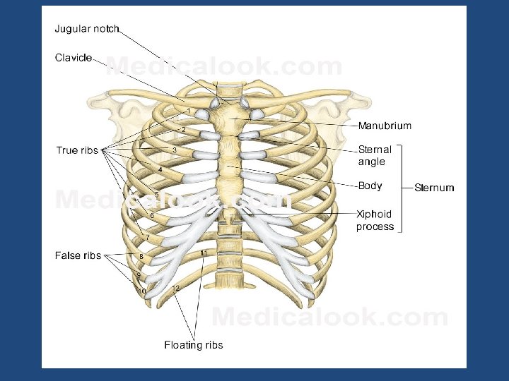
Ribs • Composed of 12 pairs (24 bones) and the Sternum • The ribs are shaped like crescents, with one end flattened and the other end rounded. The rounded ends are attached at joints to the thoracic vertebrae at the back and the flattened ends come together at the sternum, in the front. • The upper seven pairs of ribs attach to the sternum with costal cartilage and are known as “true ribs.
Ribs • The 8 th through 10 th ribs have non-costal cartilage which connects them to the ribs above. The last two ribs are called “free ribs” because they do not attach to the sternum or to other ribs and simply “hang free. ” The length of each rib increases from number one to seven and then decreases until rib pair number 12. The first rib is the shortest, broadest, flattest, and most curved.
Rib Sternal End Angle Tubercle Neck Head
Sternum Manubrium Angle Clavicular Notch Body Sternal Notch What is missing? Xiphoid Process
Appendicular Skeleton 126 bones • Pectoral Girdle & upper extremities • Clavicle 2 • Scapula 2 • Humerus 2 • Radius 2 Ulna • • • 2 Carpals 16 Metacarpals 10 Phalanges 28 64 bones
Lower Extremities 62 bones • Coxal Bones 3 Ilium, ischium and pubis • • Femur Tibia Fibula Tarsals 2 2 2 14
Terminology: • Articulating Surface: These are the areas of the bone that move in conjunction with another bone. • Condyle: Rounded projection for articulation with another bone. • Head: A portion of the bone that expands beyond its constricted neck.
• Non-articulating Surfaces: Serve as attachment sights • Projections • 1. Tuberosity : a large roughened projection • 2. Tubercle: a small rounded projection • 3. Spine: a sharp projection • 4. Crest: a ridge • 5. Process: an obvious portion that protrudes from a surface. • 6. Trochanter: a prominent process.
• • • Depressions 1. Fossa Shallow or hollow bone depression 2. Groove: long shallow depression 3. Foramen: a hole 4. Sinus: an air filled cavity 5. Meatus: a tube shaped opening
Bone Structure: • • Diaphysis: The shaft of a long bone. • Epiphysis: Two enlarge ends of the bones • Proximal Epiphysis and Distal Epiphysis • Proximal Epiphysis contains the red bone marrow in adult bones.
Pelvic Girdle
Pelvic girdle • Ilium: The larger, superior Portion of the pelvic region. ( Hip bone) • Iliac Crest: Rim of the ilium or hip. • Iliac fossa the depression of the ilium. • Ilium is fused to the Sacrum by the sacroiliac joint.
Ischium: The inferior and posterior portion of the hip. Pubis: The inferior and anterior portion of the • Subpubic angle: male is more straight thanfemal skeloton. • Obturator Foramen: filled with connective tissue. • Symphysis pubis: joins the right and left halves. • Acetabulum: piont of articulation of the lower limb and the pelvic girdle.
Sacrum Iliac Fossa Pelvic Brim Acetabulum Obturator Foraman Ischium Pubis Pubic Symphasis Pelvis
Coxal Bone Iliac Crest Posterior Superior Iliac Spine Anterior Inferior Iliac Spine Posterior Inferior Iliac Spine Acetabulum Obturator Foramen Greater Sciatic Notch Ischial Tuberosity Ischial Spine
Femur Greater Trochanter Lateral Epicondyle Head Lesser Trochanter Medial Epicondyle
Femur posterior Lesser Trochanter Head Greater Trochanter Gluteal Tuberosity Linea Aspera Medial Epicondyle Medial Condyle Lateral Epicondyle
Tibia Medial Malleolus Anterior Tibial Tuberosity Crest Patella Medial Condyle Lateral Condyle
Fibula Lateral Malleolus Head
Proximal Phalanx Middle Phalanx 5 4 3 2 1 Distal Phalanx Proximal Phalanx Tarsals Foot Metatarsals Phalanges
Cuboid Calcaneous Talus Navicular Foot - Tarsals 3 rd Cuneiform 1 st Cuneiform 2 nd Cuneiform
Bones Of the Upper Extremities. • • Humerus Scapula Clavicle Radius Ulnu Carples Metacarples Phalangees.
Posterior Scapula Supraspinous Fossa Scapular Spine Acromion Infraspinous Fossa
Anterior Scapula Coracoid Process Glenoid Cavity Subscapular Fossa
Clavicle (inferior view) Sternal End Acromial End Conoid Tubercle
Humerus – Anterior View Greater Tubercle Intertubercular Groove Lateral Epicondyle Capitulum Coronoid Fossa Trochlea Lesser Tubercle Medial Epicondyle Deltoid Tuberosity
Humerus – Posterior View Head Greater Tubercle Olecranon Fossa Medial Epicondyle Trochlea Lateral Epicondyle
Ulna Coronoid Process Olecranon Process Trochlear Notch Styloid Process Head Radial Notch
Radius Radial Tuberosity Head Styloid Process
Hand Wrist Middle Phalanx Distal Phalanx Proximal Phalanx Phalanges Distal Phalanx Metacarpals Carpals 5 4 3 2 1 Proximal Phalanx
Bone Structure • Bones are organs. Thus, they’re composed of multiple tissue types. Bones are composed of: – – – – Bone tissue (a. k. a. osseous tissue). Fibrous connective tissue. Cartilage. Vascular tissue. Lymphatic tissue. Adipose tissue. Nervous tissue.
• All bones consist of a dense, solid outer layer known as compact bone and an inner layer of spongy bone – a honeycomb of flat, needle-like projections called trabeculae. • Bone is an extremely dynamic tissue!!!! Above: Note the relationship btwn the compact and spongy bone. Below: Close up of spongy bone.
Note the gross differences between the spongy bone and the compact bone in the above photo. Do you see the trabeculae?
Bone Tissue • • Bone tissue is a type of connective tissue, so it must consist of cells plus a significant amount of extracellular matrix. Bone cells: 1. Osteoblasts • Bone-building cells. • Synthesize and secrete • • collagen fibers and other organic components of bone matrix. Initiate the process of calcification. Found in both the periosteum and the endosteum The blue arrows indicate the osteoblasts. The yellow arrows indicate the bone matrix they’ve just secreted.
Bone Structure 2. Osteocytes • • Mature bone cells. Osteoblasts that have become trapped by the secretion of matrix. No longer secrete matrix. Responsible for maintaining the bone tissue. Yellow arrows indicate osteocytes – notice how they are surrounded by the pinkish bone matrix. Blue arrow shows an osteoblast in the process of becoming an osteocyte. On the right, notice how the osteocyte is “trapped” within the pink matrix
3. Osteoclasts – – – Huge cells derived from the fusion of as many as 50 monocytes (a type of white blood cell). Cells that digest bone matrix – this process is called bone resorption and is part of normal bone growth, development, maintenance, and repair. Concentrated in the endosteum. On the side of the cell that faces the bone surface, the PM is deeply folded into a ruffled border. Here, the osteoclast secretes digestive enzymes (how might this occur? ) to digest the bone matrix. It also pumps out hydrogen ions (how might this occur? ) to create an acid environment that eats away at the matrix. What advantage might a ruffled border confer? Why do we want a cell that eats away at bone? (Hint: bone is a very dynamic tissue. )
• Here, we see a cartoon showing all 3 cell types. Osteoblasts and osteoclasts are indicated. • Note the size of the osteoclast (compare it to the osteoblast), and note the ruffled border. • Why is there a depression underneath the osteoclast? • What is the name of the third cell type shown here? • What do you think the tan material represents?
Bone Structure • Bone Matrix: – Consists of organic and inorganic components. – 1/3 organic and 2/3 inorganic by weight. • Organic component consists of several materials that are secreted by the osteoblasts: – Collagen fibers and other organic materials » These (particularly the collagen) provide the bone with resilience and the ability to resist stretching and twisting.
• Inorganic component of bone matrix – Consists mainly of 2 salts: calcium phosphate and calcium hydroxide. These 2 salts interact to form a compound called hydroxyapatite. – Bone also contains smaller amounts of magnesium, fluoride, and sodium. – These minerals give bone its characteristic hardness and the ability to resist compression. Three-dimensional array of collagen molecules. The rodshaped molecules lie in a staggered arrangement which acts as a template for bone mineralization. Bone mineral is laid down in the gaps. Note collagen fibers in longitudinal & cross section and how they occupy space btwn the black bone cells.
This bone: a. Has been demineralized b. Has had its organic component removed
Long Bone Structure • Shaft plus 2 expanded ends. • Shaft is known as the diaphysis. – Consists of a thick collar of compact bone surrounding a central marrow cavity • In adults, the marrow cavity contains fat - yellow bone marrow. • Expanded ends are epiphyses – Thin layer of compact bone covering an interior of spongy bone. – Joint surface of each epiphysis is covered w/ a type of hyaline cartilage known as articular cartilage. It cushions the bone ends and reduces friction during movement.
Bone Marrow • Bone marrow is a general term for the soft tissue occupying the medullary cavity of a long bone, the spaces amid the trabeculae of spongy bone, and the larger haversian canals. • There are 2 main types: red & yellow. • Red bone marrow = blood cell forming tissue = hematopoietic tissue • Red bone marrow looks like blood but with a thicker consistency. • It consists of a delicate mesh of reticular tissue saturated with immature red blood cells and scattered adipocytes. Notice the red marrow and the compact bone
Distribution of Marrow • In a child, the medullary cavity of nearly every bone is filled with red bone marrow. • In young to middle-aged adults, the shafts of the long bones are filled with fatty yellow bone marrow. – Yellow marrow no longer produces blood, although in the event of severe or chronic anemia, it can transform back into red marrow • In adults, red marrow is limited to the axial skeleton, pectoral girdle, pelvic girdle, and proximal heads of the humerus and the femur. Note the compact bone on the bottom and marrow on the bottom.
Bone Development • Osteogenesis (a. k. a. ossification) is the process of bone tissue formation. • In embryos this leads to the formation of the bony skeleton. • In children and young adults, ossification occurs as part of bone growth. • In adults, it occurs as part of bone remodeling and bone repair.
Formation of the Bony Skeleton • Before week 8, the human embryonic skeleton is made of fibrous membranes and hyaline cartilage. • After week 8, bone tissue begins to replace the fibrous membranes and hyaline cartilage. – The development of bone from a fibrous membrane is called intramembranous ossification. Why? – The replacement of hyaline cartilage with bone is known as endochondral ossification. Why?
Growth in Bone Length • Epiphyseal cartilage (close to the epiphysis) of the epiphyseal plate divides to create more cartilage, while the diaphyseal cartilage (close to the diaphysis) of the epiphyseal plate is transformed into bone. This increases the length of the shaft.
At puberty, growth in bone length is increased dramatically by the combined activities of growth hormone, thyroid hormone, and the sex hormones. • As a result osteoblasts begin producing bone faster than the rate of epiphyseal cartilage expansion. Thus the bone grows while the epiphyseal plate gets narrower and ultimately disappears. A remnant (epiphyseal line) is visible on X-rays (do you see them in the adjacent femur, tibia, and fibula? )
Growth in Bone Thickness • Osteoblasts beneath the periosteum secrete bone matrix on the external surface of the bone. This obviously makes the bone thicker. • At the same time, osteoclasts on the endosteum break down bone and thus widen the medullary cavity. • This results in an increase in shaft diameter even though the actual amount of bone in the shaft is relatively unchanged.
Fractures • Despite its mineral strength, bone may crack or even break if subjected to extreme loads, sudden impacts, or stresses from unusual directions. – The damage produced constitutes a fracture. • The proper healing of a fracture depends on whether or not, the blood supply and cellular components of the periosteum and endosteum survive.
Fracture Types • Fractures are often classified according to the position of the bone ends after the break: Open (compound) bone ends penetrate the skin. Closed (simple) bone ends don’t penetrate the skin. Comminuted bone fragments into 3 or more pieces. Common in the elderly (brittle bones). Greenstick bone breaks incompletely. One side bent, one side broken. Common in children whose bone contains more collagen and are less mineralized. Spiral ragged break caused by excessive twisting forces. Sports injury/Injury of abuse. Impacted one bone fragment is driven into the medullary space or spongy bone of another.
Bone Remodeling • Bone is a dynamic tissue. – What does that mean? • Wolff’s law holds that bone will grow or remodel in response to the forces or demands placed on it. Examine this with the bone on the left.
Check out the mechanism of remodeling on the right! Why might you suspect someone whose been a powerlifter for 15 years to have heavy, massive bones, especially at the point of muscle insertion? Astronauts tend to experience bone atrophy after they’re in space for an extended period of time. Why?
Nutritional Effects on Bone • Normal bone growth/maintenance cannot occur w/o sufficient dietary intake of calcium and phosphate salts. • Calcium and phosphate are not absorbed in the intestine unless the hormone calcitriol is present. Calcitriol synthesis is dependent on the availability of the steroid cholecalciferol (a. k. a. Vitamin D) which may be synthesized in the skin or obtained from the diet. • Vitamins C, A, K, and B 12 are all necessary for bone growth as well.
Hormonal Effects on Bone • Growth hormone, produced by the pituitary gland, and thyroxine, produced by the thyroid gland, stimulate bone growth. – GH stimulates protein synthesis and cell growth throughout the body. – Thyroxine stimulates cell metabolism and increases the rate of osteoblast activity. – In proper balance, these hormones maintain normal activity of the epiphyseal plate (what would you consider normal activity? ) until roughly the time of puberty.
Hormonal Effects on Bone • At puberty, the rising levels of sex hormones (estrogens in females androgens in males) cause osteoblasts to produce bone faster than the epiphyseal cartilage can divide. This causes the characteristic growth spurt as well as the ultimate closure of the epiphyseal plate. • Estrogens cause faster closure of the epiphyseal growth plate than do androgens. • Estrogen also acts to stimulate osteoblast activity.
Parathyroid Hormone • Released by the cells of the parathyroid gland in response to low blood [Ca 2+]. Causes blood [Ca 2+] to increase. • PTH will bind to osteoblasts and this will cause 2 things to occur: • The osteoblasts will decrease their activity and they will release a chemical known as osteoclast-stimulating factor. • Osteoclast-stimulating factor will increase osteoclast activity. • PTH increases calcitriol synthesis which increases Ca 2+ absorption in the small intestine. • PTH decreases urinary Ca 2+ excretion and increases urinary phosphate excretion.
Decreased Blood [Ca 2+] Increased PTH release by parathyroid gland Binds to osteoblast causing decreased osteoblast activity and release of osteoclaststimulating factor Increased calcitriol synthesis OSF causes increased osteoclast activity Decreased bone deposition and increased bone resorption Decreased Ca 2+ excretion Increased intestinal Ca 2+ absorption Increased Blood [Ca 2+]
Clinical Conditions • Osteomyelitis – Osteo=bone + myelo=marrow + itis=inflammation. – Inflammation of bone and bone marrow caused by pus-forming bacteria that enter the body via a wound (e. g. , compound fracture) or migrate from a nearby infection. – Fatal before the advent of antibiotics.
Clinical Conditions • Osteoporosis – Group of diseases in which bone resorption occurs at a faster rate than bone deposition. – Bone mass drops and bones become increasingly porous. – Compression fractures of the vertebrae and fractures of the femur are common. – Often seen in postmenopausal women because they experience a rapid decline in estrogen secretion; estrogen stimulates osteoblast and inhibits osteoclast activity. • Based on the above, what preventative measures might you suggest?
Clinical Conditions • Gigantism – Childhood hypersecretion of growth hormone by the pituitary gland causes excessive growth. • Acromegaly – Adulthood hypersecretion of GH causes overgrowth of bony areas still responsive to GH such as the bones of the face, feet, and hands. • Pituitary dwarfism – GH deficiency in children resulting in extremely short long bones and maximum stature of 4 feet.