Skeletal System Skeletal System Functions of the skeletal


 and hind (pelvic) limbs -")

 • 3 pairs of")
 - Tarsal bones - Hock")


- Slides: 40
Skeletal System
Skeletal System • Functions of the skeletal • Skeleton =made up of various forms of connective tissue system – Musculoskeletal system • 2 systems work together – Support body – Allow movement • Consists of – Bones – Joints – Cartilage –Binds together and supports structures of the body • Bone • Tendons • Ligaments • Cartilage
Bone • Red bone marrow –Forms red/white blood cells –Clotting cells • One of the hardest tissues in the body • When in embryo = skeleton is made of cartilage and fibrous membranes –Harden into bone before birth • Ossification –Formation of bone from fibrous tissue –Continues until maturity • Normal bone continuously is built up and broken down throughout animals life –Allows bone to heal and repair itself
Types of Bones • Classified by shape – Long • Found in limbs • Support body weight • Act as levers for propulsion - Short Knee and hock Absorbs shock of joint - Flat Encircle cavities containing vital organs Ribs - Unpaired Vertebrae
Makeup of Bone
Composition of Bone • Mature bone – Composition • 25% Water • 45% Mineral – Calcium = 37% – Phosphorus = 18. 5% 30% Organic matter - Structure - Compact bone Provides rigidity and strength - Spongy bone Contains bone marrow
Osteoblasts • Osteoblasts – Oste/o = bone – -blasts = immature – Immature bone cells that produce bony tissue – When mature, become osteocytes
Red and Yellow Bone Marrow • Located in spongy part of bone • Red bone marrow –Hematopoietic • Hemat/o – blood • -poietic pertaining to formation –Produces red blood cells, white blood cells • Yellow bone marrow –In adult animals, in limb bones – replaces red bone marrow –Composed mainly of fat cells –Serves as fat storage area
Periosteum • • Covers bone Thin adherent membrane Protects bone Influences growth – Damage to periosteum usually means irregular bone growth Splints Spavins Ringbone
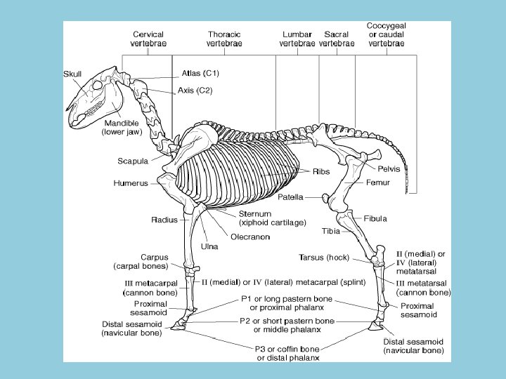
Bones • Skeleton divided into 2 parts –Axial skeleton • Includes –Skull –Vertebral column » Spine –Ribs –Sternum –Tail – Appendicular skeleton • Includes – Extremities – Shoulder – Pelvic girdle • Append = add or hang – Hang from axial skeleton
Axial Skeleton
Axial Skeleton - Skull • Cranium –Portion of skull that encloses the brain • Frontal –Forms roof of cranial cavity • “front” portion of skull • Parietal –Paired bones –Form roof of back cranial cavity • Occipital – Forms back part of cranial cavity • Where opening of spinal cord is • Temporal – Paired bones – Form sides and base of cranium – Temple is seen on outside of horse
Axial Skeleton - Skull
Axial Skeleton - Face • Zygomatic –Projections from temporal and frontal bones –Form cheekbone • Maxilla –Forms upper jaw • Mandible –Forms lower jaw • Lacrimal –Forms medial part of orbit • Incisive – Forms rostral part of hard palate and lower edge of nares • Nasal – Forms bridge of nose
Figure 5. 11
Vertebral Column – Spinal Cord • Supports head and body • Protects spinal cord • Made up of individual bones – Vertebrae • Divided into parts –Vary depending on location and function • Some vertebrae names – C 1 (cervical vertebrae #1) – Atlas - C 2 (cervical vertebrae #2) – Axis
Parts of the Vertebrae • Body –Solid portion ventral to spinal cord • Arch –Dorsal part that surrounds spinal cord • Lamina –Left or right dorsal half of the arch • Spinous process –Single projection from the dorsal part of the arch • Transverse process –Project caudolaterally from right and left sides of vertebrae • Articular Process –Paired cranial and caudal projections located at dorsum of arch • Foramen –Means opening –Opening in middle of vertebrae where spinal cord runs = vertebral foramen • Intervertebral discs – Cartilage discs that separate each vertebrae and provide cushion
Vertebrae
Vertebral Column - Five sections - Cervical - 7 vertebrae - Thoracic - 18 vertebrae - Lumbar - 6 vertebrae - 5 in Arabians - Sacral - 5 vertebrae - Fused to make sacrum - Coccygeal - Tail - 15 -21 vertebrae
Table 5. 1: Number of Vertebrae By Species Regions of Vertebral Column • Cervical “C” Neck area • Thoracic “T” Chest area • Lumbar “L” Loin area • Sacral “S” Sacrum area • Coccygeal Tail area
Thoracic Cavity - Chest cavity - Protects - Heart - Lungs - Vessels and nerves - Trachea and esophagus - Makeup - Thoracic vertebrae - Ribs - Paired bones - Attach to thoracic vertebrae - Sternum - Breastbone - Forms midline ventral portion of rib cage
JOINTS
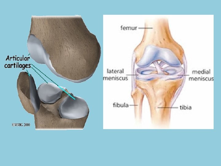
Cartilage • Form of connective tissue • Diminishes effect of concussion • More elastic than bone –Useful in more flexible parts of the skeleton • Articular cartilage –Covers joint surfaces of bone – Diminishes effect of concussion – Provides smooth surface that reduces friction • Meniscus –Curved fibrous cartilage found in some joints • Cushion from force –Canine stifle
Joints • Also called articulations –Articulate • Join so that motion can occur between parts • Connections between bones • Different types of joints based on –Function –Degree of movement
Ligaments, Tendons, and Bursa • Ligament – Band of fibrous connective tissue – Connects bone to bone • Tendon – Band of fibrous connective tissue – Connects muscle to bone • Joint Capsule – Contains bursa – Fibrous sac – Acts as cushion • Ease movement in areas of friction – Shoulder joint • Bursa where tendon passes over bone
Figure 5. 8
Synovial Membrane and Fluid • Found in bursa and synovial joints –Inner lining • Synovial membrane –Secretes synovial fluid » Acts as lubricant for smooth joint action/movement
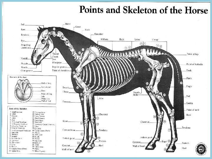
Appendicular Skeleton - Made up of: - Front (thoracic) and hind (pelvic) limbs - Thoracic girdle - Shoulder - Scapula - Clavicle - Pelvic girdle - Pelvis - Os Coxae - Ilium - Ishium - Pubis
Thoracic Bones - Bones of Upper Leg - Scapula - Shoulder blade - Clavicle - Humerus - Ulna - Radius - Bones of Lower Leg - Carpal bones - Bones of Knee
Pelvic Bones • Bones of Upper Leg – Pelvis (hip) • 3 pairs of bones –Ilium –Ischium –Pubis – Femur – Patella – Tibia – Fibula - Bones of Lower Leg - Tarsal bones - Bones of hock
Lower Limbs - Carpal bones - Knee (carpus joint) - Tarsal bones - Hock (tarsus joint) - Metacarpus bones - front limb - Metatarsus bones - hind limb - Four in horses - One major - Third metacarpus/metatarsus - Cannon bone - Two minor - Second and fourth - Splint bones Front Leg
Lower Limbs con’t - One digit - Middle finger of humans - Three phalanges - First/proximal phalanx (long pastern bone) – P 1 - Second/middle phalanx (short pastern bone) – P 2 - Third/distal phalanx (coffin or pedal bone) – P 3 - Joints - Cannon and 1 st phalanx = fetlock joint - 1 st and 2 nd phalanx = pastern joint - 2 nd and 3 rd phalanx = coffin joint - Sesamoid - Two proximal - Near fetlock - One distal (navicular) - Near coffin
The Equine Hoof
The Equine Hoof
Ligaments and Tendons Suspensory Ligament - Strong and flat - Extremely elastic - Most prone to injury - Runs along back of leg between splint bones - Splits at fetlock, attaches to sesamoids and runs towards front of joint - Joins extensor tendon and attaches to front of coffin bone
Superficial Digital Flexor Tendon - Runs down the back of the leg - Branches below the fetlock and inserts onto 1 st and 2 nd phalanx - Flexes the elbow, carpus and lower joints Check Ligament - Short and strong - Runs from upper end of suspensory to just below knee and attaches to superficial flexor tendon
Nuchal Ligament - Elastic ligament extending from poll to withers - Assists neck muscles in holding head and neck in position