MUSCLES STRUCTURE AND FUNCTION The Muscular System MUSCLES


 Muscles make")


. jpg")


 ions")

 1. Enlarged part of sarcoplasmic reticulum where")
 tubules 1. Channels that extend from surface of")
 tubules 3. Perpendicular to S. R. 4. Job")


- Slides: 58
MUSCLES STRUCTURE AND FUNCTION
The Muscular System
MUSCLES n n Myology is the study of muscles (myo = muscle) Muscles make up 40 to 50% of our body weight.
MUSCLES n THE FUNCTIONS OF MUSCLE TISSUE INCLUDE: a. movement - voluntary & involuntary b. heat - 85% of body heat c. posture
Types of muscle n Skeletal muscle Attached primarily to bones n Striated n Voluntary n Major function- movement of bones at joints, maintain posture n n (See Table 8. 2)
Types of Muscle n Cardiac muscle n Wall of the heart Striated Involuntary Autorhythmicity – the ability to contract by itself Major function - pump/move blood n (See Table 8. 2) n n
Types of Muscle n Smooth muscle n n n Located in viscera – blood vessels, skin, abdominal organs, etc. Nonstriated Involuntary May have autorhythmicity Major function - peristalsis, movement of organs (See Table 8. 2)
Characteristics of Muscle Tissue n n Excitability – the ability of muscles to respond to a stimulus Extensibility - the ability to stretch Elasticity - the ability to return to normal length Contractility - the ability to contract
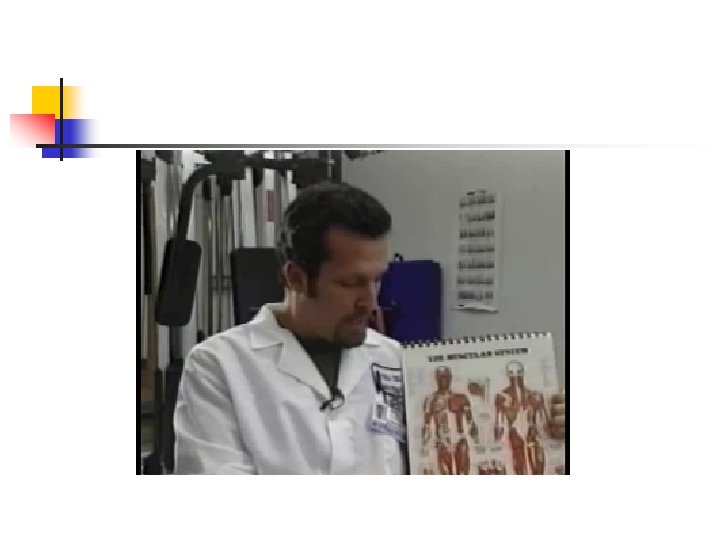
Introduction to the Muscular System SEER TRAINING MODULES - MUSCULAR SYSTEM
http: //www. infovisual. info/03/img_en/008%20 Muscles%20(anterior%20 view). jpg
MUSCLE STRUCTURE n FASCIA - TWO TYPES A. SUPERFICIAL IS JUST UNDER THE SKIN IT IS MADE UP OF ADIPOSE AND LOOSE CONNECTIVE TISSUE.
SUPERFICIAL FASCIA Used with permission 11/26/06 http: //www. integralanatomy. com/images/2_human_anatomy_gil_hedley. jpg
MUSCLE STRUCTURE n FUNCTIONS OF SUPERFICIAL FASCIA INCLUDE: 1. WATER STORAGE 2. INSULATION 3. PROTECTION
MUSCLE STRUCTURE B. DEEP FASCIA - SURROUNDS MUSCLES IT IS MADE UP OF DENSE OR FIBROUS CONNECTIVE TISSUE
DEEP FASCIA Used with permission 11/26/06 http: //www. integralanatomy. com/images/6_human_anatomy_gil_hedley. jpg
MUSCLE STRUCTURE n FUNCTIONS OF DEEP FASCIA INCLUDE: 1. HOLD MUSCLES TOGETHER 2. SEPARATE MUSCLES INTO GROUPS 3. FORM TENDONS
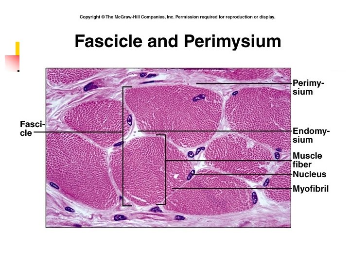
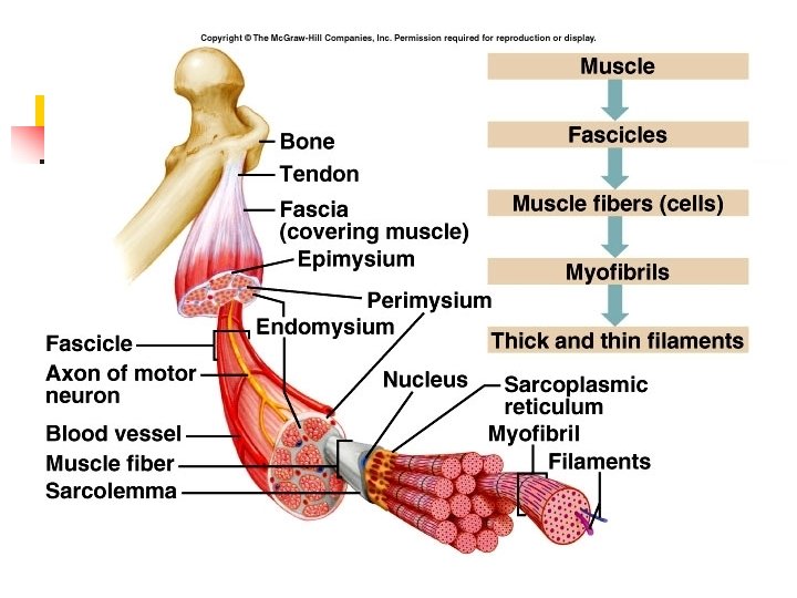
STRUCTURE OF SKELETAL MUSCLE n CONSISTS OF SKELETAL MUSCLE TISSUE, NERVOUS TISSUE, BLOOD, AND OTHER CONNECTIVE TISSUE, AND LOTS OF BLOOD VESSELS
FROM THE OUTSIDE IN
http: //training. seer. cancer. gov/module_anatomy/unit 4_2_muscle_structure. html
QUESTIONS n n n What is the difference between superficial and deep fascia? What is the function of each? A fascicle is ________. Another name for a muscle cell is _______. What is the epimysium?
Skeletal Muscle Fibers
INSIDE A MUSCLE FIBER n MYOFIBRILS RODLIKE BUNDLES OF FILAMENTS, THAT ARE PARALLEL TO ONE ANOTHER
INSIDE A MUSCLE FIBER n MYOFILAMENTS BUNDLES OF PROTEINS, GIVES MUSCLE STRIPES 1. ACTIN 2. MYOSIN 3. TROPONIN* 4. TROPOMYOSIN*
http: //www. mie. utoronto. ca/labs/lcdlab/biopic/fig/47. 07. jpg
MUSCLE FIBER HISTOLOGY SARCOLEMMA - THE CELL MEMBRANE n SARCOPLASM - THE CYTOPLASM (HAS LOTS OF NUCLEI AND MITOCHONDRIA) n
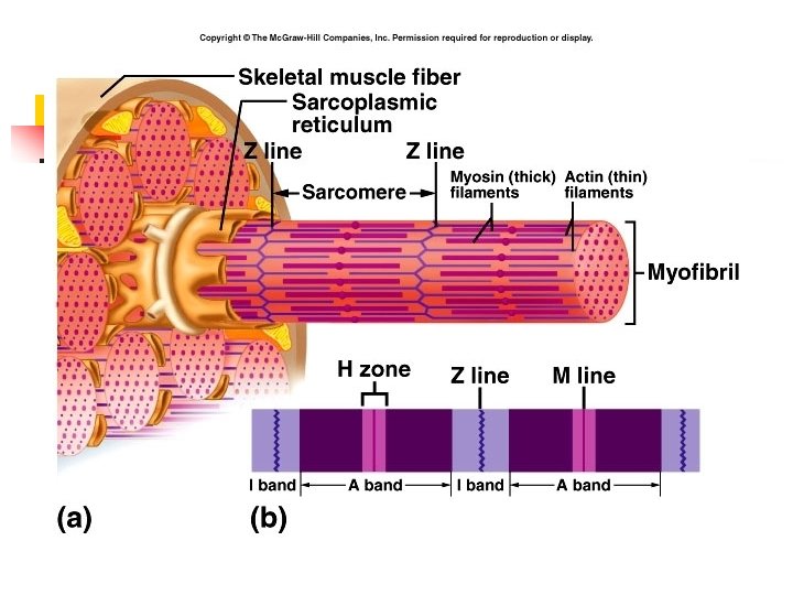
MUSCLE FIBER HISTOLOGY n SARCOPLASMIC RETICULUM A NETWORK OF CHANNELS & TUBULES THAT IS SIMILAR TO THE ENDOPLASMIC RETICULUM IN OTHER CELLS
http: //www. mhhe. com/biosci/esp/2001_gbio/folder_structure/an/m 5/s 5/assets/images/anm 5 s 5_1. jpg
MUSCLE FIBER HISTOLOGY n Sarcoplasmic reticulum functions include: 1. Storing calcium (Ca 2+) ions 2. Releasing Ca 2+ ions during muscle contraction 3. Reabsorbing Ca 2+ ions when muscle relaxes
QUESTIONS n n Myofibrils are bundles of _______. The proteins that make up myofilaments include __________, and _____. The cell membrane of a muscle fiber is the _________. What is the sarcoplasmic reticulum?
MUSCLE FIBER HISTOLOGY n Terminal cisternae (cisterns) 1. Enlarged part of sarcoplasmic reticulum where actin & myosin fibers overlap 2. Calcium storage
MUSCLE FIBER HISTOLOGY n Transverse (T) tubules 1. Channels that extend from surface of the sarcolemma into the cell 2. Found between the terminal cisterns
MUSCLE FIBER HISTOLOGY n Transverse (T) tubules 3. Perpendicular to S. R. 4. Job is to carry action potential (nerve impulse) deep into muscle fibers from the surface
http: //www. mhhe. com/biosci/esp/2001_gbio/folder_structure/an/m 5/s 5/assets/images/anm 5 s 5_1. jpg
More on Myofibrils are made up of 2 types of myofilaments. 1. Thick made of the protein myosin 2. Thin made of the proteins actin, tropomyosin & troponin 3. Arranged in compartments known as sarcomeres
Myofibril structure http: //www. lib. mcg. edu/eshuphysio/program/section 2/2 ch 2/sarconom. htm
QUESTIONS n n What is the function of the terminal cisterns? The T-tubules extend from ______ to _______ and do what? . Thick filaments are made up of _____. Thin filaments are made up of_____.
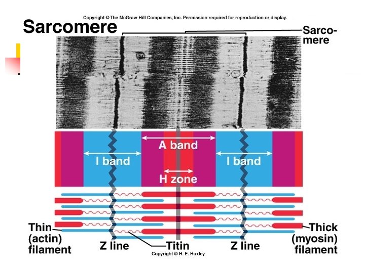
MUSCLE FIBER HISTOLOGY Sarcomere - the functional unit of muscle contraction n Separated by dense material called Z lines n Segment of myofibril that extends from Z line to Z line n
Muscle Fiber Structure Animation
Sarcomere structure n n n A - band - the dark area made up of thick filaments Found in the middle of sarcomere Darkest where thin & thick filaments overlap http: //www. sport-fitness-advisor. com/images/sarcomere. jpg
The A - band http: //www. lib. mcg. edu/eshuphysio/program/section 2/2 ch 2/aband. htm
Sarcomere structure n n n I - band - light area made up of thin filaments Found at ends of sarcomere Attached to Z-lines http: //www. sport-fitness-advisor. com/images/sarcomere. jpg
The I - band http: //www. lib. mcg. edu/eshuphysio/program/section 2/2 ch 2/iband. htm
Sarcomere structure n The H - zone is the central region of the a - band that contains only thick filaments. http: //www. lib. mcg. edu/eshuphysio/program/section 2/2 ch 2/hband. htm
QUESTIONS n n The functional unit of muscle contraction is the ______. What do Z-lines do? The area consisting of thick filaments is the _______. The area consisting of thin filaments is the ______.
Thick filament structure n n Made of myosin 2 twisted strands with globular parts that stick out like golf clubs http: //pdbdev. sdsc. edu: 48346/pdb/molecules/myosin-painting. gif
More on myosin n n The golf-club structures are called cross bridges. The cross bridges have actin binding sites (& ATP binding sites). http: //user. gru. net/clawrence/vccl/chpt 3/HEM 21. gif
Thin filament structure n n n Made up of actin 2 strands wrapped around each other Also has troponin & tropomyosin http: //www. embl-heidelberg. de/Cell. Biophys/Local. Probes/motorproteins/actinfilament. jpg
QUESTIONS n n Where do you find the H-zone? What are cross bridges and where do you find them? What is the I-band where do you find it? Besides actin, the other 2 proteins found in a thin filament are _______ & _______.
ELECTRON MICROGRAPH OF A SARCOMERE http: //www. neuro. wustl. edu/neuromuscular/mother/myosin. htm
Insect Muscle structure and function
RECAP OF MUSCLE FIBER STRUCTURE http: //www. octc. kctcs. edu/gcaplan/anat/images/Image 286. gif