The Skeletal System 1 Skeletal System Parts of


- Slides: 40
The Skeletal System 1
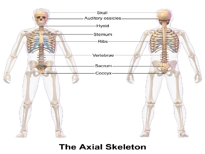
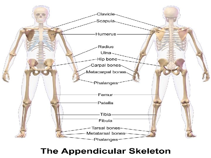
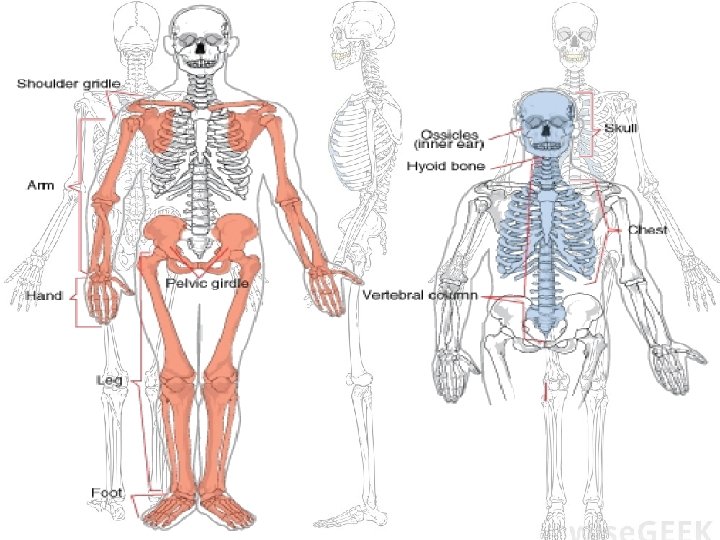
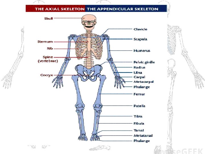
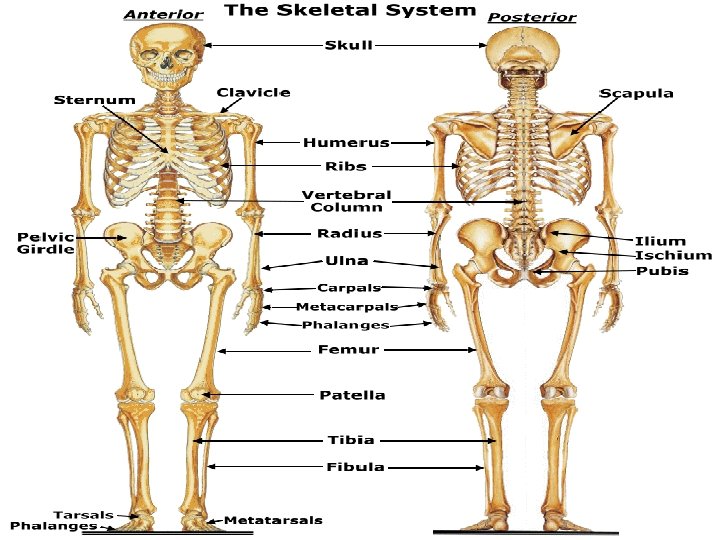
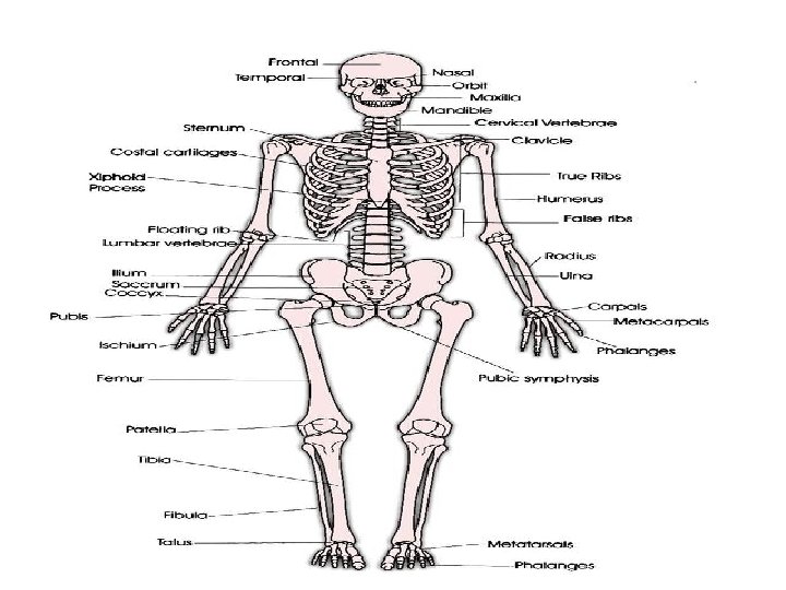
Skeletal System • Parts of the skeletal system: 1. 2. 3. 4. Bones Joints Ligaments Cartilage • Separated into 2 main divisions: 1. Axial 2. Appendicular 2
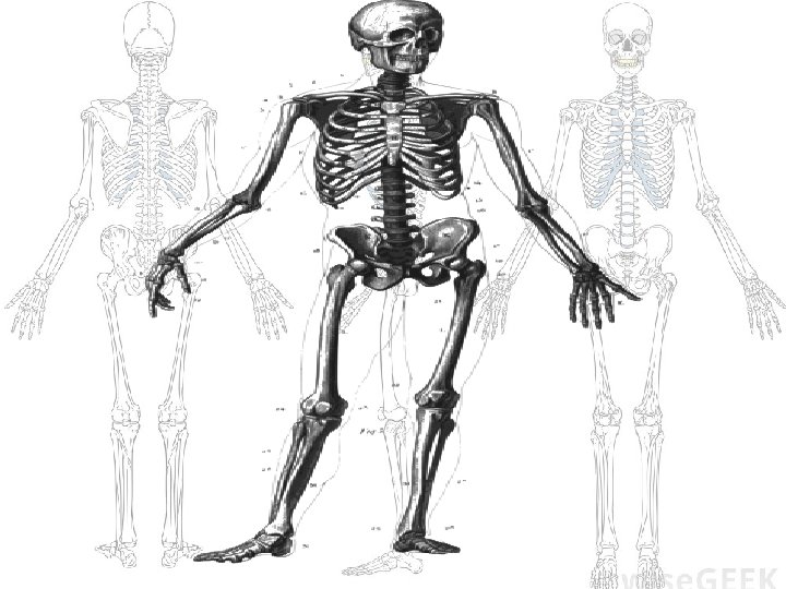
Review of the Functions of the Skeletal System 1. Support of the body 2. Protection of soft organs – Skull and vertebrae for brain and spinal cord – Rib cage for thoracic cavity organs 3. Movement due to attached skeletal muscles 4. Storage of minerals (Ca+ and P) & fats 5. Blood cell formation (hematopoiesis) 3
Bones of the Human Body • The adult skeleton has 206 bones • 2 basic types of bone tissue: 1. Compact Bone – dense, looks smooth and homogenous; provides strength and stability. 2. Spongy Bone – lattice -like pieces of bone with open spaces; provides shock absorption and flexibility Spongy bone Compact bone Figure 5. 2 b 4
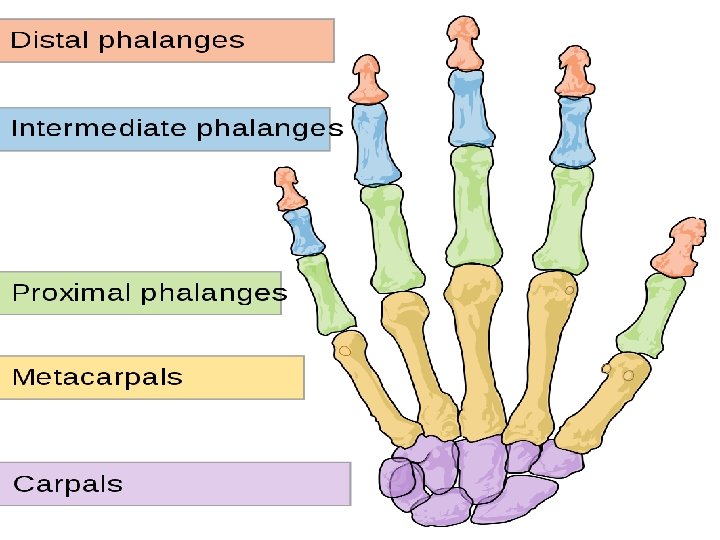
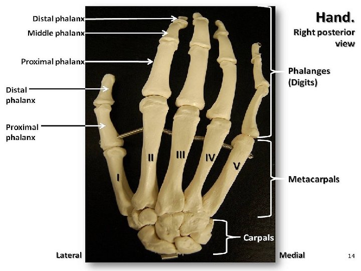
Classification of Bone Shapes • Bones are classified as: – Long – Short – Flat – Irregular Figure 5. 15
Classification of Bone Shapes 6
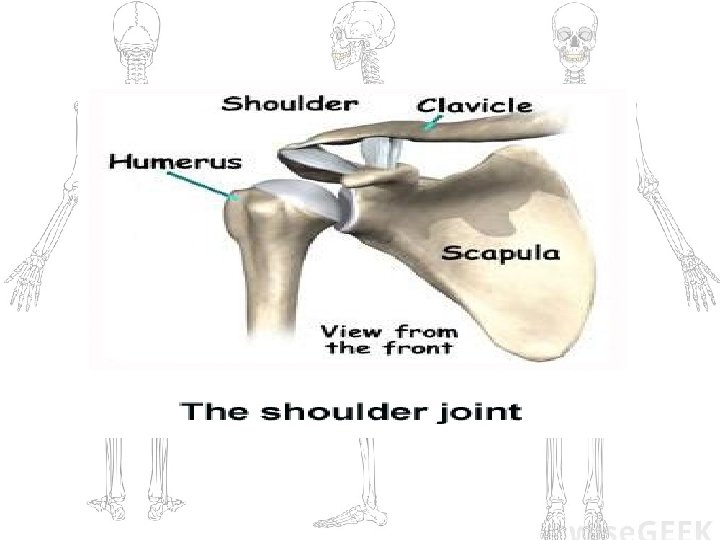
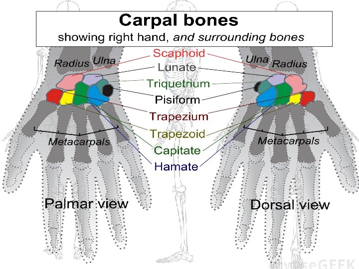
Classification of Bone Shapes 1. Long bones – longer than they are wide – – Usually shaft w/head at ends Limb bones except wrist and ankle Mostly compound bone Example: Humerus 2. Short bones – cube shaped – Mostly spongy bone – Wrist and ankle – Sesamoid bones are a type of short bone which form within tendons (ex. patella) 7
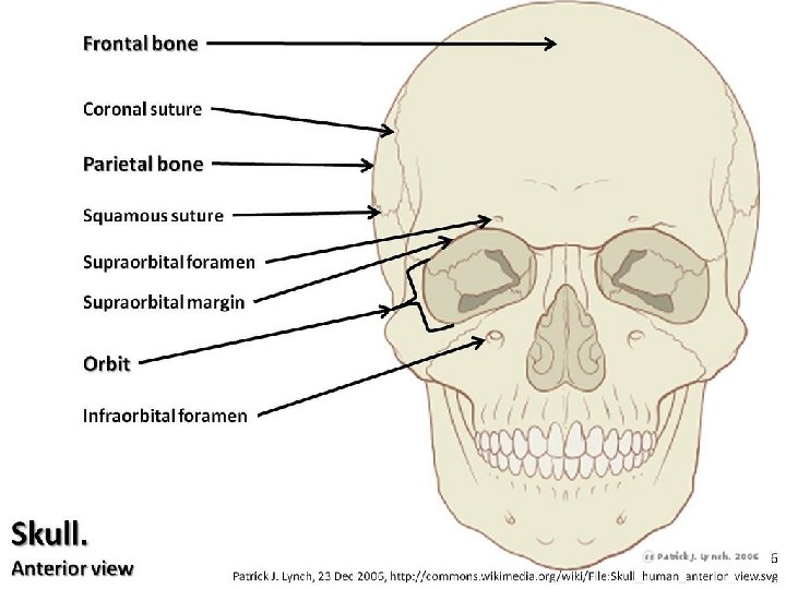
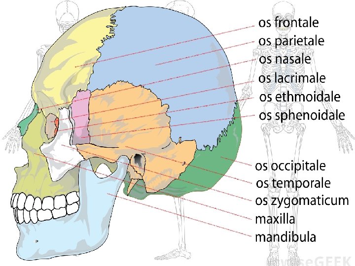
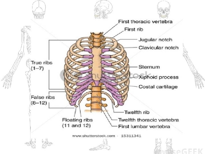
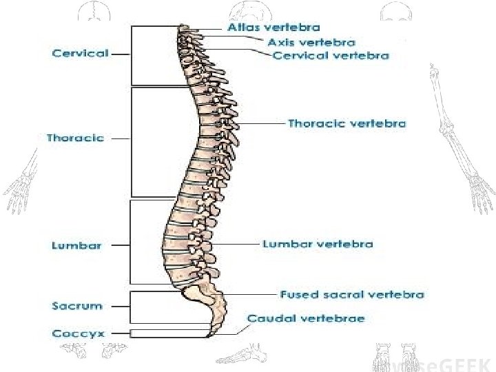
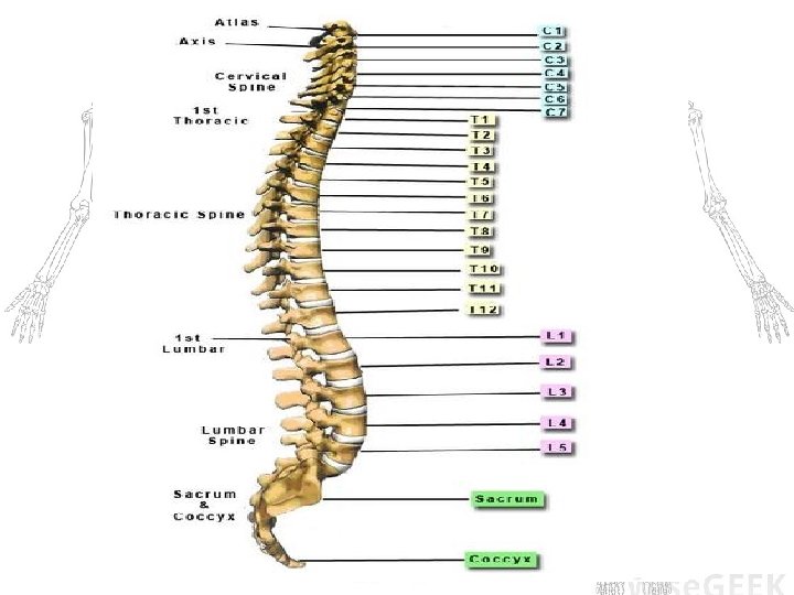
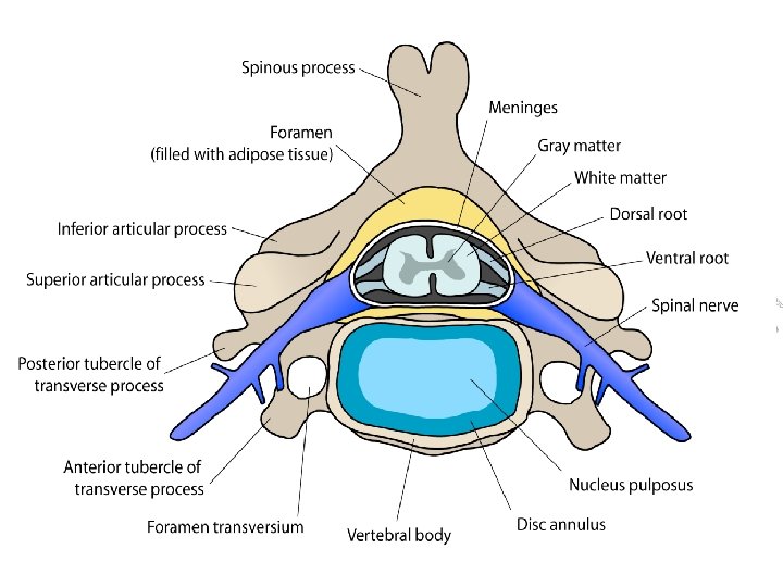
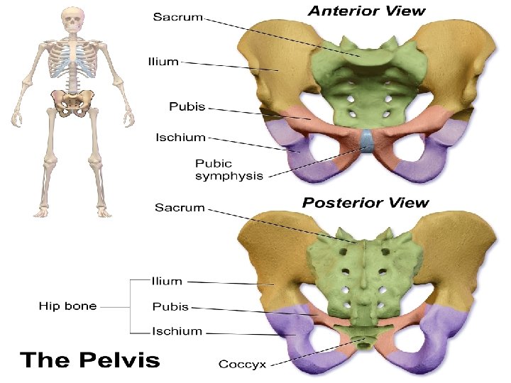
Classification of Bone Shapes 3. Flat bones – Thin, flat and usually curved – 2 thin layers of flat compound bone sandwiching spongy bone – Ex. Skull, ribs and sternum 4. Irregular bones – miscellaneous – Vertebrae, hip bones 8
Anatomy of a Long Bone Articular cartilage Proximal epiphysis • Diaphysis – shaft of bone – Compact bone – Covered by connective tissue called periosteum Diaphysis Spongy bone Epiphyseal line Periosteum Compact bone Medullary cavity (lined by endosteum) • Epiphysis – ends of bone – Compact bone surrounding spongy bone center Distal epiphysis (a Figure 5. 2 a 9
Where do Long Bones get their strength? • The diaphysis region of a long bone functions to transfer loads more evenly from weight-bearing joint surfaces throughout both the diaphysis and the epiphysis.
Anatomy of a Long Bone • Epiphyseal plate – Flat plate of hyaline cartilage seen in young, growing bone • Epiphyseal line Articular cartilage Proximal epiphysis Diaphysis Spongy bone Epiphyseal line Periosteum Compact bone Medullary cavity (lined by endosteum) – Remnant of the epiphyseal plate – Seen in adult bones Distal epiphysis (a) 11
Changes in the Human Skeleton • In embryos/fetus, the skeleton is primarily hyaline cartilage • During development, much of this cartilage is replaced by bone - ossification • Cartilage remains in isolated areas: – Bridge of the nose – Parts of ribs – Joints 12
Long Bone Formation and Growth Figure 5. 4 a 13
Types of Bone Cells Rickets – disease where bones fail to calcify, legs bow out • Osteocytes -Caused by a lack of vitamin D • Osteoblasts -Bones can also atrophy in bed ridden people – Mature bone cells – Bone-forming cells • Osteoclasts — Giant bone-destroying cells – Break down bone matrix for remodeling and release of calcium (Important for allowing bones to adapt to changes in their weight bearing capacities. ) • Bones are remodeled in response to Ca+ levels in the blood and the pull of gravity and muscles on the bones; such as occurs with activity or exercise. 14
Synovial Fluid • The principal role of synovial fluid is to reduce friction between the cartilage of two bones meeting at joints, especially during movement.
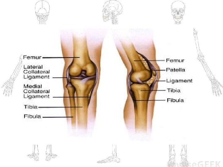
What is a Sprain? • A sprain is an injury to a ligament (the tissue that connects 2+ bones at a joint). In a sprain, one or more ligaments is either over stretched/ extended or torn.