Chapter 35 Chest Trauma National EMS Education Standard
- Slides: 159
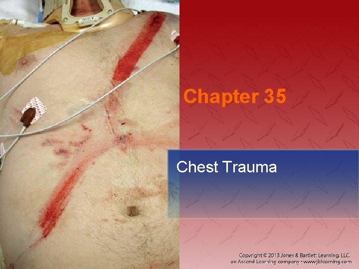
Chapter 35 Chest Trauma
National EMS Education Standard Competencies Trauma Integrates assessment findings with principles of epidemiology and pathophysiology to formulate a field impression to implement a comprehensive treatment/disposition plan for an acutely injured patient.
National EMS Education Standard Competencies Chest Trauma • Recognition and management of − Blunt vs penetrating mechanisms − Open chest wound − Impaled object
National EMS Education Standard Competencies • Pathophysiology, assessment, and management of − Blunt vs penetrating mechanisms − Hemothorax − Pneumothorax • Open • Simple • Tension
National EMS Education Standard Competencies • Pathophysiology, assessment, and management of (cont’d) − − − Cardiac tamponade Rib fractures Flail chest Commotio cordis Traumatic aortic disruption Pulmonary contusion
National EMS Education Standard Competencies • Pathophysiology, assessment, and management of (cont’d) − − Blunt cardiac injury Tracheobronchial disruption Diaphragmatic rupture Traumatic asphyxia
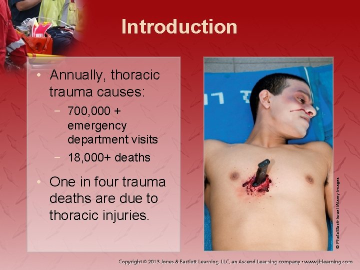
Introduction • Annually, thoracic trauma causes: • One in four trauma deaths are due to thoracic injuries. © Photo. Stock-Israel /Alamy Images − 700, 000 + emergency department visits − 18, 000+ deaths
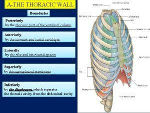
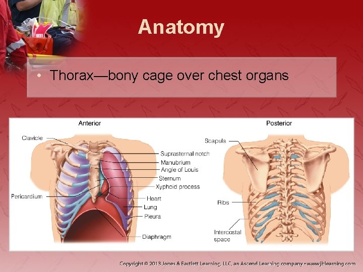
Anatomy • Thorax—bony cage over chest organs
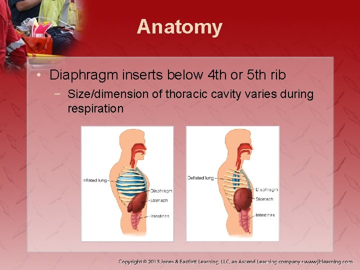
Anatomy • Diaphragm inserts below 4 th or 5 th rib − Size/dimension of thoracic cavity varies during respiration
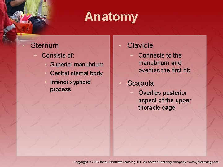
Anatomy • Sternum − Consists of: • Superior manubrium • Central sternal body • Inferior xyphoid process • Clavicle − Connects to the manubrium and overlies the first rib • Scapula − Overlies posterior aspect of the upper thoracic cage
Anatomy • 12 pairs of ribs attach to 12 thoracic vertebrae. − First 7 pairs attach directly to sternum − 8– 10 attach indirectly to sternum − 11 and 12 are “floating ribs”
Anatomy • Intercostal space houses: − Intercostal muscles − Neurovascular bundle • Mediastinum contains: − Heart and great vessels − Esophagus − Lymphatic channels − Trachea − Mainstem bronchi − Vagus and phrenic nerves
Anatomy • Heart resides inside pericardium − Anterior portion is the right ventricle − Mostly protected anteriorly by the sternum − Average cardiac output: 70 × 70 = 4, 900 m. L/min
Anatomy • Aorta: largest artery in body − Three points of attachment: • Annulus • Ligamentum arteriosum • Aortic hiatus
Anatomy • Lungs occupy most space in thoracic cavity − Lined with pleura − Pleura is separated by viscous fluid • Keeps lungs from collapsing on exhalation
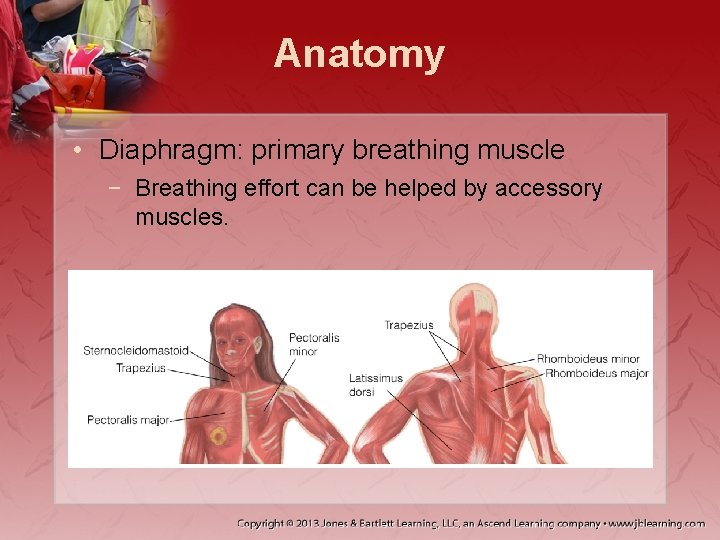
Anatomy • Diaphragm: primary breathing muscle − Breathing effort can be helped by accessory muscles.
Physiology • Primary functions of thorax: − Maintain oxygenation and ventilation. − Maintain circulation. • Breathing process includes: − Delivery of oxygen to body − Elimination of carbon dioxide
Physiology • Brain stimulates breathing via chemoreceptors. − If CO 2 is too high, respiratory rate increases. − Hypoxic drive: secondary mechanism
Physiology • Intercostal and accessory muscles pull chest wall away from the body as the diaphragm contracts downward. − Negative pressure draws air through mouth and nose to alveolar spaces. • Replaces air in the alveoli
Physiology • Blood is delivered by pulmonary circulation to capillaries adjacent to alveoli. − Has low O 2 and high CO 2 concentrations − Oxygenation process delivers O 2 to blood
Physiology • Ventilation: how CO 2 leaves the body − CO 2 diffuses down its concentration gradient and enters air within the alveoli. − Positive pressure is created within the thorax and air is exhaled.
Physiology • Proper heart function is essential. − Ability to pump blood depends on: • A functional pump • Adequate blood volume • Lack of resistance to pumping mechanisms − Factors determine cardiac output.
Pathophysiology • Traumatic injury to chest may compromise: − Ventilation − Oxygenation − Circulation
Pathophysiology • Two mechanisms of injury: − Blunt − Penetrating • Two basic injury patterns − Closed − Open
Pathophysiology • Blunt trauma may lead to: − − − Fracture of ribs, sternum, areas of chest wall Bruise of lungs and heart Damage to aorta Broken ribs lacerating intrathoracic organs Organs torn from attachment
Pathophysiology • Blast injuries may be blunt or penetrating. − Primary shock wave (blunt trauma) − Secondary shock wave (penetrating trauma)
Pathophysiology • Thoracic trauma may impair cardiac output. − − Blood loss Pressure change Vital organ damage Combination of these
Pathophysiology • Ventilatory impairments can be rapidly fatal. − Shallow breathing reduces minute volume. − Air entering pleural space compresses lungs and decreases tidal volume. − Blood collection prevents full lung expansion.
Pathophysiology • Other complications include: − Atelectasis reduces area for gas exchange. − Bruised lung tissue may cause hypoxemia. − Tearing or rupture of respiratory structures prevents O 2 from reaching alveoli.
Scene Size-Up • Be sure scene is safe to enter. • Follow standard precautions. • After identifying number of patients: − Triage patients and request resources. − Determine MOI if possible.
Primary Assessment • Form a general impression. − Assess level of consciousness. − Perform a rapid scan. − Observe the neck for: • Accessory muscle use during breathing • Extended or engorged external jugular veins
Primary Assessment • Airway and breathing − Assess while performing spinal immobilization. − Airway compromise presentation depends on: • Severity of impairment • Duration • Associated injuries
Primary Assessment • Airway and breathing (cont’d) − Signs of obstruction may include: • Stridor • Hoarseness • Alterations in mental status − Abnormal findings may include: • Tachypnea • Hemoptysis • Retractions
Primary Assessment • Airway and breathing (cont’d) − Immediate manage airway impairment: • Manually immobilize patient. • Use jaw-thrust maneuver. − Identify and manage impairment of oxygenation and ventilation.
Primary Assessment • Airway and breathing (cont’d) − Inspect chest wall. − Underlying injury may be indicated by: • Signs of soft-tissue injury • Retractions • Impaled objects
Primary Assessment • Airway and breathing (cont’d) − Address life-threats first. − Apply occlusive dressing to penetrating injuries. − Assess ventilation and oxygenation.
Primary Assessment • Airway and breathing (cont’d) − Apply O 2 with nonrebreathing mask at 15 L/m. • Evaluate skin circulation. • Decreasing O 2 saturation may indicate hypoxia. • Watch for impending tension pneumothorax signs.
Primary Assessment • Airway and breathing (cont’d) − Palpate for: • • • Point tenderness Bony instability Crepitus Subcutaneous emphysema Edema Tracheal position
Primary Assessment • Airway and breathing (cont’d) − Percuss for: • Hyperresonance • Dullness − Auscultate for: • Adventitious lung sounds • Confirmation of lung sounds
Primary Assessment • Circulation − Begin by checking mental status for: • • • Restlessness Agitation Confusion Irrationality Comatose
Primary Assessment • Circulation (cont’d) − Check pulses. • Tachycardia is not always associated with hypovolemia. • Low heart rate does not rule out hypovolemia or shock. • Thready or weak pulse may suggest volume loss.
Primary Assessment • Circulation (cont’d) − Irregular pulse suggests: • Hypoxia • Hypoperfusion • Serious underlying injuries or shock
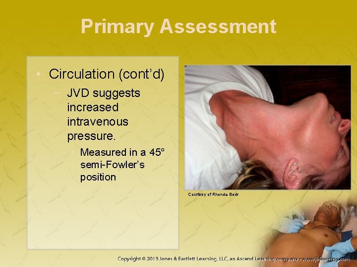
Primary Assessment • Circulation (cont’d) − JVD suggests increased intravenous pressure. • Measured in a 45° semi-Fowler’s position Courtesy of Rhonda Beck
Primary Assessment • Circulation (cont’d) − Auscultate the heart. • Note if heart sounds are easily heard or muffled. − If shock suggested, may not be from thorax. • Obtain history and complete physical.
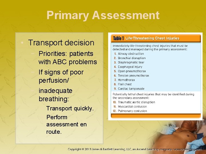
Primary Assessment • Transport decision − Priorities: patients with ABC problems − If signs of poor perfusion/ − inadequate breathing: • Transport quickly. • Perform assessment en route.
History Taking • May need to be done en route • Obtain relevant patient history (SAMPLE). • Questions about event should focus on MOI.
Secondary Assessment • Includes a head-to-toe assessment − Check for injuries that may compromise ABCs. • Obtain full set of vitals. • Monitoring equipment can aid assessment
Secondary Assessment • If chest injury is isolated with limited MOI, focus on: − Isolated injury − Patient’s complaint − Body region affected
Secondary Assessment • If significant trauma, assess head-to-toe. − − Skin (ecchymosis, other trauma) Identify all wounds, control bleeding. Note location and extent of injury. Assess underlying systems.
Secondary Assessment • If significant trauma affects multiple systems: − − Perform full body scan using DCAP-BTLS. Inspect region for deformities. Palpate for tenderness. Check for lacerations and swelling.
Reassessment • Obtain repeated assessments of: − − Vital signs Oxygenation Circulation Breath sounds
Reassessment • If pneumothorax is suspected, patient should be considered unstable. − Reassess at least every 5 minutes. • Maintain a high degree of suspicion during on-scene treatment and transport.
Emergency Medical Care • Management focuses on: − − Maintaining airway Ensuring oxygenation and ventilation Supporting circulatory status Expeditious transport
Emergency Medical Care • Airway management same, except: − Jaw-thrust maneuver instead of head tilt–chin lift • Avoid nasal airways if signs of facial injury − Use ET intubation instead. • Reconsider if possible partial tracheal tear.
Emergency Medical Care • Ensure oxygenation and ventilation. • Assess circulatory system’s ability to provide oxygenation and ventilation. − If compromised, provide supportive measures.
Emergency Medical Care • Pharmacologic agents are limited. − Medications for airway management − Pain management, limited by: • Local protocols • Short transport times • Clinical state of patient
Emergency Medical Care • Nonpharmaceutical approaches: − Splinting injury − Cold pack application − Careful handling • Transport to appropriate facility.
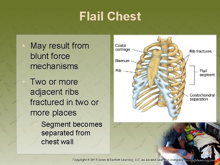
Flail Chest • May result from blunt force mechanisms • Two or more adjacent ribs fractured in two or more places − Segment becomes separated from chest wall
Flail Chest • Location and size affects degree that chest wall and air movement are impaired. − Flail sternum (most extreme) • Underlying pressure causes paradoxical movement of segment and rest of chest wall.
Flail Chest • May not be initially apparent − Palpate for rib cage fractures and crepitus. • Management may involve: − Positive-pressure ventilation − Positive end-expiratory pressure
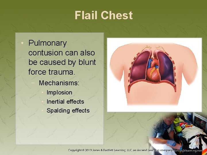
Flail Chest • Pulmonary contusion can also be caused by blunt force trauma. − Mechanisms: • Implosion • Inertial effects • Spalding effects
Flail Chest • Pneumothorax or hemothorax may occur if bone fragments are driven into the body. − Pain may prevent adequate tidal volume. • Limits the ability to compensate for flail segment
Flail Chest • Assessment and management − Palpation may reveal: • Crepitus • Tenderness • Dissection of air into tissue − Auscultation may reveal: • Decreased or absent breath sounds
Flail Chest • Assessment and management (cont’d) − Associated findings include: • Tenderness • Shallow breathing • Tachycardia − Signs and symptoms include: • Hypoxia • Hypercarbia • Pain
Flail Chest • Assessment and management (cont’d) − Poses a threat to patient’s ability to breathe • Intubation and positive-pressure ventilation may be needed. − Stabilization of flail segment is controversial.
Rib Fractures • Most common thoracic injury − Pain contributes to: • • Inadequate ventilation Self-splinting Atelectasis Pneumonia from inadequate respiration
Rib Fractures • Palpate chest for subcutaneous emphysema. • Blunt trauma may result in fracture at: • Point of impact • Edge of object • Posterior angle of rib
Rib Fractures • May indicate associated injuries − Ribs 4– 9 • • Aortic injury Tracheobronchial injury Pneumothorax Vascular injury − Ribs 9– 11 • Intra-abdominal injury
Rib Fractures • Assessment and management − Patients report: • Pleuritic chest pain • Mild dyspnea − Exam shows: • Chest wall tenderness • Soft-tissue injury • Crepitus • Subcutaneous emphysema
Rib Fractures • Assessment and management (cont’d) − Management focuses on: • ABCs and evaluating for other injuries − Administer supplemental O 2 − Gently splint chest wall. − Consider intravenous analgesic.
Sternal Fractures • One in 20 patients with blunt thoracic trauma − Associated with other injuries, including: • Myocardial contusions • Flail sternum • Intra-abdominal injuries
Sternal Fractures • Assessment and management − Patient reports pain over anterior part of chest. − Palpation may reveal: • Tenderness • Crepitus • Possible flail segment
Sternal Fractures • Assessment and management (cont’d) − Perform an ECG rhythm analysis. − Supportive treatment only • ABCs • Manage associated injuries. • Analgesics
Clavicle Fractures • One of the most common fractures • Assessment and management − Patient will: • Report pain in the shoulder • Usually hold arm across front of body
Clavicle Fractures • Assessment and management (cont’d) − Splint fracture with a sling and swathe. • Apply gentle upward support to the olecranon process of the ulna. • Knot should be tied on the side of the neck.
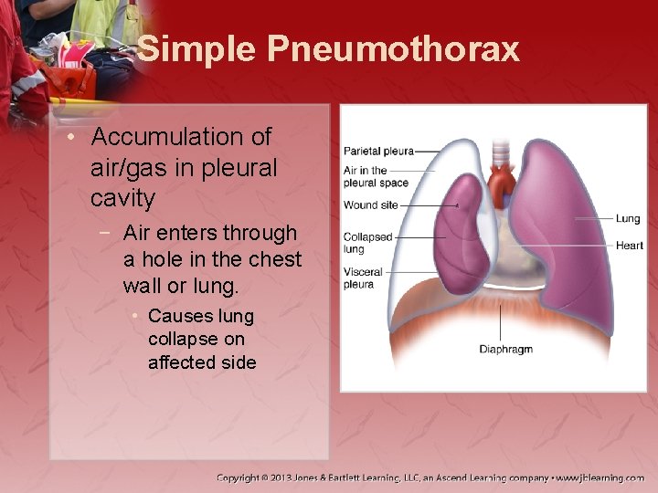
Simple Pneumothorax • Accumulation of air/gas in pleural cavity − Air enters through a hole in the chest wall or lung. • Causes lung collapse on affected side
Simple Pneumothorax • The larger the hole, the faster the collapse. • Delayed or improper treatment may lead to a tension pneumothorax. • Some low-velocity wounds may heal themselves.
Simple Pneumothorax • Assessment and management − Small pneumothorax • Mild dyspnea and pleuritic chest pain • Diminished or unequal breath sounds • Hyperresonance − Larger pneumothorax • Increasing dyspnea • Signs of serious respiratory compromise and hypoxia
Simple Pneumothorax • Assessment and management (cont’d) − Cover large open wounds immediately. • Nonporous dressing secured on three sides − Maintain ABCs, provide high-concentration O 2. • If tension develops, dressing may need to be removed to release trapped air.
Open Pneumothorax • Occurs when chest wall defect allows air into thoracic space − Results from penetrating chest trauma − Negative pressure draws air into pleural space. − As size increases, lung loses ability to expand.
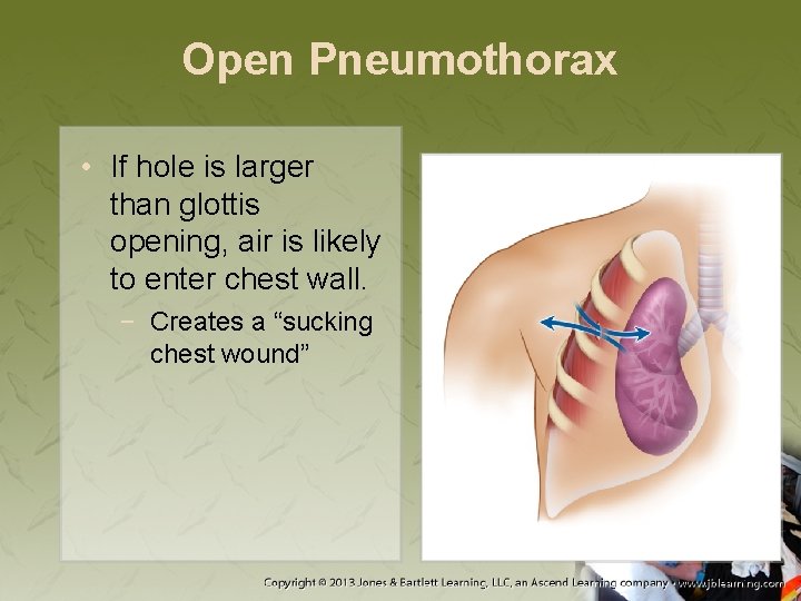
Open Pneumothorax • If hole is larger than glottis opening, air is likely to enter chest wall. − Creates a “sucking chest wound”
Open Pneumothorax • If pulmonary vasculature on involved side remains intact: − Heart will continue to perfuse the collapsed lung. − Pneumothorax prevents ventilation.
Open Pneumothorax • Assessment and management − Physical assessment shows: • • • Chest wall defect Impaled object Sucking chest wound Bubbling wound Subcutaneous emphysema
Open Pneumothorax • Assessment and management (cont’d) − Signs and symptoms may include: • Tachycardia and tachypnea • Restlessness − As pneumothorax increases, breath sounds decrease on affected side.
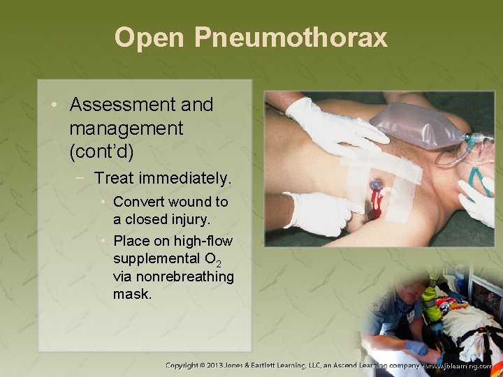
Open Pneumothorax • Assessment and management (cont’d) − Treat immediately. • Convert wound to a closed injury. • Place on high-flow supplemental O 2 via nonrebreathing mask.
Open Pneumothorax • Assessment and management (cont’d) − If progression to tension pneumothorax, remove dressing or seal to allow a vent through opening. • If ineffective, treat for tension pneumothorax.
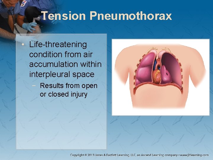
Tension Pneumothorax • Life-threatening condition from air accumulation within interpleural space − Results from open or closed injury
Tension Pneumothorax • As air accumulates, pressure builds against surrounding tissue. − Compresses the lung, which diminishes: • Ability to oxygenate blood • Ability to eliminate CO 2
Tension Pneumothorax • Pressure causes eventual lung collapse and mediastinum to shift. − May exceed pressure in major venous structures − If venous return decreases, the body will increase heart rate.
Tension Pneumothorax • Assessment and management − Classic signs may include: • Absence of breath sounds on affected side • Pulsus paradoxus • Tracheal deviation
Tension Pneumothorax • Assessment and management (cont’d) − Tachycardia is induced because blood cannot return to heart. • Accumulates in great vessels • Pressure pushes blood into jugular vein.
Tension Pneumothorax • Assessment and management (cont’d) − During normal inspiration: • Negative pressure decreases blood return. • Preload and systolic blood pressure decrease. − In tension pneumothorax, effect is magnified. • Pulsus paradoxus
Tension Pneumothorax • Assessment and management (cont’d) − Jugular veins are distended when engorged 1 to 2 cm above the clavicle. − May show trachea deviation from affected side
Tension Pneumothorax • Assessment and management (cont’d) − May have diminished breath sounds − May have pleuritic chest pain and dyspnea − Hypotension is a late finding.
Tension Pneumothorax • Assessment and management (cont’d) − Administer immediate high-flow supplemental O₂. − Inspect the chest. − Cover open wounds with dressing. − If elevated pressure is suggested: • May need to perform a needle decompression
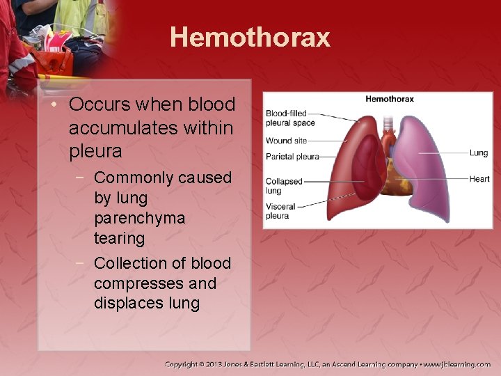
Hemothorax • Occurs when blood accumulates within pleura − Commonly caused by lung parenchyma tearing − Collection of blood compresses and displaces lung
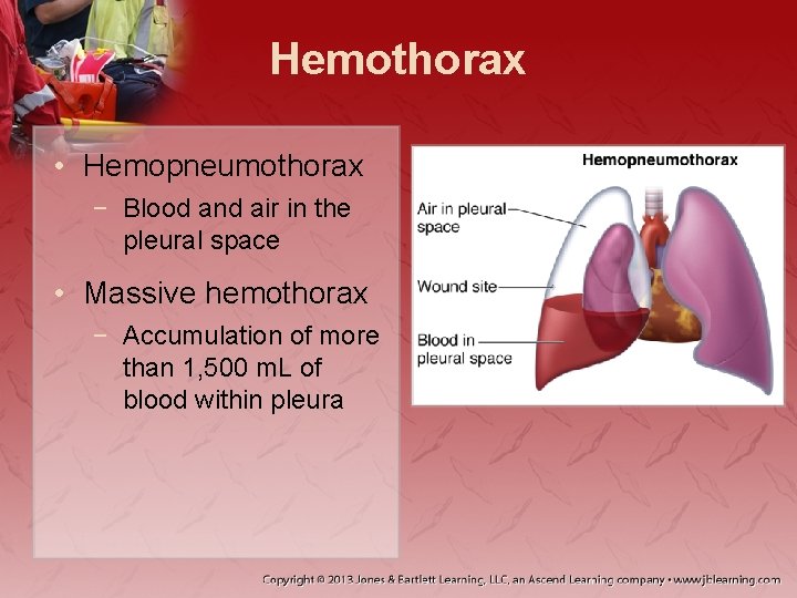
Hemothorax • Hemopneumothorax − Blood and air in the pleural space • Massive hemothorax − Accumulation of more than 1, 500 m. L of blood within pleura
Hemothorax • Assessment and management − Signs include: • Ventilatory insufficiency • Hypovolemic shock
Hemothorax • Assessment and management (cont’d) − Findings that differentiate from other injuries: • • • Lack of tracheal deviation Possible hemoptysis Dullness on percussion Flat neck veins with hypovolemia Distended neck veins with increased pressure
Hemothorax • Assessment and management (cont’d) − Prehospital management: • • Supportive care Rapid transport High-flow supplemental O 2 Two large-bore peripheral IV
Pulmonary Contusion • Alveolar and capillary damage results from lung tissue compression against chest wall. − Injury may lead to: • Loss of fluid and blood into involved tissues • White blood cell migration into area • Local tissue edema
Pulmonary Contusion • Local surfactant in alveoli is diluted. − Causes atelectasis • Delivery of O 2 is reduced, causing hypoxia • Attempt to shunt blood from injury further worsens hypoxemia.
Pulmonary Contusion • Assessment and management − May not initially show presence or severity − Hypoxia and CO 2 retention may cause: • Respiratory distress • Tachycardia • Agitation
Pulmonary Contusion • Assessment and management (cont’d) − Evidence of overlying injury: • • Contusions Tenderness Crepitus Paradoxical motion − Auscultation may reveal: • • Wheezes Rhonchi Rales Diminished lung sounds
Pulmonary Contusion • Assessment and management (cont’d) − Treatment includes: • Managing airway • Using caution when administering IV fluids • Administering small amounts of analgesics for pain
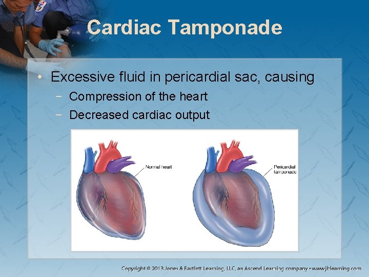
Cardiac Tamponade • Excessive fluid in pericardial sac, causing − Compression of the heart − Decreased cardiac output
Cardiac Tamponade • Hemodynamic effects determined by: − Size of pericardium perforation − Rate of hemorrhage − Chamber of heart involved
Cardiac Tamponade • Mortality varies. • Can occur in both medical and trauma − Medical—slow fluid collection − Trauma—bleeding is rapid.
Cardiac Tamponade • Continued bleeding increases pressure in the pericardium. − Atria and vena cavae compress. − Preload delivery is drastically reduced. − Heart increases rate to compensate.
Cardiac Tamponade • Assessment and management − 30% diagnosed will have Beck triad: • Muffled heart tones • Hypotension • JVD − Classic finding: electrical alternans in ECG strip
Cardiac Tamponade • Assessment and management (cont’d) − Findings typical of shock, including: • Weak or absent peripheral pulses • Cyanosis • Tachycardia or tachypnea
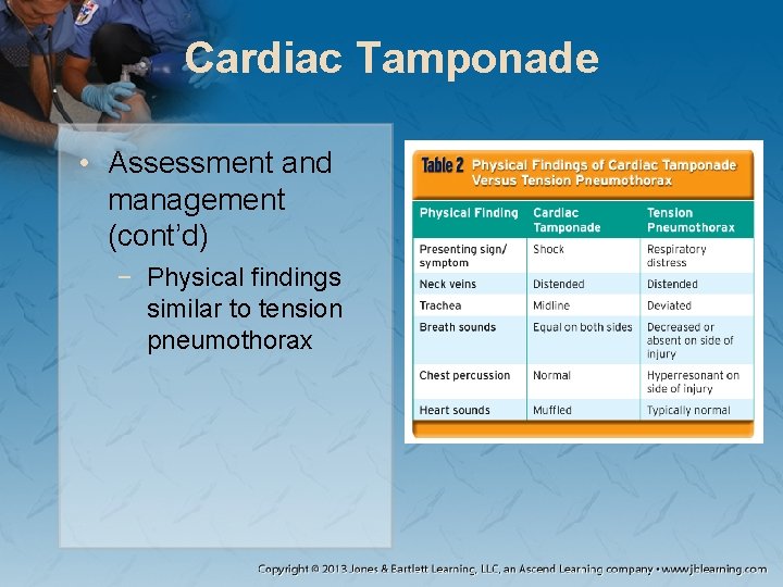
Cardiac Tamponade • Assessment and management (cont’d) − Physical findings similar to tension pneumothorax
Cardiac Tamponade • Assessment and management (cont’d) − Treatment includes: • • Assess and manage ABCs. Ensure adequate O 2 and establishing IV access. Provide a rapid fluid bolus. Rapidly transfer to trauma center.
Myocardial Contusion • Sudden deceleration of chest wall may cause collision of heart to sternum. − Characterized by: • Local tissue contusion and hemorrhage • Edema • Cellular damage within myocardium
Myocardial Contusion • Damage to myocardial tissues may cause: − Ectopic activity − Reentry pathways − Dysrhythmias • Structural changes may include: − Ventricular septal defect − Myocardial rupture or aneurysm − Coronary artery occlusion
Myocardial Contusion • Assessment and management − Signs and symptoms include: • Sharp, retrosternal chest pain • Soft-tissue or bony injury in area • Crackles or rales on auscultation − Often an abnormal ECG
Myocardial Contusion • Assessment and management (cont’d) − Treatment includes: • Nonspecific supportive care • Fluid resuscitation • Consulting with medical control before administering antidysrhythmic agents
Myocardial Rupture • Acute perforation of: − − − − Ventricles Atria Intraventricular septum Intra-atrial septum Chordae Papillary muscle Valves
Myocardial Rupture • Assessment and management − Patients present with: • Acute pulmonary edema • Signs of cardiac tamponade
Commotio Cordis • Cardiac arrest caused by a direct blow to the thorax during the repolarization period − Result of chest wall impact directly over heart − Second most common cause of sudden cardiac death in young male athletes
Commotio Cordis • Assessment and management − Signs and symptoms may include: • Unresponsiveness • Cyanotic • Tonic-clonic seizures
Commotio Cordis • Assessment and management − Survival rates increased because of: • Increased awareness • CPR preparation • Accessibility of AED at sporting events
Traumatic Aortic Disruption • Dissection or rupture of the aorta • Usually caused by crashes and falls − Aorta is injured at fixed points by shearing forces. − Impact causes the aortic arch to swing forward. − Tension and area rotation cause aorta to rupture at point of attachment.
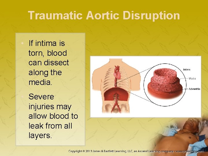
Traumatic Aortic Disruption • If intima is torn, blood can dissect along the media. • Severe injuries may allow blood to leak from all layers.
Traumatic Aortic Disruption • Assessment and management − Symptoms vary and may include: • • Tearing pain behind sternum or in the scapula Hypovolemic shock Dyspnea Altered mental state
Traumatic Aortic Disruption • Assessment and management (cont’d) − With hematoma, presentation may include: • • Dysphagia Stridor Hoarseness Difficulty swallowing
Traumatic Aortic Disruption • Assessment and management (cont’d) − Recognition often from suspicion based on MOI • Assessment of extremity pulses is important − Expect associated injuries, may include: • Multiple rib fractures • Pericardial tamponade • Clavicle fracture
Traumatic Aortic Disruption • Assessment and management (cont’d) − − Assess and manage ABCs. Gradual IV hydration to treat hypotension Do not use pressor agents. Expedite transport to trauma center.
Great Vessel Injury • Great vessels protected by bony structures and other tissues (except for aorta) − Injuries more likely with penetrating trauma − Injuries may result in occlusion or artery spasm.
Great Vessel Injury • Presentation may include: − − − Pain Pallor Paresthesias Pulselessness Paralysis
Great Vessel Injury • Assessment and management − If bleeding not prevented, presentation includes: • Hypovolemic shock • Hemothorax • Cardiac tamponade
Great Vessel Injury • Assessment and management (cont’d) − Procedures for acute blood loss • Establish IV for hydration during transport. • Treat pericardial tamponade immediately. • Do not use pneumatic antishock garment.
Diaphragmatic Injuries • Occurs in a small percentage of trauma • Most occur on left side − Liver protects right side. • Recovery is inhibited by pressure differences between abdominal and thoracic cavities.
Diaphragmatic Injuries • Three phases: − Acute—begins at injury; ends with recovery from other injuries − Latent—entrapment of abdominal contents − Obstructive—abdominal contents herniate through defect
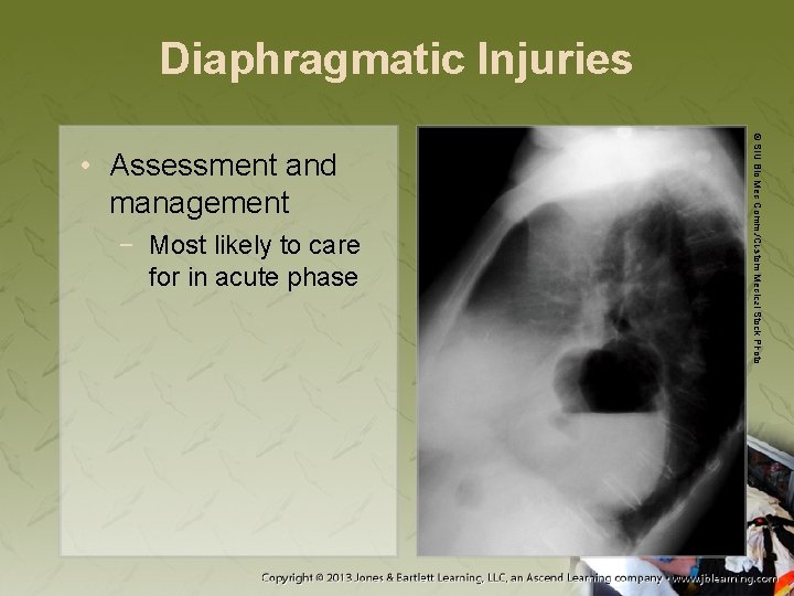
Diaphragmatic Injuries • Tension gastrothorax − Herniation of abdominal contents into thoracic cavity, causing pressure to: • Compress lung on affected side. • Compromise circulatory function.
Diaphragmatic Injuries − Most likely to care for in acute phase © SIU Bio Med Comm. /Custom Medical Stock Photo • Assessment and management
Diaphragmatic Injuries • Assessment and management (cont’d) − Acute phase: • Hypotension • Tachypnea • Bowel sounds in chest • Chest pain • Absent breath sounds − Obstructive phase: • Nausea and vomiting • Abdominal pain • Constipation • Dyspnea • Abdominal distension
Diaphragmatic Injuries • Assessment and management (cont’d) − Management focus: maintaining oxygen and providing rapid transport • Elevate head of backboard. • Positive-pressure ventilation • NG tube placement (if allowed by protocol)
Esophageal Injuries • Rapidly fatal injury in GI system • Assessment and management − Signs and symptoms include: • Pleuritic chest pain • Subcutaneous emphysema • Associated tracheal injury
Esophageal Injuries • Assessment and management (cont’d) − No specific therapy in the prehospital setting − Do not give anything orally.
Tracheobronchial Injuries • Rare, typically caused by penetrating injuries − High mortality rate − Allows for rapid movement of air into pleural space, causing a pneumothorax • May progress to tension pneumothorax
Tracheobronchial Injuries • Assessment and management − Presentation varies and may include: • Hoarseness • Dyspnea and tachycardia • Respiratory distress
Tracheobronchial Injuries • Assessment and management (cont’d) − Focuses on ABCs • Bag-bask ventilation instead of intubation • Avoid high ventilatory pressure.
Traumatic Asphyxia • Induced by traumatic injury that forcefully compresses the thoracic cavity − Causes pressure to major veins of the head, neck, and kidneys • Causes rupture of the capillary beds
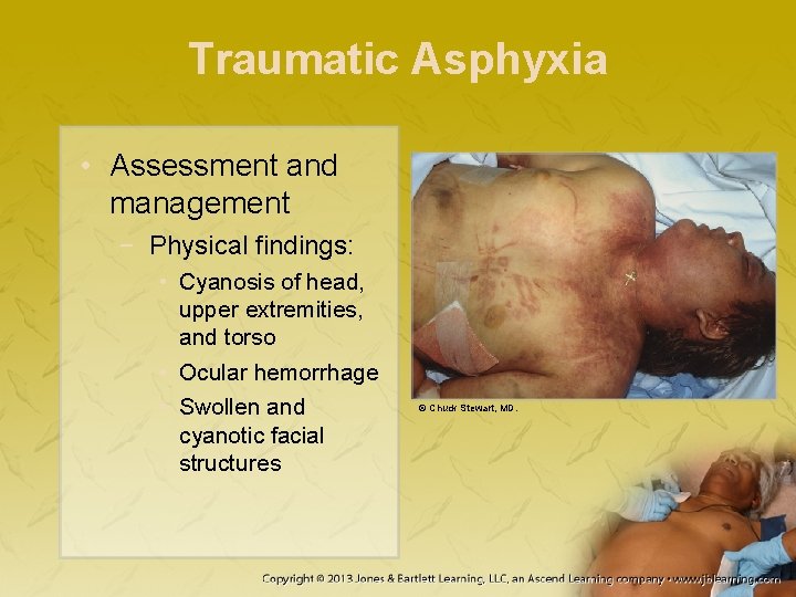
Traumatic Asphyxia • Assessment and management − Physical findings: • Cyanosis of head, upper extremities, and torso • Ocular hemorrhage • Swollen and cyanotic facial structures © Chuck Stewart, MD.
Traumatic Asphyxia • Assessment and management (cont’d) − − Provide high-flow supplemental O 2. Take cervical spine precautions. Obtain IV access with two large-bore IV lines. Transport to nearest trauma center.
Summary • Thorax contains ribs, thoracic vertebrae, clavicle, scapula, sternum, heart, lungs, diaphragm, great vessels, esophagus, lymphatic channels, trachea, mainstem bronchi, and nerves. • Oxygenation, ventilation, and some aspects of circulation take place in the thorax. • Thoracic injuries can cause air or blood in the lungs or prevent organs from moving properly.
Summary • A thoracic trauma assessment begins with scene size-up and ABCs assessment. • When assessing breathing, note any injury to the thorax, which may indicate underlying injuries. • Consider ventilation and oxygenation adequacy. • Always consider spine stabilization.
Summary • Managing chest injuries includes maintaining airway, ensuring oxygenation and ventilation, supporting circulation, and transporting quickly. • Chest wall injuries include flail chest, rib fractures, sterna fractures, and clavicle fractures. • In flail chest, two or more ribs are broken in two or more places, which can result in a free-floating rib segment.
Summary • Flail chest management includes airway management and possible positive-pressure ventilation. • Rib fractures cause significant pain and may prevent adequate ventilation. • Rib fracture management should focus on the ABCs and gentle splinting of the chest.
Summary • Lung injuries include simple pneumothorax, open pneumothorax, tension pneumothorax, hemothorax, and pulmonary contusion. • In a pneumothorax, air leaks into the pleural space from an opening in the chest or the surface of the lung. • Management of a pneumothorax starts with the ABCs and high-concentration oxygen administration.
Summary • A tension pneumothorax results from air collection in the pleural space, and is a lifethreatening condition. • Patients with a tension pneumothorax should be placed on high-flow supplemental oxygen via a nonrebreathing mask. Cover open wounds with a nonporous or occlusive dressing.
Summary • A hemothorax is the accumulation of blood between the parietal and visceral pleura. • If a patient with a hemothorax does not require airway intervention, place the patient on high-flow supplemental oxygen via a nonrebreathing mask. • A hemopneumothorax is the collection of both blood and air in the pleural space.
Summary • A pulmonary contusion occurs from compression of the lung, resulting in alveolar and capillary damage, edema, and hypoxia. • If a patient has a pulmonary contusion or cardiac tamponade, assess and manage the ABCs and consider administering IV fluids. • Myocardial injuries include cardiac tamponade, myocardial contusion, myocardial rupture, and commotio cordis.
Summary • Cardiac tamponade occurs when excessive fluid builds up in the pericardial sac around the heart, which compresses the heart and compromises stroke volume. • Treating cardiac tamponade begins by managing the ABCs, ensuring adequate oxygen delivery, and establishing IV access. Pericardiocentesis is the ultimate treatment option.
Summary • Myocardial contusion is blunt trauma to the heart, and may cause hemorrhage, edema, and cellular damage. • Management for myocardial contusion is supportive, but should also include cardiac monitoring and establishing IV access. • Myocardial rupture is perforation of one or more elements of the anatomy of the heart, occurring from blunt or penetrating trauma.
Summary • Management for myocardial rupture should include supportive care and rapid transport to a trauma center for a thoracotomy. • Commotio cordis occurs from a direct blow to the chest during a critical portion of the heart’s repolarization period. • ALS treatment for commotio cordis must follow standard ACLS guidelines for sudden cardiac arrest.
Summary • Vascular injuries include traumatic aortic disruption and great vessel injury. • Traumatic aortic disruption is the ripping of the aorta. • Management for traumatic aortic disruption focuses on symptom control. • Other injuries include diaphragmatic injuries, esophageal injuries, tracheobronchial injuries, and traumatic asphyxia.
Credits • Chapter opener: Courtesy of ED, Royal North Shore Hospital/NSW Institute of Trauma & Injury • Backgrounds: Blue–Jones & Bartlett Learning. Courtesy of MIEMSS; Gold–Jones & Bartlett Learning. Courtesy of MIEMSS; Green–Courtesy of Rhonda Beck; Red–© Margo Harrison/Shutter. Stock, Inc. • Unless otherwise indicated, all photographs and illustrations are under copyright of Jones & Bartlett Learning, courtesy of Maryland Institute for Emergency Medical Services Systems, or have been provided by the American Academy of Orthopaedic Surgeons.