Peripheral Nervous System Chapters 14 Anatomy 32 The


 it inervates the superior oblique muscles of the eye.")
- it has three branches that carry sensory")
,")


 controls muscles of swallowing extends beyond the face and")

 runs below the tongue and innervates the")


- Slides: 47
Peripheral Nervous System Chapters 14 Anatomy 32
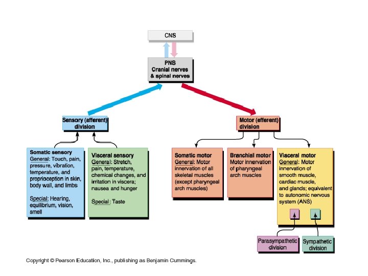
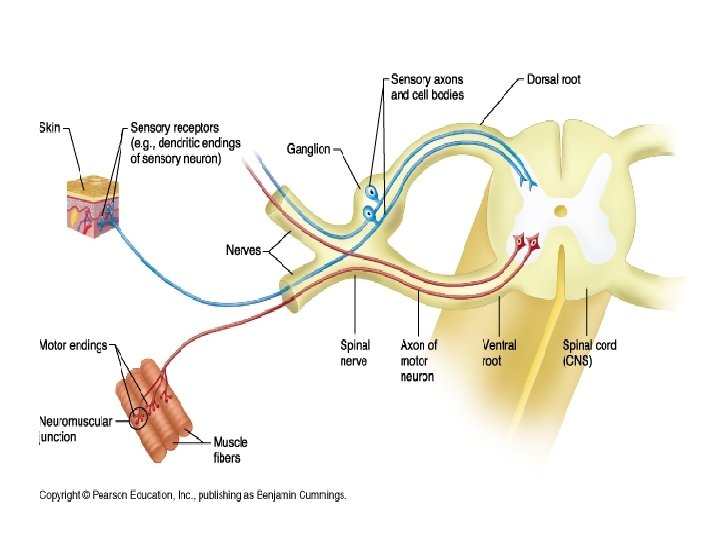
The peripheral nervous system and its subdivisions The PNS includes all nervous structures found outside the CNS. It requires Three components: sensory receptors (pick up stimuli and send signals to CNS), motor endings (axons synapse with effectors like muscle and glands), and nerves and ganglia (neurons and cell bodies outside of CNS). Note that almost all nerves are mixed, meaning theycarry both sensory and motor neurons. Receptors can be classified by location, stimulus detected, or structure I. Peripheral sensory receptors- Receptors are either dendritic ends of sensory neurons that monitor general sensory information over wide areas, Receptors may also associate with cells that transfer the signal to a sensory neuron (ex. merkel cells) and monitor specialized senses by existing in localized regions. A. Classification by location of sensory receptors 1. Receptors that receive stimulus from the environment are sensitive to pressure, pain, smell, sight, and hearing are called exteroceptors. 2. Receptors that are sensitive to the body’s internal changes, either visceral or chemical changes, taste, and temperature are called interoceptors. 3. Receptors found within skeletal muscles, tendons, joints, and ligaments monitor the amount of stretching and send input on movement. They are called proprioceptors.
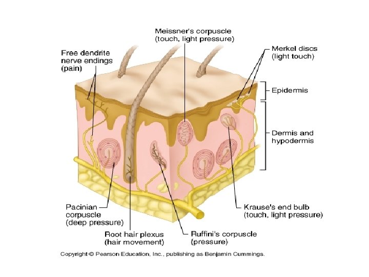
B. Classification by stimulus detected 1. Mechanoreceptors are stimulated by mechanical forces such as touch, pressure, ect. 2. Thermoreceptors are stimulated by temperature changes 3. Chemoreceptors respond to chemicals changes in blood, taste, or smell. 4. Photoreceptors respond to light such as those found in the eye. C. Classification by structure- the specialized sense organs have receptors with special structures for detecting certain stimuli, the ones listed below are general sensory receptors found throughout the body that detect mechanical forces. 1. Free dendritic endings- naked dendritic endings are in every tissue and most often in the integument. They sense pain and temperature changes and some sense light touch. Itch receptors (newly discovered) are also free dendritic ends but are very thin in diameter. 2. Encapsulated dendritic endings- these are nerve endings wrapped in connective tissue and serve as mechanoreceptors. Meissner’s corpuscles- detect light touch in the skin Krause’s End bulbs- detect fine touch in mucus membranes Pacinian Corpuscles- found in deep connective tissue (hypodermis) respond to vibration and briefly respond to deep pressure. Ruffini’s Corpuscles- found in dermis respond to continuous pressure
Proprioceptors- found in muscle tissue, tendons, and ligaments, they monitor stretch in locomotory organs
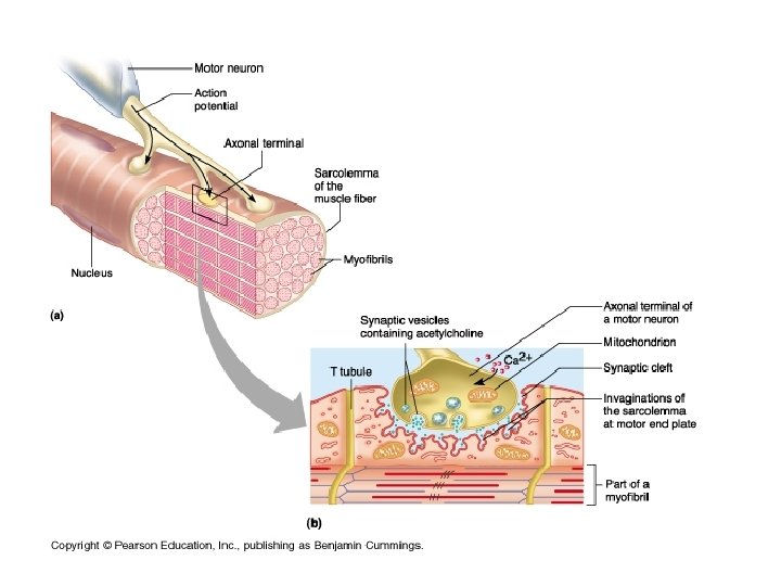
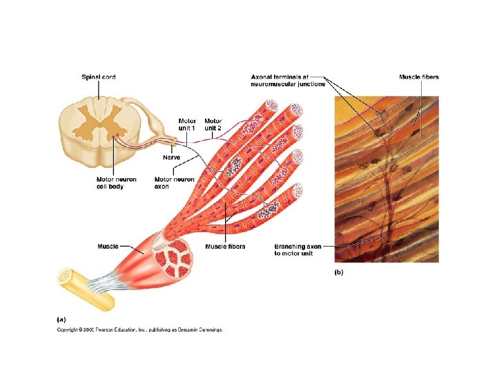
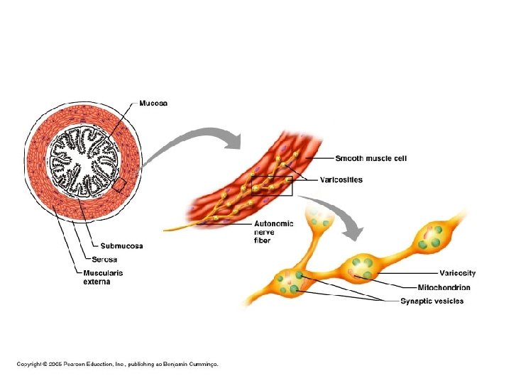
II. Peripheral motor endings- Axons that synapse with either muscle or glands to activate them. A. Innervation of skeletal muscles- each muscle fiber is associated with a neuromuscular junction (synapse between a motor axon and muscular tissue). A motor neuron branches in order to innervate individual skeletal muscle fibers and is called a motor unit. At the synapse the axon releases synaptic vesicles filled with a neurotransmitter called acetylcholine which depolarizes the sarcolemma. The basal lamina of the sarcolemma grooves releases acetylcholinesterase, an enzyme that immediately breaks down acetylcholine to prevent further stimulation of muscle twitch. The motor unit signal causes an overall contraction of the muscle. The finer the motor movement the more motor neurons and each innervates less muscular fibers. B. Innervation of visceral muscle and glands- the visceral motor axon forms a row of axon knobs (varicosities) and have a larger distance in the synaptic cleft. The response is slower because of the time it takes for the neurotransmitter to diffuse. The heart is also innervated by these motor neurons, but there are no varicosities.
III. Cranial Nerves- These nerves serve the head and neck; they originate in the brain and most of them exit the skull through cranial foramina not vertebral foramina. They are numbered I – XII rostrally to caudally.
• A. Olfactory nerve I- sensory nerve for smell, it runs below the frontal lobe, purely sensory, cerebrum
– B. Optic nerve II- a brain tract exiting through the optic chiasma, it sends signals of images to the brain, purely sensory, cerebrum
C. Oculomotor nerve III- caudal to optic chiasma it innervates internal eye muscles to move the eye (superior, inferior, lateral, medial rectus) and eyelids. It adjust the pupil and lens. Motor nerve, visceral motor, and proprioceptive, midbrain
D. Trochlear nerves IV- (pulley) it inervates the superior oblique muscles of the eye. Motor nerve, midbrain
E. Trigeminal nerves V – (three fold)- it has three branches that carry sensory information from the face (superficial and internal) and motor information for chewing muscles. Mixed, pons
F. Abducens nerves VI- innervates a muscle that abducts the eye ( lateral rectus), motor nerve, pons
G. Facial nerves VII- innervates muscles of facial expression, activates facial glands, conveys sensory from taste buds. Mixed and visceral motor, pons
H. Vestibulocochlear nerves VIII- sensory nerve for hearing and equilibrium, purely sensor, medulla oblongata
I. Glossopharyngeal nerves IX. innervates the tongue and pharynx, controls a muscle used for swallowing, activates salivary gland, conducts taste, and other facial sensory. Mixed and visceral motor, medulla oblongata
J. Vagus Nerves X- (wanders) controls muscles of swallowing extends beyond the face and neck into the thorax and abdomen to innervate internal organs for motor and sensory impulses, some sensory near area. Mixed and visceral motor, medulla oblongata.
K. Accessory nerves XI- accessory for the vagus nerve- it joins it, and controls muscles that moves the head and some of the same as the vagus. Motor and visceral motor, medulla oblongata.
L. Hypoglossal nerves XII- (below the tongue) runs below the tongue and innervates the tongue muscles, motor nerves, medulla oblongata.
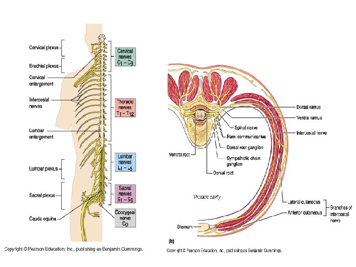
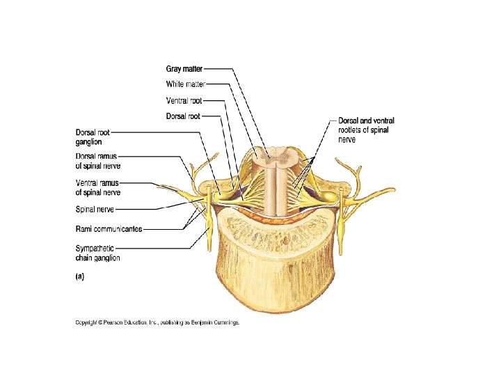
III. Spinal Nerves- There are 31 pairs of nerves exiting the spinal column: 8 cervical, 12 thoracic, 5 lumbar, 5 sacral, and 1 coccygeal. They innervate the body in sections as seen on page 426. Each nerve has a dorsal (sensory) and ventral root (motor) that attach to the spinal cord at the rootlets. Each spinal nerve also has dorsal and ventral ramus that carries motor and sensory nerves. The ventral ramus connects to rami commicantes that connect to symphathetic chain ganglia. The dorsal rami supplies the posterior parts of the body and ventral rami supplies the lateral and anterior sides of the body. • A. Innervation of the back- the nerves follow a neat and simple pattern. B. Innervation of the anterior thoracic an abdominal wall- supply intercostals muscles, skin or anterior and lateral thorax and abdomen.
C. Introduction to nerve plexuses- these are networks of nerve clusters formed by ventral rami from different spinal nerves. Plexuses serve the limbs and are designed to prevent paralysis of a limb muscle by the distruction of just one spinal nerve. 1. The cervical plexus and innervation of the neck- formed by C 1 -C 4 nerves, most branches are cutaneous nerves and anterior neck muscles and diaphragm.
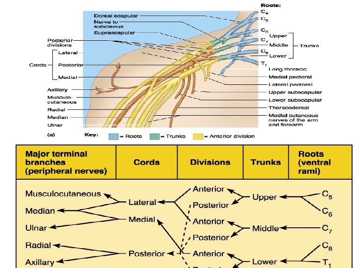
2. The brachial plexus and innervation of the upper limb- formed by C 5 -C 8 nerves, it supplies the upper limbs. The plexus’ extremely complex lies between the cervical and axillary regions. Roots run deep of the sternocleidomastoid, they unite to form trunks which divide into anterior and posterior divisions that break into lateral, medial, and posterior cords that divide into the terminal branches (around axilla) that innervate the arm Pg 430. The nerves of the arms are: axillary nerve, musculocutaneous nerve, median nerve, ulnar nerve, and radial nerves.
3. The lumbar plexus and innervation of the lower limb- formed by L 1 -L 4 nerves the main branches innervate the anterior thigh via the femoral nerve. Medial thigh and adductor muscles are innervated by the obturator nerve.
4. The sacral plexus and innervation on the lower limbformed by L 4 -S 4 nerves, its many branches innervate the buttock, lower limb, pelvis, and perineum. The largest branch is the sciatic nerve that supplies lateral and posterior limb regions. It branches into the tibial and fibial nerve.
5. Innervation of joints of the body- As a health professional you need to know the nerves that innervate the joints. Use Hilton’s Law: any nerve that innerates a muscle producing movement at a joint also innervates the joint itself and the skin over it. Example: Knee joint is surrounded by anterior and posterior thigh muscles that are innervated by femoral, obturator, and branches of the sciatic nerves.
6. Innervation of the skin: dermatomeseach spinal nerves innervates a skin zone as shown in page 436
Chapter 15: Introduction to the autonomic nervous system IV. nerves that innervate smooth muscle, cardiac muscle, and glands. The stimulus they send is not voluntarily controlled. It is the visceral motor division
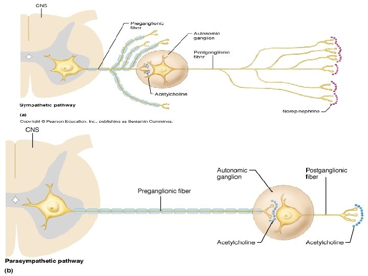
A. Comparison of autonomic and somatic motor systems- the motor unit of autonomic system includes two motor neurons: preganglionic neuron (myelinated) has an axon that synapses with the postganglionic neuron (unmyelinated) in autonomic ganglion, then the axon of the postganglionic neuron synapses with the viscera. A somatic motor unit contains one myelinated axon per innervated muscle cell. **Note that autonomic ganglia is motor not sensory.
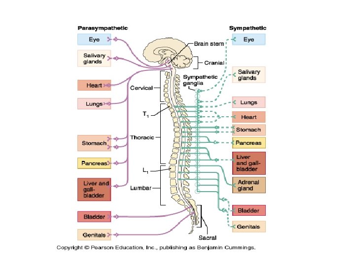
• B. Divisions of the autonomic NS- both divisions have motor units that cause opposite effects of the visceral organs they innervate. • Symphatheic division- mobilizes the body, responsible for “fight or flight response”: increase heart rate, breathing rate, higher blood pressure, dilate pupils, vasoconstriction, etc. Nerves arise from thoracic and lumbar regions. It releases norepinephrine (noradrenaline). Innervates with greater branching. • Parasymphathetic division- relaxes or unwinds the body, is active when body is at rest, it conserves body energy, directs digestion and waste elimination, opposite effects of fight or flight response. Nerves arise from the brain and sacral regions. It releases acetylcholine (cholinergic). Innervates with less branching.
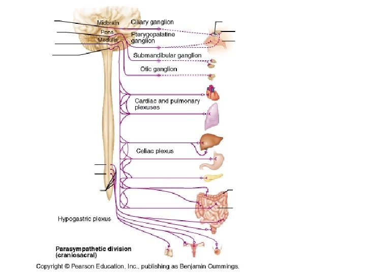
• V. The parasympathetic division- (all summarized in figure 15. 5, 15. 6, and table 15. 2 • A. Cranial outflow- these are ANS nerves belonging to the parasympatehtic division (relaxing) that innervate the organs in the head, neck, thorax, and abdomen. 1. Oculomotor outflow—smooth muscles in the eye (pupil /lens adjustment) • • 2. Facial nerve outflow-stimulate secretion of glands of the head (tears, mucus, saliva) • 3. Glossopharyngeal outflow- stimulates secretion of a large salivary= parotid gland – 4. Vagus nerve outflow- innervate visceral organs of the thorax (heart and lungs) and most of the abdomen (liver, gallbladder, pancreas, stomach, small intestine, and stops half way through large intestine) through the autonomic nerve plexuses. • • B. Sacral outflow- This section runs from s 2 -s 4 and innervates the abdominal and pelvic organs the vagus nerve outflow did not innervate, including large intestine, rectum, bladder, and reproductive organs.
• VI. The sympathetic division- This division innervates more organs and is more complex than the parasympathetic. • A. basic organization- The sympathetic system innervates the integument: its glands and the arrector pili in addition to internal organs and blood vessels. There is also more glanglia due to the preganglionic and postganglionic cell bodies.
• 1. Sympathetic trunk ganglia. There are 22 -24 sympathetic trunk ganglia that run along the sides of the vertebral column (next to the centrum) in a vertical direction from neck to pelvis. The ganglia connect to long synaptic chains called sympathetic trunks so the overall appearance a “beaded”chain. The sides of the ganglia (contains cell bodies) connect so spinal nerves (running horizontally) by gray and white rami communicantes. Thus the axon of the postganglionic neuron joins the spinal nerves. • 2. Prevertebral ganglia- Some ganglia does not lie on the sides of the vertebral column but anterior to it along a large abdominal artery (abdominal aorta). They are not paired and occur only in the abdomen and pelvis.
• B. Symphathetic pathways- Typically the preganglionic neuron sends the axon from the spinal cord out through the ventral root to spinal nerve to the white ramus communicantes into the sympathetic trunk where it synapses with the postganglionic neuron that sends its axon through the gray communicantes out through the spinal nerves and finally to the organ it innervates.
• C. Role of the adrenal medulla in the sympathetic division- A gland that sits on the superior aspect of the kidney. It releases norepinephrine (noradrenaline) and epinephrine (adrenaline) into the blood stream to excite the body in time of “fight, flight, or fright” response.
• • VII. Central control of the autonomic nervous system- The CNS controls the activity of the ANS A. Brain stem and spinal cord- medulla oblongata regulates heart rate, blood pressure, and digestive activity. The midbrain controls the sympathetic response to fear. The spinal cord control visceral reflexes (defication and urination reflex can be voluntarily controlled). • B. Hypothalamus and amydala- The hypothalamus is the main integration center of ANS and the amygdala coordinates sympathetic learned fear responses • C. Cerebral cortex- Voluntary control of ANS may happen via meditation when deep thoughts of relaxation activate the limbic system which in turns activates the hypothalamus to activate the parasympathetic NS. Thoughts of frightful experiences may stimulate the amygdala to activate the hypothalamus that activates the sympathetic NS. VIII. Visceral sensory neurons- Receptors in the viscera are free dendritic ends that send afferent signals caused by stretching, temperature and chemical changes, and irritation. Integration translates these signals into hunger, fullness, pain, or nausea. Visceral sensation may be hard to localize. Sometime pain is referred pain, a problem with an organ like the heart may send pain down the arm (not an area where the heart is located. ) IX. Visceral reflexes- These are also called peripheral reflexes because they may not involve the CNS a visceral sensory branches may synapse with postganglionic motor neurons in sympathetic ganglia. This makes the system independent of CNS control. • •
• A map of referred pain: these are skin or body regions that present pain when there is visceral pain. The organ and site of referred pain are innervated by the same nerve.