THE NERVOUS SYSTEM Central Nervous System Peripheral Nervous


 • carry impulses from sensory neurons to motor neurons SYNAPSE •")


 • DURA MATER – outer brain covering, lines the")


 increase in memory loss, difficulty recognizing")


- Slides: 35
THE NERVOUS SYSTEM Central Nervous System Peripheral Nervous System Autonomic Nervous System Allied Health I
Central Nervous System • Communication and coordination system of the body • Seat of intellect and reasoning • Consists of the brain, spinal cord, and nerves
NEURON • • • Nerve cell Transmits a message from one cell to the next Has a nucleus, cytoplasm, and cell membrane DENDRITES • • Nerve cell processes that carry impulse to cell body May be one or many
AXON • Carries impulse away from cell body • Only one on a neuron NEURILEMMA (MYELIN SHEATH) • Covering that speeds up the nerve impulse along the axon • Myelin is a fatty substance that protects the axon SENSORY NEURONS (AFFERENT) – emerge from the skin or sense organs, carry impulses to spinal cord and brain MOTOR NEURONS (EFFERENT) – carry messages from the brain and spinal cord to muscles and glands
ASSOCIATIVE NEURONS (INTERNEURONS) • carry impulses from sensory neurons to motor neurons SYNAPSE • space between neurons, messages go from one cell to the next
Nerve impulse – A STIMULUS creates an IMPULSE. The impulse travels into the neuron on the dendrite(s) and out on the axon. At the end of the axon, a NEUROTRANSMITTER is released that carries the impulse across the SYNAPSE, to the next dendrite. Divisions of the Nervous System 1. CENTRAL NERVOUS SYSTEM – brain and spinal cord
2. PERIPHERAL NERVOUS SYSTEM – cranial nerves and spinal nerves
3. AUTONOMIC NERVOUS SYSTEM – includes peripheral nerves and ganglia, supplies heart muscle, smooth muscle and secretory glands, involuntary action
The Brain
• 3 lb mass of soft nervous tissue • 100 billion neurons • Protected by skull, three membranes called meninges, and cerebrospinal fluid • Adequate blood supply is needed, brain tissue will die in 4 -8 minutes, without O 2 • Divided into 4 major parts: cerebrum, diencephalon, cerebellum, brain stem
Coverings of the Brain (MENINGES) • DURA MATER – outer brain covering, lines the inside of the skull, tough dense fibrous connective tissue. • SUBDURAL SPACE – between dura and arachnoid • ARACHNOID – middle layer, resembles fine cobweb, • PIA MATER – covers the brain’s surface, comprised of blood vessels held together by connective tissue • SUBARACHNOID SPACE - between arachnoid and pia mater, filled with CEREBROSPINAL FLUID – acts as a liquid shock absorber and source of nutrients for the brain.
Ventricles of the Brain • Brain contains four cavities filled with cerebrospinal fluid called CEREBRAL VENTRICLES. 1. Right and left lateral ventricles 2. Third ventricle – behind and below the lateral ventricles 3. Fourth ventricle is below the 3 rd, in front of the cerebellum and behind the pons and medulla oblongata
CHOROID PLEXUS – network of blood vessels lining the ventricles which helps in the formation of cerebrospinal fluid CEREBROSPINAL FLUID • Forms inside ventricles of the brain • Serves as a liquid shock absorber protecting the brain and spinal cord BLOOD-BRAIN BARRIER – choroid plexus capillaries prevent substances (like drugs) from penetrating brain tissue – this makes infections, like meningitis, difficult to cure (blood brain barrier)
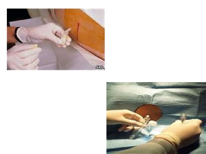
LUMBAR PUNCTURE – removal of CSF from spinal
Lumbar Puncture
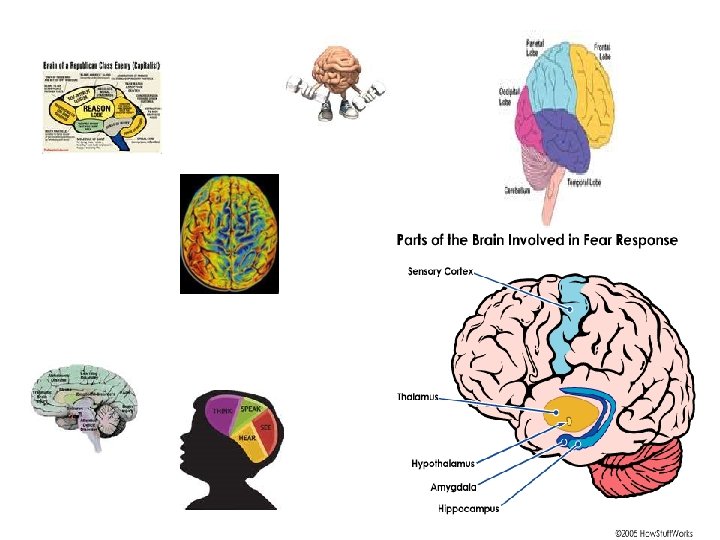
CEREBRUM • Largest part of the brain • Divided into R and L hemispheres by deep groove (longitudinal fissure) CONVOLUTIONS – elevated folds on the surface of the cerebrum, they increase the surface area of the brain SULCI – fissure or grooves separating cerebral convolutions Divided into four lobes – 1. FRONTAL, 2. PARIETAL, 3. OCCIPITAL 4. TEMPORAL
Cerebral function: Conscious thought, judgment, memory, reasoning, and will power. DIENCEPHALON • Located between cerebrum and midbrain • Composed of THALAMUS and HYPOTHALAMUS Vital functions of the hypothalamus: 1. Autonomic nervous control 2. Temperature control 3. Appetite control 4. Emotional state 5. Sleep control
CEREBELLUM • Located behind the pons and below the cerebrum • Composed of two hemispheres • Controls all body functions related to skeletal muscles, including: 1. Balance 2. Muscle tone 3. Coordination of muscle movements BRAIN STEM • Made up of PONS, MEDULLA and MIDBRAIN
• Pathway for ascending and descending tracts Pons – in front of cerebellum, between midbrain and medulla – contains center that controls respiration Midbrain – vision and hearing Medulla oblongata – bulbshaped structure Between pons and spinal cord, inside the cranium above foramen magnum. Responsible for: 1. Heart rate 2. Blood pressure
Teratoma
SPINAL CORD • Begins at foramen magnum and continues down to 2 nd lumbar vertebrae • White and soft, in spinal canal • Surrounded by cerebrospinal fluid Functions as: 1. Reflex center 2. Conduction pathway to and from the brain
PERIPHERAL NERVOUS SYSTEM • All of the nerves of the body and ganglia • Autonomic nervous system is specialized part of PNS NERVES • Bundle of nerve fibers enclosed by connective tissue • Sensory nerves carry impulses to brain and spinal cord • Motor nerves carry impulses to muscles or glands • Mixed nerves contain both sensory and motor fibers
CRANIAL NERVES • • • 12 pairs Begin in the brain Designated by number and name I Olfactory II IV V VI VIII Optic Oculomotor Trochlear Trigeminal Abducens Facial Vestibulocochlear IX Glossopharyngeal
X XI XII Vagus Accessory Hypoglossal SPINAL NERVES • Originate at spinal cord and go through openings in vertebrae • 31 pairs of spinal nerves • All are mixed nerves • Named in relation to their location on the spinal cord
AUTONOMIC NERVOUS SYSTEM • Regulates activities of visceral organs • Not subject to conscious control SYMPATHETIC NERVOUS SYSTEM – the “fight or flight” system – when the body perceive danger, SNS sends message to adrenal medulla to secrete adrenaline – heartbeat increases PARASYMPATHETIC NERVOUS SYSTEM – counters effects of SNS, decreases heart rate
REFLEX • Unconscious and involuntary • In a simple reflex, only a sensory nerve and motor nerve involved example, “knee-jerk” reflex
DISORDERS OF THE NERVOUS SYSTEM MENINGITIS • Inflammation of the lining of the brain and spinal cord • May be bacterial or viral Symptoms – headache, fever and stiff neck • In severe form, may lead to paralysis, coma and death • If bacterial, may be treated with antibiotics • EPILEPSY • Seizure disorder of the brain, characterized by • recurring and excessive discharge from neurons
• Seizures believed to be result of spontaneous, uncontrolled electrical activity of neurons • Cause – uncertain • Victim may have hallucinations and seizures Grand mal – severe, convulsive seizure Petit mal – milder ALZHEIMER’S DISEASE • Progressive disease that begins with problems remembering • Nerve endings in cortex of brain degenerate and block signals that pass between nerve cells • Abnormal fibers build up creating tangles • Cause – unknown • First stage (2 -4 years) involves confusion, shortterm memory loss, anxiety, poor judgement
• 2 nd stage (2 -10 years) increase in memory loss, difficulty recognizing people, motor problems, logic problems, and loss of social skills • 3 rd stage (1 -3 years) inability to recognize oneself, weight loss, seizures, mood swings and aphasia • PARALYSIS – loss of power of motion or sensation • HEMIPLEGIA – paralysis on one side of the body
Cerebral Vascular Accident • • Stroke or CVA Interruption of blood and O 2 to brain Tissue death Third leading cause of death in USA Risk Factors • • Smoking Hypertension Heart disease Family history Causes of CVA • 90% caused by blood clots
• 90% caused by blood clots • Clots lodge in carotid arteries, blocking the flow of blood to the brain • 10% caused by ruptured blood vessels in the brain Symptoms • • Hemiplegia on opposite side of the body Sudden, severe headache Dizziness Sudden loss of vision in one eye Aphasia Dysphasia Coma Possible death
Treatment • Get to the hospital immediately!! • CT done to determine etiology • If a clot, treatment aimed at dissolving clot Prevention • • If TIAs – one aspirin a day Stop smoking Exercise and lose weight Control hypertension
The End