The Nervous System Chapter 7 The Nervous System


 1. Sensory (afferent) Division- consists of nerve fibers that convey impulses")
 Division –carries impulses from the CNS to effector organs, muscles, and")
 • Somatic nervous system: (aka voluntary nervous system) – Consciously or")


- Slides: 59
The Nervous System Chapter 7
The Nervous System • Every thought, action, or emotion you have is controlled by your nervous system • Communicates with your body through a series of specific and rapid electrical impulses
Functions • Uses sensory receptors to monitor internal and external changes (stimuli and sensory input) • Processes and interprets the sensory input and makes decisions about what should be done (integration) • Effects a response by activating muscles or glands (motor output)
Structural Classification Central Nervous System= the brain + spinal cord. • • Occupies the dorsal body cavity Acts as the integrating and command center of the nervous system.
Structural Classification Peripheral Nervous System= spinal nerves + cranial nerves Consists mainly of nerves that extend from the brain and spinal cord. • Links all the parts of the body by carrying impulses from the sensory receptors to the CNS and from the CNS to appropriate glands and muscles. •
Functional Classification (PNS) 1. Sensory (afferent) Division- consists of nerve fibers that convey impulses to the CNS. -Somatic sensory fibers – from the skin, skeletal muscle and joints - Visceral sensory fibers- from the visceral organs.
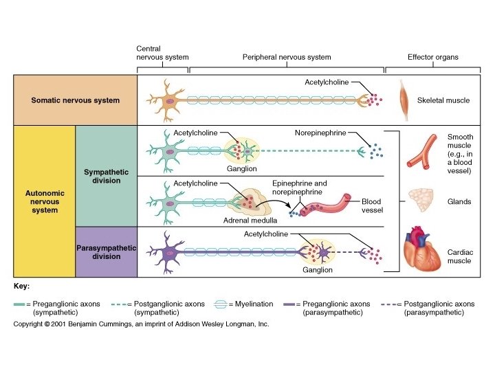
2. Motor (efferent) Division –carries impulses from the CNS to effector organs, muscles, and glands. a. Somatic/voluntary nervous system b. Automatic/involuntary nervous system
Functional Classification (PNS) • Somatic nervous system: (aka voluntary nervous system) – Consciously or voluntarily control our muscles – Not necessarily all muscles: ex: reflexes • Autonomic nervous system: (aka involuntary nervous system) – Activity of smooth or cardiac muscle
Nervous Tissue • Even though nervous tissue is complex, it is made up of just two principal types of cells: 1. supporting cells 2. neurons.
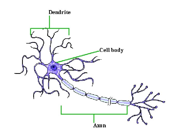
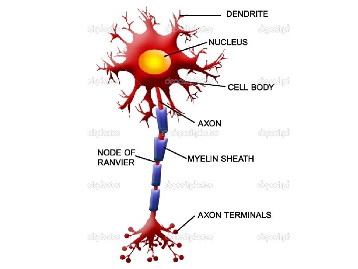
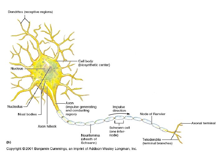
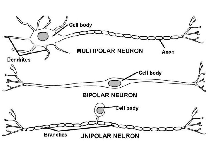
Nervous Tissue • Neurons • • • Cell body Nissl Substance/Neurofibrils Dendrites Axon Terminals Myelin Sheath Neurilemma Nodes of Ranvier Ganglia Tracts/Nerves
Nervous Tissue • Cell body- metabolic center of the neuron containing organelles. • Nissl substance- rough ER • Neurofibrils- intermediate filaments important in maintaining cell shape. • Dendrites- convey incoming messages toward the cell body. • Axons – generate nerve impulses and conduct them away from the cell body.
Nervous Tissue • Axon Terminals- contain hundreds of tiny vesicles that contain neurotransmitters • Myelin Sheath – protects and insulates the axon and increases the transmission rate of nerve impulses. • Neurilemma- external to myelin sheath • Nodes of Ranvier- gaps or indentations on myelin sheath
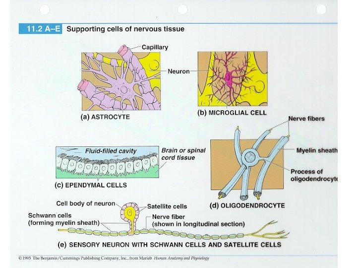
Nervous Tissue Supporting Cells of the CNS – Neuroglia • Astrocytes- abundant star-shaped cells that account for nearly half of neural tissue. – Make exchanges between capillaries and neurons
• Microglia- spiderlike phagocytes that dispose of debris, including dead brain cells and bacteria.
• Ependymal Cells- line cavities of brain and spinal cord. – Circulate cerebralspinal fluid.
• Oligodendrocytes- produce myelin sheaths.
Nervous Tissue supporting cells and neurons • Supporting Cells of the PNS 1. Schwann Cells 1. Satellite Cells
Functional Classification of Neurons • Groups neurons according to the direction the nerve impulse is traveling • Sensory or afferent neurons – carry impulses from sensory receptors to the CNS. Their dendrites are associated with specialized receptors. Their cell bodies are in ganglion outside the CNS. • Motor or efferent neurons – carry impulses form the CNS to the viscera and/or muscles and glands. Their cell bodies are in the CNS.
Structural Classification of Neurons • Based on the number of processes extending from the cell body. • Multipolar • Bipolar • Unipolar
Physiology of Neurons Irritability and Conductivity Irritability – the ability to respond to a stimulus and convert it to a nerve impulse. • A resting neuron is polarized. There are more Na+ outside and less K+ inside. • Stimuli causes the permeability of the plasma membrane to change, changing the polarity. • This depolarization activates the neuron to initiate and transmit an action potential. • Repolarization allows the cell to conduct the next impulse.
Physiology of Neurons Irritability and Conductivity – the ability to transmit the impulse to other nerve cells, muscles, or glands.
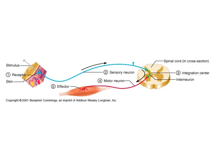
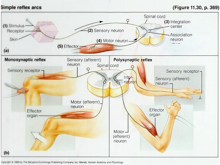
Reflexes • Rapid, predictable and involuntary responses to stimuli that occur over neural pathways called reflex arcs. • Autonomic Reflexes – regulate the activity of smooth muscle, the heart, and glands. • Somatic Reflexes – includes all reflexes that stimulate the skeletal muscles.
Reflex Arcs • All reflex arcs have a minimum of five elements 1. Sensory receptor 2. Afferent neurons 3. CNS integration center 4. Efferent neurons 5. Effector organ
Reflex Arcs • There is always a delay at the synapse. • Some reflexes involve only the spinal cord neurons to function. • Some reflexes require that the brain become involved because many different types of information need to be evaluated in order to arrive at the correct response.
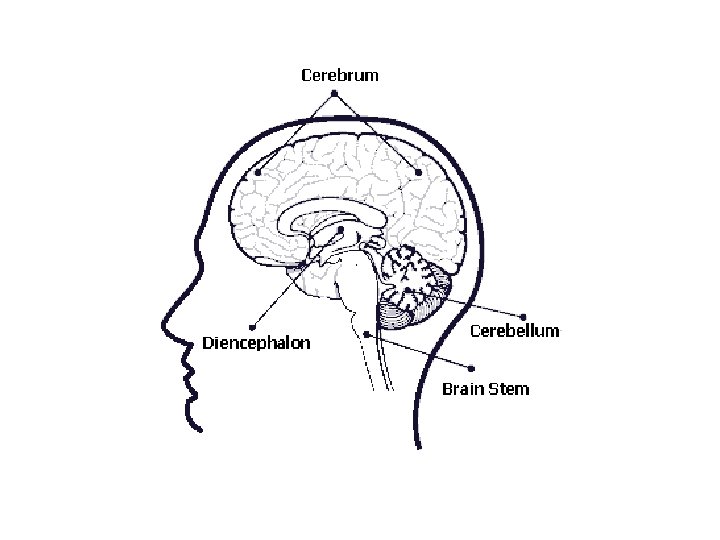
Central Nervous System the brain • The brain is commonly discussed in terms of 4 major regions… 1. Cerebral Hemisphere 2. Diencephalon 3. Brain Stem 4. Cerebellum
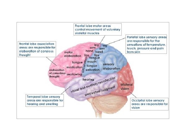
Cerebral Hemispheres • The pair of cerebral hemispheres are the most superior and largest part of the brain. • The entire surface of these hemispheres has elevated ridges called gyri, separated by shallow grooves called sulci. • Deeper grooves called fissures separate the brain into lobes, named for the cranial bone that lies over them.
Cerebral Hemispheres Lobes 1. 2. 3. 4. Parietal Lobe Occipital Lobe Temporal Lobe Frontal Lobe Functional Areas 1. 2. 3. 4. 5. 6. 7. 8. Somatic Sensory Area Specialty Senses Primary Motor Area Corticospinal Tract Broca’s Area Speech Area Corpus Callosum Basal Nuclei
Diencephalon • The diencephalon sits atop the brain stem and is enclosed by the cerebral hemispheres. 1. Thalamus 2. Hypothalamus 3. Epithalamus
Brain Stem • Provides a pathway for ascending and descending tracts and has many small gray matter areas. 1. Midbrain 2. Pons 3. Medulla Oblongata 4. Reticular Formation
Cerebellum • It projects dorsally from under the occipital lobe. • The cerebellum provides the precise timing for skeletal muscle activity and controls our balance and equilibrium. • Because of it’s activity, our body movements are smooth and coordinated.
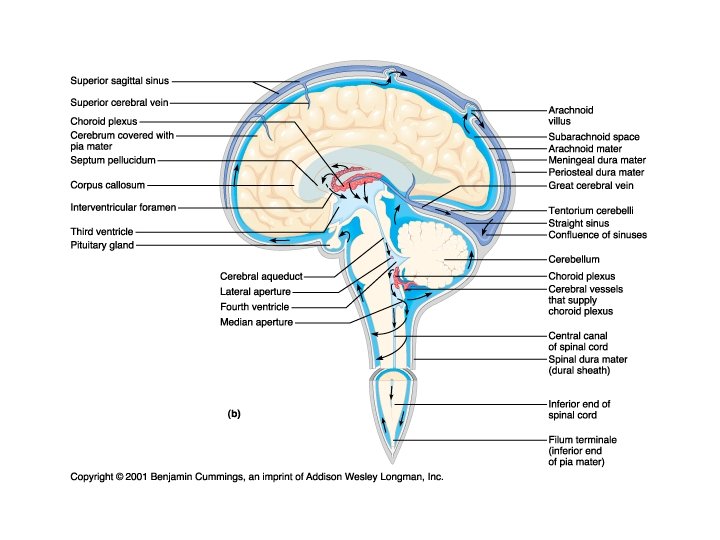
Protection of the CNS • The CNS is protected by bone (skull and vertebral column) , membranes (the meninges), and a watery cushion (cerebrospinal fluid). • Protection from toxins in the blood is provided by the blood-brain barrier.
Protection of the CNS meninges • The three connective tissue membranes that cover and protect the CNS structures.
Protection of the CNS cerebrospinal fluid • A watery broth similar to the make up of the blood plasma from which it is continually formed. • The CSF is formed in clusters of capillaries that hang in the diencephalon called the choroid plexuses. It circulates through the brain ventricles and returns to the blood, constantly draining as new CSF forms, keeping the overall volume and pressure relatively constant
Protection of the CNS blood-brain barrier • The brain is absolutely dependent on a constant internal environment. • It could not function if exposed to the types of fluctuations of hormones, ions, and nutrients that continually occur elsewhere in the body, especially after eating and exercising. • Neurons are kept separate from blood borne substances by the BBB. • Remember how nerve impulses are initiated
Peripheral Nervous System • The PNS consists of nerves and scattered groups of neuronal cell bodies, called ganglia, found outside the CNS
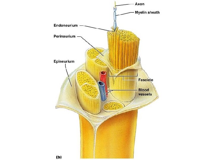
What is a “Nerve”? - nerve structure • A nerve is a a bundle of neuron fibers found outside the CNS. Neuron fibers are wrapped in protective tissue coverings. (which should look very familiar) • Like neurons, nerves are classified according to the direction in which they transmit impulses. (afferent- sensory, efferent-motor, or mixed –both directions)
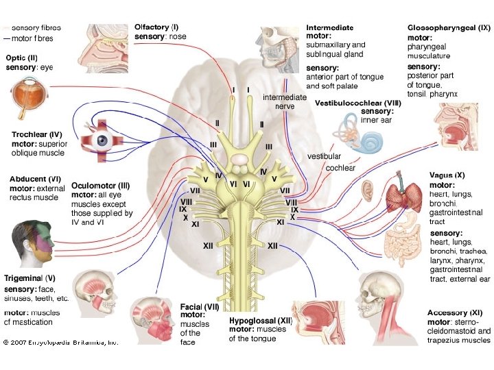
PNS- Cranial Nerves • There are 12 pairs of cranial nerves primarily serve the head and neck. Only one pair extends to the thoracic and abdominal cavities. • The cranial nerves are mostly mixed nerves (except for those associated with special senses) and are numbered in order and given a name that revels their major function.
PNS- Spinal Nerves and Nerve Plexuses • The 31 pairs of spinal nerves are formed by a combination of the ventral and dorsal roots of the spinal cord. • They are named for the region of the cord from which they arise. (Cervical, Thoracic, Lumbar, Sacral) • Almost immediately after they are formed they divide into dorsal and ventral rami (branches)
PNS- Spinal Nerves and Nerve Plexuses • Dorsal Rami serve the posterior body trunk • The Ventral Thoracic Rami serve the anterior and lateral trunk. • The other ventral rami form the 4 nerve plexuses which service the limbs. – Cervical – Brachial – Lumbar – Sacral
PNS- Autonomic Nervous System • The ANS is a division of the PNS that regulates cardiac muscle, smooth muscle and glands automatically. • The ANS is the major force in maintaining homeostasis. • It is divided into two main arms which counterbalance each other. The sympathetic mobilizes the body during extreme situations while the parasympathetic allows us to “unwind”.
Developmental Aspects of the Nervous System • Formed during the first month of embryonic development. • Any maternal infection in early pregnancy can have harmful effects on the nervous system of the fetus.
Developmental Aspects of the Nervous System • Maternal measles- causes deafness and CNS damage. • Radiation • Drugs (alcohol, cocaine, opiates) • Lack of oxygen cause the death of neurons. – Example: smoking decreases amount of oxygen to blood.
Cerebral Palsy • Neuromuscular disability in which voluntary muscles are poorly controlled and spastic. • Can be caused by a temporary lack of oxygen.
Congenital Malformations • Anacephaly- failure of cerebrum to develop • Hydrocephaly- abnormal accumulation of cerebrospinal fluid on brain • Spina bifida- vertebrae form incompletely
Developmental Aspects continued • One of last areas to mature- hypothalamus – Babies have issues regulating body temperature • No more neurons formed after birth, but growth and maturation of nervous system continues through childhood. • Young adult- when brain reaches maximum weight.
Developmental Aspects continued • Over next 60 years neurons are damaged and die. • They cannot reproduce themselves. • However, we can continue to learn through life