Medical Genetics LECTURE Klinefelter Turner Down Syndrome Muhammad


 is the failure of chromosome pairs")


 • Occurrence")
 v Monosomy of sex chromosome (only one X")

,")


 • Sex chromosome: – 47 XXY")

 in Maternal Blood")
 DNA from")

 can produce")

- Slides: 34
Medical Genetics LECTURE Klinefelter, Turner & Down Syndrome Muhammad Faiyaz-Ul-Haque, Ph. D, FRCPath
Lecture Objectives: By the end of this lecture, the students should be able to: • Define non-disjunction and describe its consequences for meiosis and mitosis. • Classify chromosomal abnormalities • Understand the common numerical chromosomal disorders: mono and trisomy • Understand the common numerical sex chromosome disorders: Turner`s & Klinefelter`s syndromes
Stages of Mitosis & Meiosis Mitosis Meiosis
Stages of Meiosis
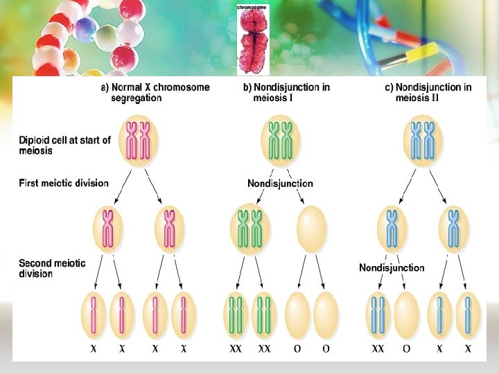
Non-disjunction in Meiosis v Nondisjunction ("not coming apart") is the failure of chromosome pairs to separate properly during meiosis stage 1 or stage 2. v Can affect each pair of chromosomes v Is not a rare event v As a result, one daughter cell has two chromosomes or two chromatids, and the other has none. v The result of this error is a cell with an imbalance of chromosomes (Aneuploidy)
MEIOSIS MITOSIS
Down Syndrome Down syndrome, also called Trisomy 21, is a condition in which extra genetic material causes delays in the way a child develops, both mentally and physically.
Down’s Syndrome § Three copies of chromosome 21
3 copies = trisomy 1 copy = monosomy
Down syndrome, trisomy 21 Karyotype: 47, XY, +21 • The incidence of trisomy 21 rises sharply with increasing maternal age • Most cases arise from non disjunction in the first meiotic division • The father contributing the extra chromosome in 15% of cases (i. e. Down syndrome can also be the result of nondisjunction of the father's chromosome 21) • A small proportion of cases are mosaic and these probably arise from a non disjunction event in an early zygotic division
Down Syndrome ØShort, broad hands ØStubby fingers ØRough skin ØImpotency in males ØMentally retarded ØSmall round face ØProtruding tongue ØShort lifespan
Features of Down Syndrome • • Low muscle tone Head and facial malformations Abnormalities of the extremities Developmental delays Heart malformations Increased risk of infectious disease Early death
Down Syndrome
Turner’s Syndrome Genetic defect in which affected women have only one X chromosome, causing developmental abnormalities and infertility
Turner Syndrome • Monosomy of sex chromosome (only one X chromosome present) • Occurrence – 1 in 2500 live female births
Turner’s syndrome (Monosomy X: 45, XO) v Monosomy of sex chromosome (only one X chromosome present) v Occurring in 1 in 2500 phenotypic females v The only viable monosomy in humans v Characteristics: Webbed neck, Individuals are genetically female, not mature sexually, Sterile, Short stature, Broad chest, Low hairline, Streak ovaries, Normal intelligence, Normal life span
Features of Turner Syndrome • • • Short stature Lack of ovarian development Neck abnormalities Skeletal disorders Increased risk of osteoporosis, cardiovascular constriction, diabetes, and kidney and thyroid problems • No developmental delays • Turner syndrome is commonly treated with growth hormones, and estrogen replacement therapy. • Additionally, reproductive technology can help women with Turner syndrome become pregnant.
XO – Turner Syndrome 96 -98% do not survive to birth Turner Syndrome (XO), Incidence: 1 in 2500 female births
Turner’s Syndrome Cardiovascular Bicuspid aortic valve Coarctation of the aorta Thoracic aortic aneurysm (aortic root dilatation) Skeletal Short stature Short fourth metacarpal/matatarsal bone may be unusually short (+/- short 3 rd and 5 th). Osteoporosis (due to lack of estrogen) Scoliosis Reproductive Women with Turner syndrome are almost universally infertile.
Klinefelter’s Syndrome Klinefelter's Syndrome is a genetic condition that only affects males. The condition is present from birth and is due to an extra X chromosome (genotype XXY)
Klinefelter’s Syndrome 1 in 1, 100 births 47 chromosomes XXY only 47, XXY #23 Trisomy Nondisjunction
Klinefelter Syndrome: 47, XXY males • Male sex organs; unusually small testes which fail to produce normal levels of testosterone breast enlargement (gynaecomastia) and other feminine body characteristic • Patients are taller and thinner than average and may have a slight reduction in IQ but generally they have normal intelligence • No spermatogenesis sterile • Very rarely more extreme forms of Klinefelter syndrome occur where the patient has 48, XXXY or even 49, XXXXY karyotype. These individuals are generally severely retarded. • Klinefelter Syndrome (XXY), Incidence: 1: 1000 male births
Klinefelter’s Syndrome Scarce beard Longer fingers and arms Sterile Delicate skin Low mental ability Normal lifespan Brown spots (nevi)
Features of Klinefelter Syndrome • Tall, sexually underdeveloped & infertile, though in some case testicular function is preserved • Sparse facial and body hair • Delays in speech and motor skills as well as deficits in attention, auditory processing and social skills. • Increased risk of autoimmune disorders, breast cancer, osteoporosis, leg ulcers, depression, and dental problems • Treatment for these problems includes testosterone therapy and assisted learning
Sex chromosome unbalance is much less deleterious 47, XYY May be without any symptoms. Males are tall but normally proportioned. 10 - 15 points reduction in IQ compared to sibs. XXX females It seems to do little harm, individuals are fertile and do not transmit the extra chromosome. They do have a reduction in IQ comparable to that of Kleinfelter's males
When to do a chromosomal test • Prenatal: maternal age>37 yrs; USS changes; Family history Triple test = increased risk • Postnatal: Learning & developmental disability; growth retardation • Infertility: Recurrent miscarriage, primary infertility
Aneuploidy • Autosomal: – Trisomy 21 (Down syndrome) • Sex chromosome: – 47 XXY (Klinefelter syndrome) – 45 X (Turner syndrome)
Rapid Aneuploidy Screening by FISH • • • Available on amniocentesis sample Uncultured amniocytes FISH probes for X, Y, 21 Result in 24 -48 hours Proceed onto full karyotype (11 -14 days)
Cell Free Fetal DNA (cff DNA) in Maternal Blood
Non-invasive prenatal testing-NIPT 1. TEST : Testing of cff (Cell Free Fetal) DNA from maternal blood during pregnancy for trisomy 21 (Down syndrome), Chr. 18 (Edwards syndrome), Chr. 13 (Patau syndrome). Also sex of the fetus is determined. 3. TIMING: > week 10 4. TURNAROUND TIME: < 2 weeks 5. RELIABILITY: > 99% for trisomy 21 6. INDICATIONS: Although NIPT can be performed in every pregnancy, it is especially indicated: • If the triple test or first trimester screening indicates an increased risk • Advanced maternal age • Anxiety for invasive procedures 7. CONTRAINDICATIONS: NIPT is not the test of choice when there is : • Fetal anomalies on ultrasound • Severely elevated NT (nuchal translucency < 3. 5 mm) • A triplet pregnancy, vanishing twin • If NIPT indicates that an abnormality is present that suggests birth defect, further testing will be necessary to confirm this. The next step will most likely be CVS or amniocenteses.
Next-Generation DNA Sequencing Massively parallel chemistry and detection – Faster – Cheaper • Revolutionizing Clinical Molecular Testing Comprehensively evaluates the genes & able to detects all known common and rare disease-causing mutations
Not all chromosomal mutations are harmful. • Polyploidy (extra sets of chromosomes) can produce stronger and larger plants. • Important crop plants are produced this way, including bananas!
Take home message • Chromosome abnormalities can be numerical or structural. • Numerical abnormalities include aneuploidy and polyploidy. • In mono or trisomy, a single extra chromosome is absent or present, usually as a result of non-disjunction in the 1 st or 2 nd meiotic division. • Structural abnormalities include translocations, inversions, deletions, isochromosome & rings.