Valvular heart disease and prosthetic valve Surface anatomy


- Slides: 72

Valvular heart disease and prosthetic valve

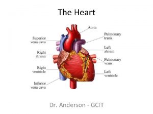
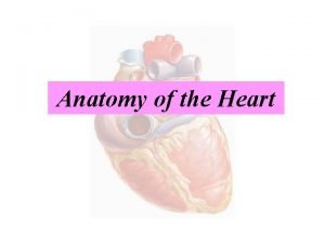
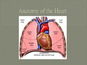
Surface anatomy n MV: behind the Lt ½ of the sternum opp. the 4 th coastal cartilage n AV: behind the Lt ½ of the sternum opp. The 3 rd ICS n TV: behind the Rt ½ of the sternum opp. The 4 th ICS n PV: behind the medial end of the 3 rd LT CC & adjoining part of the sternum


Anatomy n MV: n n n 2 Cusps, Anterior and posterior The Ant is the larger Intervenes bet. A-V and aortic orifice n AV: n 3 semilunar cusps, ant (RT), post. Wall (LT and post) n TV; n 3 cusps, ant, septal , post. n PV; n 3 semilunar cusps one post. (lt) tow ant( ant and rt)

Figure 3. The relationships of the mitral valve are important. Fedak P W et al. Circulation. 2008; 117: 963 -974 Copyright © American Heart Association, Inc. All rights reserved.

Aortic stenosis Aetiology Infants, children, adolescents n n n Congenital aortic stenosis Congenital subvalvular aortic stenosis Congenital supravalvular aortic stenosis Young adults to middle aged Calcification and fibrosis of congenitally bicuspid valve Rheumatic aortic disease Middle aged to elderly Calcification of bicuspid valve Senile degenerative aortic stenosis

AS

AVS tricuspid and bicuspid calcifications


Pathophysiolgy of AS n n Except in the congenital forms, AS develops slowly The LV becomes increasingly hypertrophied, and coronary blood flow may become inadequate n The fixed outflow obstruction limits the increase in C. O required on exercise. n The progressive LV outflow obstruction results in increased LV mass.

Symptoms of AS Exertional dyspnoea n Angina n Pulmonary edema n Exertional syncope n Sudden death n

Signs of AS Ejection systolic murmur n Slow rising carotid pulse n Reduce pulse pressure n LV hypertrophy n Signs of LV failure (crepitations, pulmonary edema) n

Investigations ECG n CXR n ECHO n CATH n

ECG in AS n n LVH with strain (slightly wide QRS in I, III and have increased amplitude) Large S in V 2 and large R in V 6 with T wave inversion in V 6

CXR in AS n AORTIC STENOSIS , dilated ascending aorta, normal heart size

ECHO criteria for assessment of aortic stenosis severity Mean gradient(mmhg) Aortic valve area (cm 2) mild <25 >1. 5 moderate 25 -45 1 -1. 5 severe >45 <1 critical >70 <0. 7

Management n Medical; Medical treatment essentially is reserved for patients who have complications of AS such as heart failure, infective . endocarditis, or arrhythmias n Surgical; The primary management of symptomatic patients with valvular AS is interventional

Aortic regurgitation Aetiology n Congenital n Bicuspid valve, or disproportionate cusps n Acquired n Rheumatic disease n Infective endocarditis n Trauma n Aortic dilatation: marfan syndrome, atheroma, syphilis, ankylosing

pathophysiology n The stroke output of the LV may be doubled or trebled n LV dilated and hypertrophied n In acute AR, The LV poorly accommodates the abrupt increase in end-diastolic volume, and diastolic filling pressure increases rapidly. The rise in LV filling pressure is transmitted to the LA, pulm. veins, and pulm. capillaries, leading to pulm. edema and congestion.

Clinical features symptoms : n Mild AR ; n n asymptomatic palpitations n Severe AR ; n Symptoms of heart failure angina n

Signs of AR n n n Large volume or ‘collapsing’ pulse Bounding peripheral pulses Early diastolic murmur Systolic murmur of increased stroke volume Signs of heart failure

Investigations ECG n CXR n MRI , CT scan n ECHO n CATH n

ECG in AR n LVH with strain (slightly wide QRS in I, III and have increased amplitude Large S in V 2 and large R in V 6 with T wave inversion in V 6 n Left atrial enlargement Left axis deviation n

CXR in AR n n Enlarged thoracic aorta cardiomegaly

ECHO in AR Dilated LV n Hyperdynamic ventricle n Fluttering anterior mitral leaflet n Doppler detects reflux n

Treatment of AR n Medical n n n Vasodilator therapy. Treat asymptomatic patients with chronic severe AR and dilated but normal LV systolic function medically, and monitor their cases for development of indications for AVR. Patients with mild AR and normal LV size require no therapy other than endocarditis prophylaxis The treatment of choice for acute AR is AVR. Medical therapy can be used as a bridge to surgery but should not replace it.

Treatment of AR n Surgical n n Surgical treatment of AR almost always requires replacement of the diseased valve with a prosthetic valve AVR is indicated when AR is beginning to cause sx or when an enlarging heart or progressive ECG changes give evidence of increasing LV overload

Surgical treatment of AR n Asymptomatic patients with evidence of LV systolic dysfunction (EF <0. 50) should undergo AVR. n Asymptomatic patients with severe AR and normal LV function but with severe LV dilatation (enddiastolic dimension >75 mm or end-systolic dimension >55 mm) should undergo AVR. .

Prosthetic heart valve n n n The two main prosthetic valve designs include: mechanical bioprosthetic(tissue) heart valves

Mechanical valves ball and cage bileaflet

Bioprosthetic Valves Aortic homograft n Human tissue valves n autograft n homograft n Animal tissue valves n Heterograft or xenograft

Animal Tissue Valves n The most commonly used animal tissues are: porcine, which is valve tissue from a pig, and bovine pericardial tissue, which is from a cow. n The leaflet valve tissue of the animals is inspected, and the highest quality leaflet tissues are then preserved. They are then stiffened by a tanning solution, most often glutaraldehyde.

Transcatheter Aortic Valve Intervention

Recently, percutaneous valve replacement n has been developed. TAVI is a reasonable alternative to surgical AVR in patients at high surgical risk. In Jordan, few cases were done because of the high cost.

images


How to choose a valve n n Mechanical valve in patients < 65 years. Tissue valves in patients > 65 years Tissue valves in patients whose life expectancy is < 10 year Tissue valve in patients who have problems which are likely to cause life threatening bleeding.

Valve types Bioprosthetic/Tissue No lifetime warfarin Less durability n Mechanical valve Need for warfarin Better durability n


Mitral stenosis n Aetiology n Isolated MS accounts for 25% of all rheum. Heart dis. , and an additional 40% have mixed MS and MR n 2/3 of cases occurs in women n Acquired MS is almost entirely rheum. in origin

Aetiology of MS n Acquired MS results from long-term damage to the mitral valve and its supporting structures. : n In rheumatic heart disease SLE Amyloidosis Postsurgical acquired MS, such as MS occurring after mitral valve annuloplasty for severe MR. n n n


Sever MS FISH MOUTH (RHD) MS

Pathophysiology of MS n The normal adult mitral valve orifice crosssectional area is 4 -6 cm 2. n When reduced to 2 cm 2, hemodynamically significant MS occurs. WHEN <1 cm 2 it is critical n As a compensating mechanism, pulmonary vasoconstriction develops, causing pulmonary hypertension. n Severe MS results in decreased cardiac

MS Pathophysiology Progressive Dyspnea (70%): LA dilation pulmonary congestion (reduced emptying) worse with exercise, fever, tachycardia, and pregnancy n n Increased Transmitral Pressures: Leads to left atrial enlargement and atrial fibrillation. Right heart failure symptoms: due to n Pulmonary venous HTN Hemoptysis: due to rupture of bronchial n vessels due to elevated pulmonary pressure n

Signs of MS n n AF Loud 1 st heart sound, opening snap, mid-diastolic murmur Signs of raised pulm capillary pressure (crepitations, pul edema, effusions) Signs of pul HTN.

Investigations of MS n ECG n LA hypertrophy if not in AF n Left atrial enlargement is illustrated by increased P wave duration in lead II, top ECG, and by the prominent negative P terminal force in lead V 1, bottom tracing

Investigations of MS n ECG n RVH

CXR n Chest radiograph of a patient with mitral stenosis shows pulmonary hypertension, mild cardiomegaly and enlargement of the left atrium (arrow) and pulmonary artery

ECHO n n n Thickened immobile cusps Reduced rate of diastolic filling Reduced valve area


Treatment of MS medical n n Asymptomatic patients with mild MS require yearly follow-up For the patient with signs or symptoms of CHF, diuretics may provide benefit RX of Tachyarrhythmias Electrophysiologic ablation of atrial fibrillation or flutter circuits may be performed in the catheterization laboratory

Percutaneous mitral balloon valvuloplasty n n Indications for this procedure are similar to those for surgery, including CHF unresponsive to medical management n asymptomatic patients with a pulmonary artery (PA) systolic pressure of 50 mm Hg or greater. n In some centers, the procedure is successful in 80 -90% of selected cases. The procedural mortality rate is 1 -2%.

Treatment surgical n Indications: n Symptomatic mitral stenosis especially if peripheral emboli Mitral valve area less than 1 cm 2 n n Mitral valvotomy n Commissurotomy consists of an incision of fused mitral valve commissures and shaving of thickened mitral valve leaflets n Fused chordae tendineae and papillary muscles can be divided to relieve subvalvular stenosis. n Supravalvular tissue contributing to the MS should be resected.

Treatment surgical n Mitral valve replacement with mechanical valve or bioprosthesis


MVR

Mitral Regurgitation n Aetiology n Acute MR : n Ruptured chordae or papillary muscle due to acute myocardial infarction or trauma n Perforation of the mitral valve leaflet n Acute failure of a prosthetic valve

Mitral Regurgitation Aetiology n Chronic MR : n n n Mitral valve prolapse Rheumatic heart disease Coronary artery disease Connective-tissue disorder Prosthetic valves

MR

Pathophysiology n In chronic MVR, the distensibility of the LA and LV are increased over time. n This dilatation of the left atrium decreases left atrial pressures, thus increasing preload. n The left ventricle dilatates and, hypertrophied generates a larger stroke volume without a significant rise in wall stress.

CLINICAL Symptoms n Acute MR n Sx of acute pulm edema and reduced CO n Chronic progressive MR n Exertional dyspnea, nocturnal dyspnea, palpitations(AF, atrial flutter, increased stroke volume) Sx of pulm edema Sx of diminished CO Sx of right sided HF n n n

Signs of MR n n n AF/ Flutter Cardiomegaly- displaced hyperdynamic apex beats Apical systolic murmur, thrill Signs of raised pulm capillary pressure (crepitations, pulm edema, effusions) Signs of pulm HTN

Investigations for MR n ECG n LAH (if not in AF) LVH n CXR n n Enlarged LA, LV Signs of pulm venous HTN Signs of pulm edema if acute n ECHO n n n Dilated LA, LV Dynamic LV(UNLESS AF PREDOMINATE) Regurgitation detected on Doppler

CXR MR n n n Marked cardiomegaly Pulm venous HTN LA appendage enlargement

TREATMENT of MR Medical Any patient with acute or chronic mitral valve regurgitation with hemodynamic compromise should be evaluated for acute myocardial infarction. n Afterload-reducing agents n If atrial fibrillation is encountered, digitalis therapy is considered n Prophylactic antibiotics are administered prior to any interventional treatment

TREATMENT OF MR SURGICAL n Indications for surgical Intervention n Acute MR with congestive heart failure or cardiogenic shock n Acute endocarditis n Class III/IV symptoms (ie, patient symptomatic while at rest or with minimal activity)

MITRAL RECONSTRUCTIVE SURGERY REPAIR TECHNIQUES LEVEL MANEUVER ANNULUS LEAFLETS REDUCTION CHORDS RESECTION SHORTENING TRANSPOSITION REPLACEMENT COMMISSURES SPLITTING RESECTION PAPPILARY MUSCLES SPLITTING SHORTENING REPOSITIONING RESECTION ENLARGEMENT


 Pathophysiology of valvular heart disease
 Mitral stenosis severity
 Upper lobe diversion
 Causes of valvular heart disease
 Pathophysiology of valvular heart disease
 Pathophysiology of valvular heart disease
 Pathophysiology of valvular heart disease
 Causes of valvular heart disease
 Site:slidetodoc.com
 Servo valve types
 Fibroserous
 Trabeculae carneae
 Traslape valvular
 Communicable disease and non communicable disease
 Prosthetic group
 Artificial limb
 Prosthetic group
 Prosthetic
 Classification of prosthodontics
 Prosthetic group example
 Nicks procedure
 Ileocecal reflex
 Cyanotic vs acyanotic
 Heart disease and stroke are the world's biggest killers
 Pulmonary valve sheep heart
 Heart mechanical valve
 Endocardium sheep heart
 Anatomy and physiology of gastritis
 Heart disease symptoms
 Randi sokol
 Ronaldo pictures
 Tetralogy of fallot murmur
 Tga cxr
 Heart disease cat
 Heart disease data
 Ie
Ie 5 terrible ts
 Pathophysiology of ischemic heart disease
 Rheumatic heart disease
 Congenital heart disease pda
 Fish mouth buttonhole stenosis
 Rhuematic fever
 Coronary heart disease
 Ischemic heart disease classification
 Strepsore
 Rheumatic heart disease causes
 Heart disease
 Lvh criteria
 Nyha class
 Chronic rheumatic heart disease
 Curved surface area and total surface area of cone
 Formula for surface area of prism
 Base and apex of the heart
 Reinforcement: heart anatomy
 Sheep heart diagram
 Anatomy of the human heart
 Base of the heart
 Correctly label the following internal anatomy of the heart
 Upper border of heart
 Borders of heart
 Gross anatomy of heart
 Applied anatomy of heart
 Types of blood vessels
 Name the valve
 Cardiac anatomy
 O que é punção capilar
 Surface anatomy
 Lung surface anatomy
 Costal pleura
 Root of lung anatomy
 Surface anatomy lower limb
 Normal liver span
 Subcostal plane
