THE MUSCULAR SYSTEM You got tickets Naming Muscles


 controlled")

 controlled • Microscopically the")


")


 1. Nerve impulse travels")


")


 Fast Twitch (Type 2) Amt. of")


 on a muscle stays")
 on a muscle increases")

")
")
")
")
")
")


")
")


- Slides: 70
THE MUSCULAR SYSTEM “You got tickets…? ”
Naming Muscles 1. Location – structure near which the muscle is found Frontalis Frontal bone Femoris Femur Gluteus Posterior of hip/thigh Oculi Eye Oris Mouth Radialis Radius Ulnaris Ulna Brachialis Arm
Naming Muscles 2. Function – body movement when the muscle is contracted Abductor Moves part away from body Adductor Moves part toward body Depressor Lowers a part Extensor Extends a part Flexor Flexes a part Levator Elevates a part Rotator Rotates a part
Naming Muscles 3. Shape – relative shape of the muscle Deltoid Shaped like delta ∆ Orbicularis Circular Serratus Saw-toothed Quadratus Square Rhomboideus Diamond-shaped Trapezius Trapezoidal
Naming Muscles 4. Size – relative size of the muscle Brevis Short Longus Long Magnus Large Maximus Largest Medius Moderately sized Minimus Smaller
Naming Muscles 5. Fiber direction – relative to the midline Oblique Diagonal to midline Rectus Parallel to midline Sphincter Circling an opening Transversus Right angle to midline
Naming Muscles 6. Number of heads/origins Bicep Two heads Tricep Three heads Quadricep Four heads
Naming Muscles Muscle names can be any combination of the options listed!
Practice • How are the following muscles named? 1. Biceps femoris 2. Flexor carpi ulnaris 3. Gluteus maximus 4. Abductor magnus 5. Rhomboid major
Answers • How are the following muscles named? 1. Biceps femoris - # of heads and location 2. Flexor carpi ulnaris – action and location 3. Gluteus maximus – location and size 4. Abductor magnus – action and size 5. Rhomboid major – shape and size
Functions of the Muscular System • “Transform energy into motion” • i. e. Produce movement! Of all types! Examples: 1. Speaking 2. Facial expression 3. Propel blood through blood vessels 4. Bring air into and out of lungs 5. Digest food 6. Produce body heat
Muscular System • Muscles are responsible for all types of body movement • 3 basic muscle types are found in the body • Skeletal muscle • Smooth muscle • Cardiac
Cardiac Muscle • Makes up the myocardium of the heart • Involuntarily (unconsciously) controlled • Microscopically the tissue appears striated • Cells are short, branching, and have a single nucleus • Cells connect to each other at intercalated discs
Smooth Muscle • Makes up the walls of organs and blood vessels • Involuntarily (unconsciously) controlled • Microscopically the tissue appears non-striated • Cells are short, spindle-shaped, and have a single nucleus • Tissue is extremely extensible while still retaining its ability to contract
Skeletal Muscle • Attached to the skeleton • Voluntarily (consciously) controlled • Microscopically the tissue appears striated • Cells are long, cylindrical, & multi-nucleated
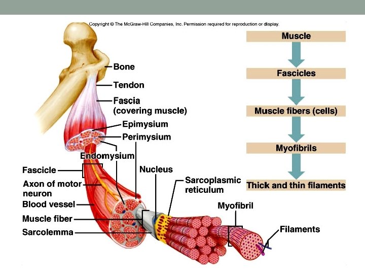
Muscle Attachment • Sites of muscle attachment • Bones • Cartilage • Connective tissue coverings • Muscle fibers blend into a connective tissue attachment • Tendon – cordlike structure that attaches the muscle to bone • Aponeurosis – sheet-like structure
Characteristics of Muscle Tissue 1. Excitability - can receive and respond to stimulation 2. Contractility - can shorten to exert a pull 3. Extensibility - can lengthen 4. Elasticity - returns to original length after contraction
Muscle Fiber Arrangement Parallel Convergent Pennate Bipennate Sphincter
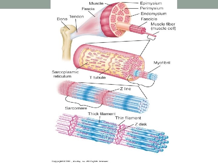
Muscle Fiber – individual muscle cell Endomysium – connective tissue that surrounds each muscle fiber Fascicle – group of muscle fibers Perimysium – connective tissue that surrounds each fascicle Epimysium – connective tissue that surrounds entire skeletal muscle Fascia – connective tissue that surrounds entire skeletal muscle (over the epimysium) and eventually forms the tendon
Structure of Skeletal Muscle epimysium tendon perimysium Muscle Fascicle Skeletal muscle Surrounded by epimysium Surrounded by perimysium endomysium Skeletal muscle fiber (cell) Surrounded by endomysium
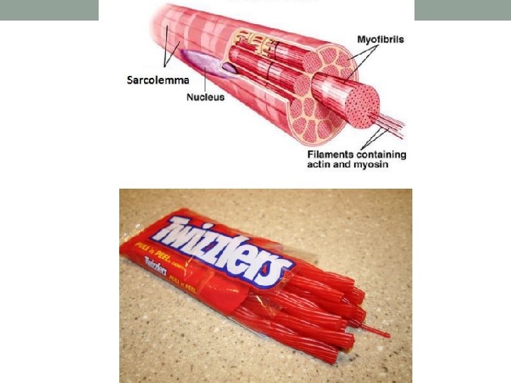
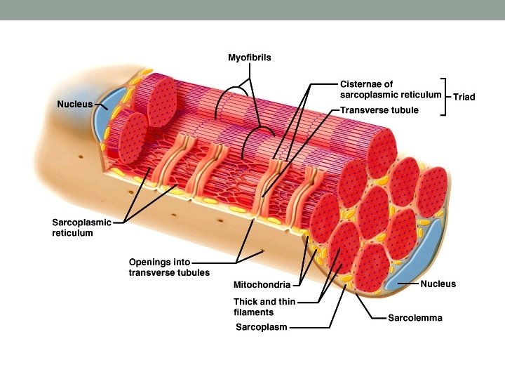
Sarcolemma – cell membrane Sarcoplasm - cytoplasm Myofibrils – long protein bundles, store glycogen and O 2 Sarcoplasmic Retiuclum – smooth ER, surrounds each myofibril, stores Ca+2 Myofilaments – Thick and thin filaments that make up a myofibril T-tubules – allow electrical impulses to travel into cell Actin Thin filament Myosin Thick filament
Skeletal Muscle Contraction • Role of Myosin and Actin • Myosin consists of two twisted strands with cross-bridges projecting outward
Skeletal Muscle Contraction • Role of Myosin and Actin • Actin is a protein with binding sites that the myosin cross-bridges attach to • Contains 2 proteins – troponin and tropomyosin
Skeletal Muscle Contraction
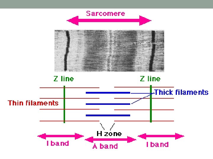
Skeletal Muscle Fibers • The sarcoplasm of each muscle fiber/muscle cell contains myofibrils (rod-like units of muscle) • Myofibrils are separated into compartments called sarcomeres that contain thick filaments and thin filaments • Thick filaments – made of protein called myosin • Thin filaments – made of protein called actin • The organization of these thick and thin filaments produces striations.
Skeletal Muscle Fibers • A sarcomere extends from Z line to Z line • I bands (light bands) are anchored to the Z lines and are length of actin filaments • A bands (dark bands) are the length of myosin filaments
Z line I band Z line A band Length of actin (thin, light stripes) Length of myosin heads (wide, dark stripes)
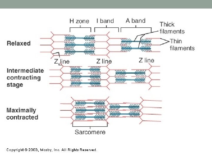
Sliding Filament Theory
Sliding Filament Theory • Myosin heads attach to actin molecules at a binding site • Cross-bridge bends and pulls actin, causing the actin filament to slide across the myosin, toward the center of the sarcomere, causing it to shorten (Z lines move closer together). • Myosin releases… attaches to new binding site and pulls again • Energy is provided from the conversion of ATP to ADP • Releases a ton of energy!! • Energy is needed to properly position myosin cross-bridge so that it can bind with actin • Cycle of “grab and release, grab and release” will continue as long as ATP is available
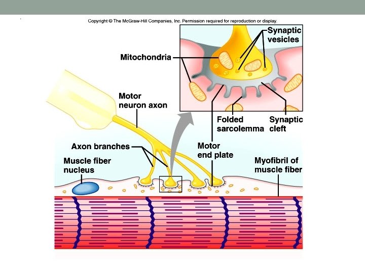
Neuromuscular Junction • Motor neurons – the cells that cause muscle fibers to contract • What type of cells are these? What do they look like? • Root word “Neuro”? • The axon of the motor neuron extends outward from the brain or spinal cord. The muscle fiber will only contract when stimulated by the motor neuron. (motor neuron)
Neuromuscular Junction • Neuromuscular Junction - The site where the motor neuron and muscle fiber meet • Synaptic Cleft – Small gap between the motor neuron and the muscle fiber • Neurotransmitter – chemical messenger • Relays the messages from the motor neuron to the muscle fiber • Crosses the synaptic cleft
Neuromuscular Junction • A motor neuron and the muscle fibers that it controls make up a motor unit • A motor neuron may connect to as few as 4 muscle fibers and as many as 1000 s of muscle fibers • When stimulated to do so, the muscle fibers of the motor unit contract all at once
Stimulus for Contraction In skeletal muscle, neurotransmitter = Acetylcholine (ACh) 1. Nerve impulse travels down motor neuron to the neuromuscular junction, causing ACh to be released from synaptic vesicles 2. ACh crosses the synaptic cleft and binds to Ach receptors on the motor end plate which generates a muscle impulse 3. Muscle impulse travels down the T-tubule (through the muscle fiber) and eventually reaches the sarcoplasmic reticulum, causing calcium ions to be released.
Stimulus for Contraction 4. Calcium ions bind to troponin and change its shape and the position of tropomyosin is altered, allowing binding sites to be exposed 5. Myosin heads bind to the actin and form crossbridge, pulling the actin filament across the myosin filament and causing the sarcomere to shorten 6. New ATP binds to myosin, which cause actin and myosin to break apart 7. ATP splits into ADP and Pi (phosphate), which provides energy for the myosin head to go back to original position
Relaxation 1. 2. 3. Acetylcholinesterase – rapidly decomposes Ach remaining in the synapse Muscle impulse stops Stimulus to sarcolemma and muscle fiber membrane ceases 4. Calcium moves back into sarcoplasmic reticulum 5. Myosin and actin binding prevented 6. Muscle fiber relaxes
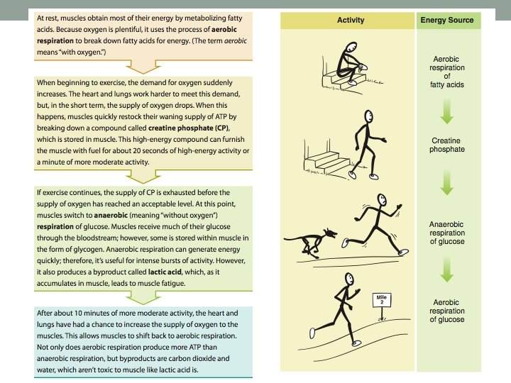
Energy Sources for Contraction • Energy for contraction comes from ATP molecules • Muscle cells have a limited amount of ATP • A couple seconds of activity will deplete the ATP in a muscle fiber • ATP must constantly be made available So where does it come from? !?
Where does ATP come from? Source/Proc Molecule that is ess broken down When the process is used Effect on the body Aerobic Respiration (Cellular Respiration) Fatty acids Glucose When oxygen is present Nothing major – ATP is released, with extra CO 2 and H 2 O Utilization of Creatine Phosphate When oxygen supply first drops 20 sec high energy 1 min moderate energy Anaerobic Respiration Glucose When there is not enough oxygen Produces lactic acid present and CP which can lead to muscle has been fatigue exhausted
Oxygen Debt • During rest or moderate activity, there is enough oxygen to supply the muscle • Cells can use aerobic respiration to produce energy • Nothing bad comes out of it – just CO 2, H 20, and energy! • During strenuous activity, oxygen deficiency may develop • Cells have to use anaerobic respiration to produce energy • Lactic acid is a product – causes muscle fatigue
Oxygen Debt • The liver will attempt to convert the lactic acid back into glucose, however, during strenuous activity, more lactic acid is being created than the liver can keep up with • As a result, lactic acid builds up in the muscle tissue • When lactic acid accumulates in the muscle, the muscle loses its ability to contract, which is referred to as fatigue. • Following strenuous exercise, the liver continues to work to convert the lactic acid into glucose. However the process requires a lot of oxygen. The amount of oxygen required is known as oxygen debt. • Leads to breathing heavily following a hard workout
Skeletal Muscle Actions • Origin – muscle attachment that remains fixed (does not move) • Insertion – muscle attachment that moves on contraction • Action – body movement that a muscle produces • Flexion, extension, abduction, adduction, etc.
Does NOT move when muscle contracts hen w ove racts m t s Doe cle con mus
Muscles Working Together • For muscles to create movement, they can only pull • Muscles in the body rarely work alone & are usually arranged in groups surrounding a joint • Agonist/Prime mover – a muscle that contracts to perform the desired action • Synergist - A muscle that helps the agonist • Antagonist - A muscle that opposes the action of the agonist, therefore undoing the desired action
Muscle Growth and Size • Hypertrophy – enlargement of muscle • Due to strenuous activity • More actin and myosin filaments are formed (NOT muscle fibers!) • Muscle fiber diameter increases in size • Atrophy – decrease in muscle size • With lack of exercise • Loss of actin and myosin filaments • Muscle fiber diameter decrease in size
Types of Muscle Fibers Slow Twitch (Type 1) Fast Twitch (Type 2) Amt. of Mitochondria Lots Less Blood Supply Good (aerobic activity) Poor (anaerobic activity) Speed of Contraction Slow Fast Time until Fatigue Long time (longer to fatigue) Short time (fatigue easily) Type of Athlete/Sport Endurance based (Marathon runners) Speed/Strength based (Weight lifters/Sprinters)
Muscle Fiber Types and Performance • Power Athletes • Sprinters • Have a high % of fast fibers • Endurance Athletes • Distance runners • Have a high % of slow fibers • Others • Nonathletes • 50% slow fibers and 50% fast fibers
Types of Muscle Contractions • When skeletal muscles contract, they may produce two types of contractions: • Isotonic Contraction • Isometric Contraction
Types of Muscle Contractions Isotonic Contraction • The tension (load) on a muscle stays constant during movement (a) Muscle contracts with force greater than resistance and shortens (concentric contraction) • Length of muscle changes, usually resulting in movement of a joint Movement • Two Types • Concentric – shortening a contraction • Eccentric – lengthening a contraction (b) Muscle contracts with force less than resistance and lengthens (eccentric contraction)
Types of Muscle Contraction Isometric Contraction • The tension (load) on a muscle increases • Length of muscle does not change • Example: holding a baby at arms length, pushing against a closed door • Necessary in everyday life to counteract effects of gravity • Example: postural muscles keeping head up
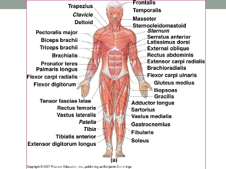
Muscles of the Head, Face, and Neck Figure 7 -12(a)
Muscles of the Head, Face, and Neck Figure 7 -12(c)
Muscles of the Chest, Back, and Shoulder Figure 7 -17(a)
Muscles of the Chest, Back, and Shoulder Figure 7 -18(a)
Muscles of the Chest, Back, and Shoulder Figure 7 -17(b)
Muscles of the Chest, Back, and Shoulder Figure 7 -18(b)
Muscles of the Arm and Forearm Figure 7 -19
Muscles of the Hip, Leg, Foot
Muscles of the Hip, Leg, Foot Figure 7 -22(a)
Muscles of the Hip, Leg, Foot Figure 7 -22(b)
Muscles of the Hip, Leg, Foot
Muscles of the Hip, Leg, Foot