Chest Cases 1 25 Case directory 1 2

- Slides: 117
Chest Cases 1 -25
Case directory 1 2 3 4 5 6 7 8 9 10 11 12 13 14 15 16 17 18 19 20 21 22 23 24 25
Case 1
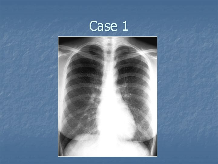
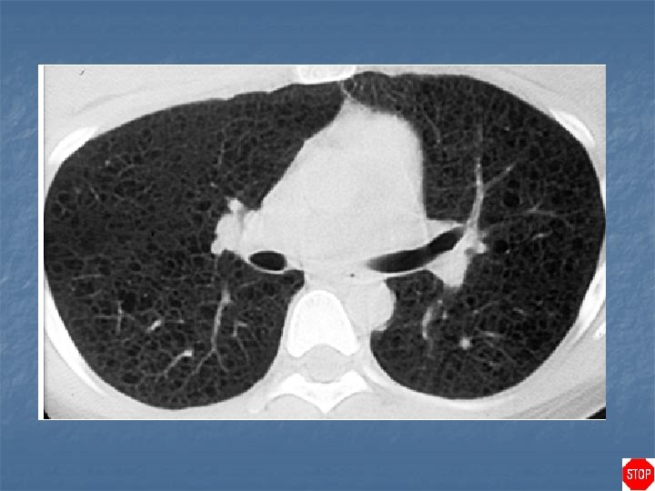
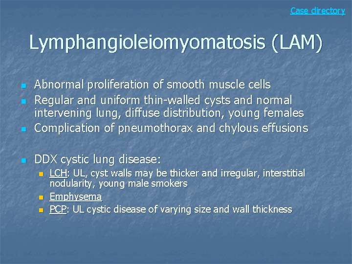
Case directory Lymphangioleiomyomatosis (LAM) n Abnormal proliferation of smooth muscle cells Regular and uniform thin-walled cysts and normal intervening lung, diffuse distribution, young females Complication of pneumothorax and chylous effusions n DDX cystic lung disease: n n n LCH: UL, cyst walls may be thicker and irregular, interstitial nodularity, young male smokers Emphysema PCP: UL cystic disease of varying size and wall thickness
Case 2
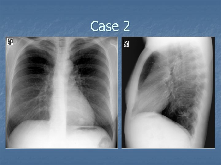
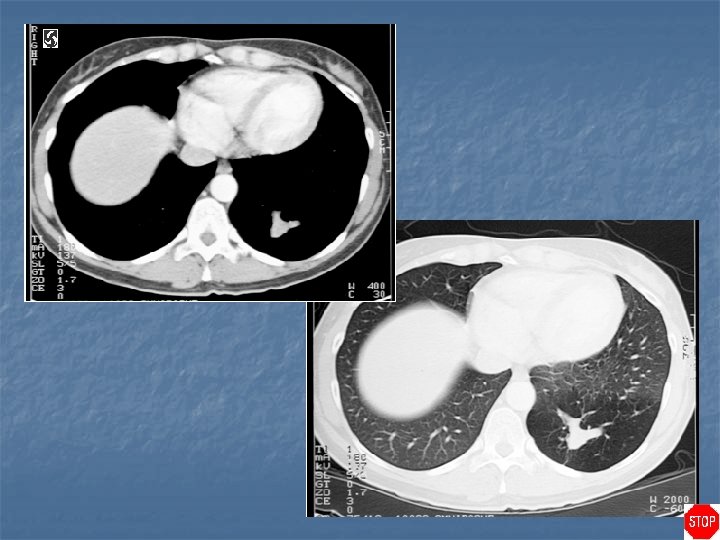
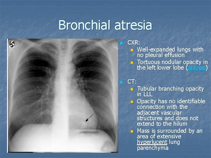
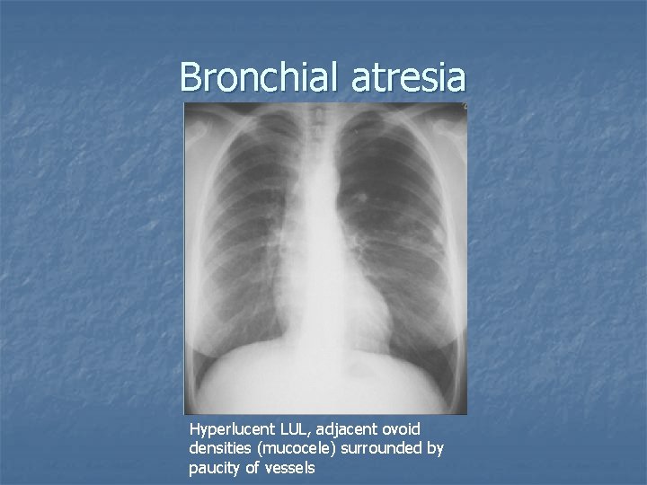
Bronchial atresia n n CXR: n Well-expanded lungs with no pleural effusion n Tortuous nodular opacity in the left lower lobe (arrows) CT: n Tubular branching opacity in LLL n Opacity has no identifiable connection with the adjacent vascular structures and does not extend to the hilum n Mass is surrounded by an area of extensive hyperlucent lung parenchyma
Bronchial atresia n n n Segmental bronchus does not communicate with central airways Sequelae of vascular insult to the lung parenchyma during early fetal development Congenital malformation of the bronchial tree 2 nd MC bronchopulmonary malformation after pulmonary sequestration MC asymptomatic MC apical posterior segment of LUL, next LLL and RML
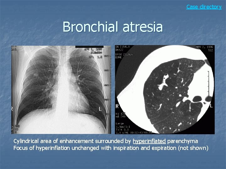
Bronchial atresia n Classic: Branching, tubular mass (mucoid impaction of a bronchus) surrounded by hyperinflated lung with decreased vascular markings n CT: segmental emphysematous changes distal to mucoid impacted bronchus n
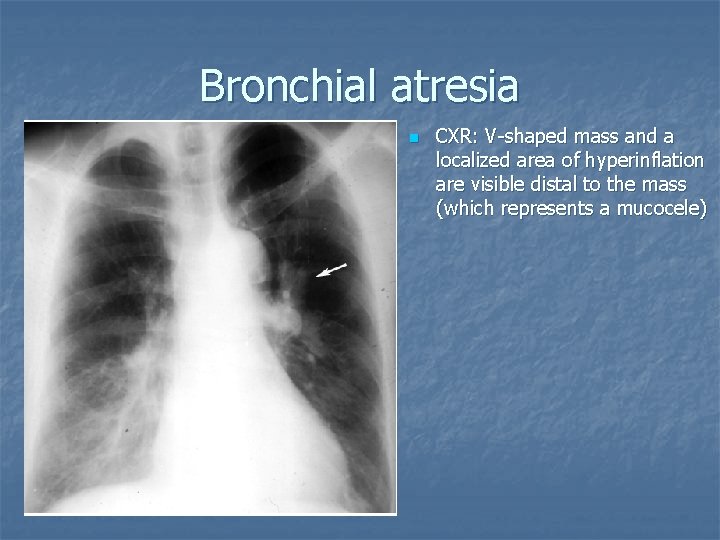
Bronchial atresia n CXR: V-shaped mass and a localized area of hyperinflation are visible distal to the mass (which represents a mucocele)
Bronchial atresia Hyperlucent LUL, adjacent ovoid densities (mucocele) surrounded by paucity of vessels
Case directory Bronchial atresia Cylindrical area of enhancement surrounded by hyperinflated parenchyma Focus of hyperinflation unchanged with inspiration and expiration (not shown)
Case 3
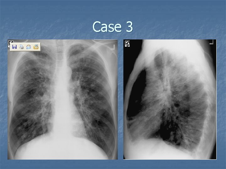
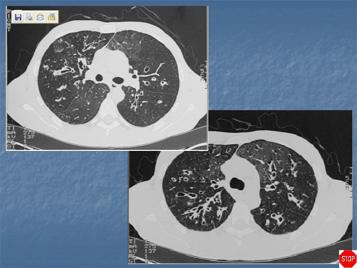
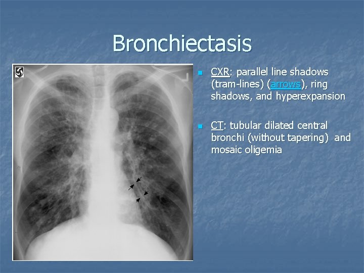
Bronchiectasis n n CXR: parallel line shadows (tram-lines) (arrows), ring shadows, and hyperexpansion CT: tubular dilated central bronchi (without tapering) and mosaic oligemia
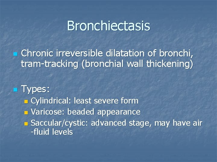
Bronchiectasis n n Chronic irreversible dilatation of bronchi, tram-tracking (bronchial wall thickening) Types: Cylindrical: least severe form n Varicose: beaded appearance n Saccular/cystic: advanced stage, may have air -fluid levels n
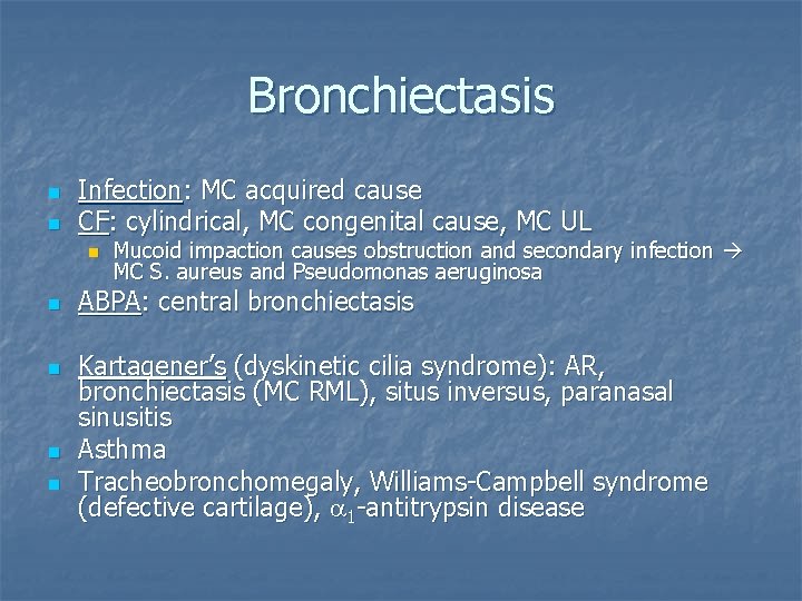
Bronchiectasis n n Infection: MC acquired cause CF: cylindrical, MC congenital cause, MC UL n n n Mucoid impaction causes obstruction and secondary infection MC S. aureus and Pseudomonas aeruginosa ABPA: central bronchiectasis Kartagener’s (dyskinetic cilia syndrome): AR, bronchiectasis (MC RML), situs inversus, paranasal sinusitis Asthma Tracheobronchomegaly, Williams-Campbell syndrome (defective cartilage), a 1 -antitrypsin disease
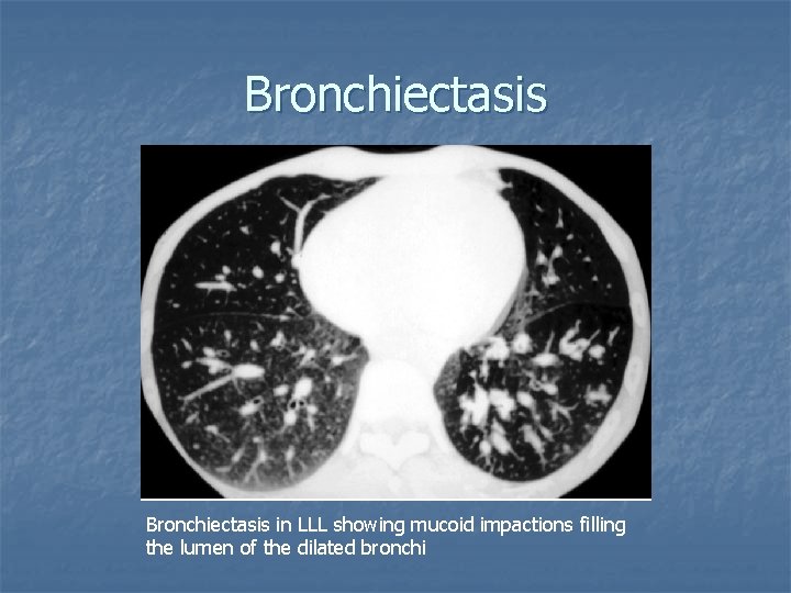
Bronchiectasis in LLL showing mucoid impactions filling the lumen of the dilated bronchi
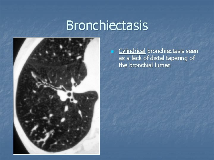
Bronchiectasis n Cylindrical bronchiectasis seen as a lack of distal tapering of the bronchial lumen
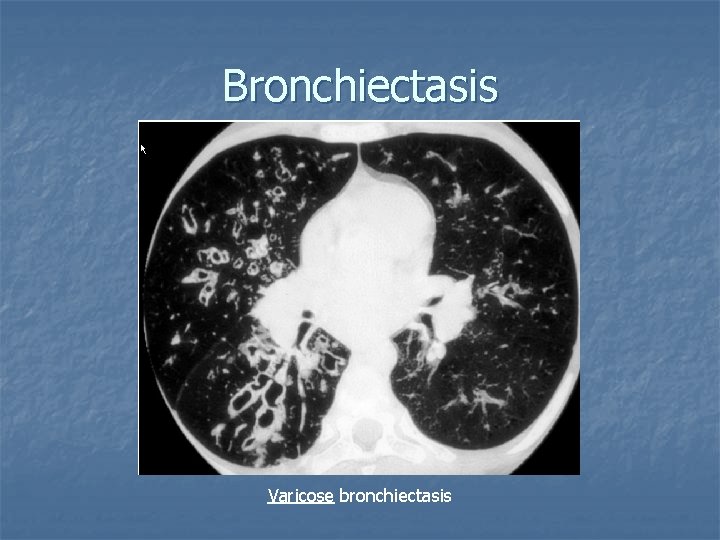
Bronchiectasis Varicose bronchiectasis
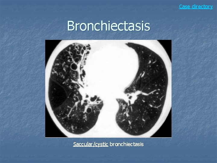
Case directory Bronchiectasis Saccular/cystic bronchiectasis
Case 4
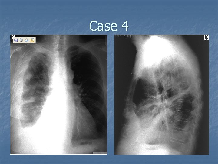
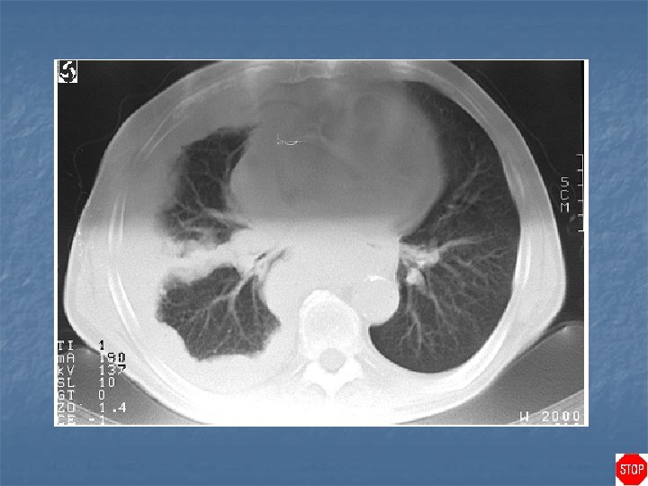
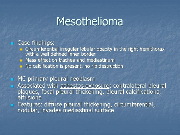
Mesothelioma n Case findings: n n n Circumferential irregular lobular opacity in the right hemithorax with a well defined inner border Mass effect on trachea and mediastinum No calcification is present, no rib destruction MC primary pleural neoplasm Associated with asbestos exposure: contralateral pleural plaques, focal pleural thickening, pleural calcifications, effusions Features: diffuse pleural thickening, circumferential, nodular, invades mediastinal surface
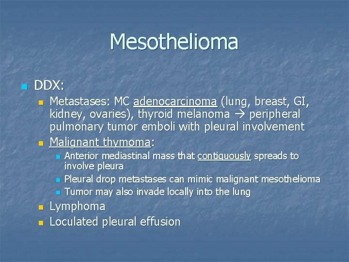
Mesothelioma n DDX: n n Metastases: MC adenocarcinoma (lung, breast, GI, kidney, ovaries), thyroid melanoma peripheral pulmonary tumor emboli with pleural involvement Malignant thymoma: n n n Anterior mediastinal mass that contiguously spreads to involve pleura Pleural drop metastases can mimic malignant mesothelioma Tumor may also invade locally into the lung Lymphoma Loculated pleural effusion
Case directory Mesothelioma
Case 5
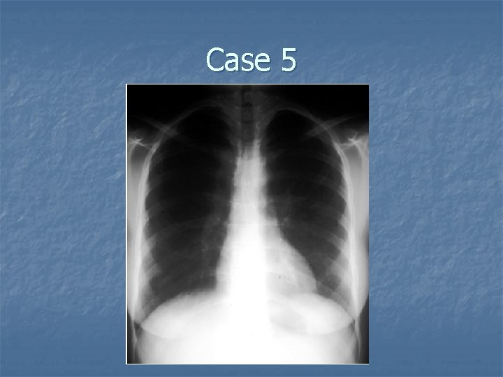
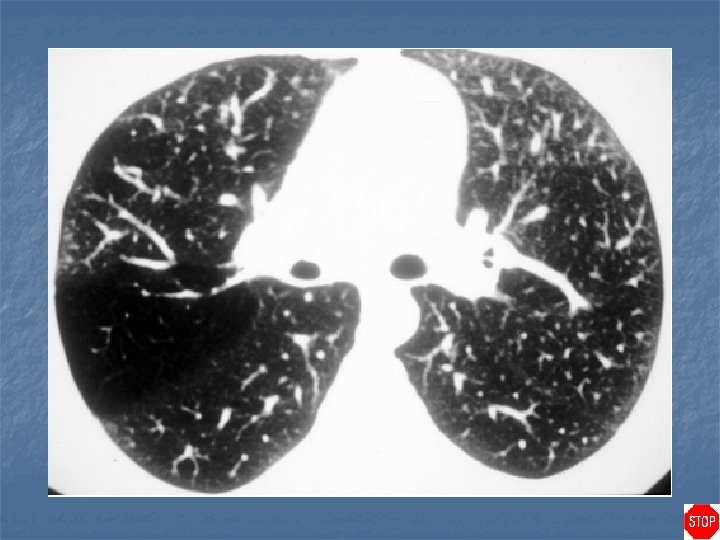
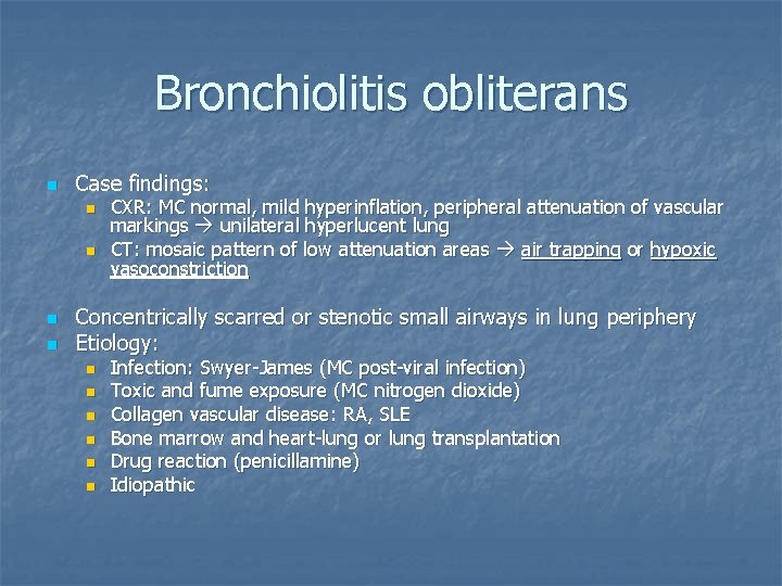
Bronchiolitis obliterans n Case findings: n n CXR: MC normal, mild hyperinflation, peripheral attenuation of vascular markings unilateral hyperlucent lung CT: mosaic pattern of low attenuation areas air trapping or hypoxic vasoconstriction Concentrically scarred or stenotic small airways in lung periphery Etiology: n n n Infection: Swyer-James (MC post-viral infection) Toxic and fume exposure (MC nitrogen dioxide) Collagen vascular disease: RA, SLE Bone marrow and heart-lung or lung transplantation Drug reaction (penicillamine) Idiopathic
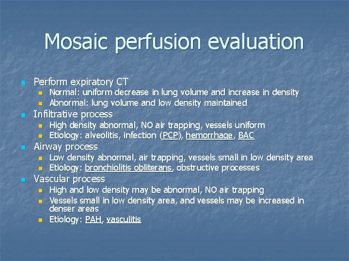
Mosaic perfusion evaluation n Perform expiratory CT n n n Infiltrative process n n n High density abnormal, NO air trapping, vessels uniform Etiology: alveolitis, infection (PCP), hemorrhage, BAC Airway process n n n Normal: uniform decrease in lung volume and increase in density Abnormal: lung volume and low density maintained Low density abnormal, air trapping, vessels small in low density area Etiology: bronchiolitis obliterans, obstructive processes Vascular process n n n High and low density may be abnormal, NO air trapping Vessels small in low density area, and vessels may be increased in denser areas Etiology: PAH, vasculitis
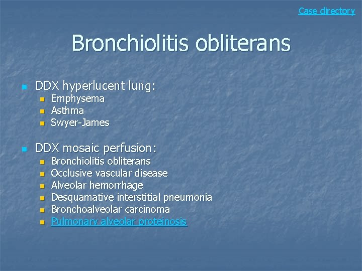
Case directory Bronchiolitis obliterans n DDX hyperlucent lung: n n Emphysema Asthma Swyer-James DDX mosaic perfusion: n n n Bronchiolitis obliterans Occlusive vascular disease Alveolar hemorrhage Desquamative interstitial pneumonia Bronchoalveolar carcinoma Pulmonary alveolar proteinosis
Case 6
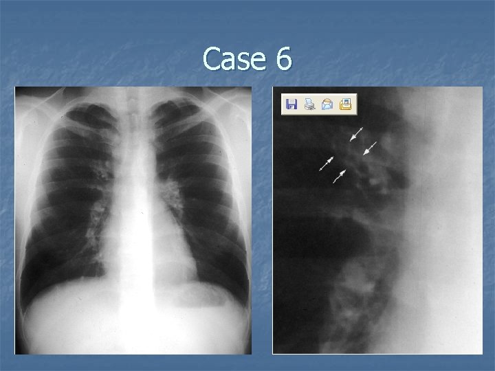
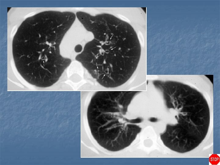
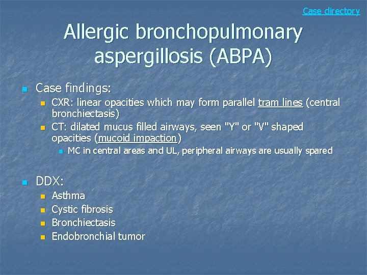
Case directory Allergic bronchopulmonary aspergillosis (ABPA) n Case findings: n n CXR: linear opacities which may form parallel tram lines (central bronchiectasis) CT: dilated mucus filled airways, seen "Y" or "V" shaped opacities (mucoid impaction) n n MC in central areas and UL, peripheral airways are usually spared DDX: n n Asthma Cystic fibrosis Bronchiectasis Endobronchial tumor
Case 7
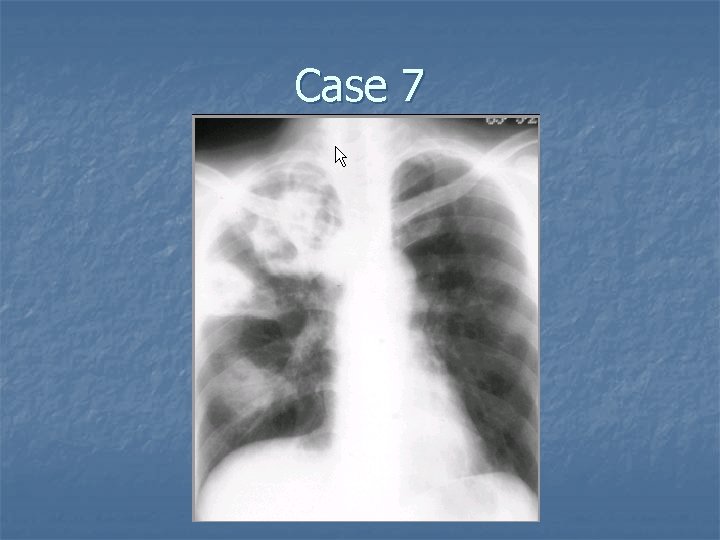
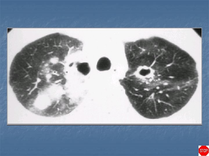
Wegener's granulomatosis n n Autoimmune condition involving the upper and lower respiratory tract and kidneys Vasculitis involving the medium-sized vessels, with formation of granulomas and necrosis of lung parenchyma Lung abscess: development of a cavity within preexisting consolidation Features: n n n Mid and lower zone opacities, which are diffuse, and both alveolar and interstitial Nodules, which may cavitate CT: diffuse, ill-defined perivascular opacities
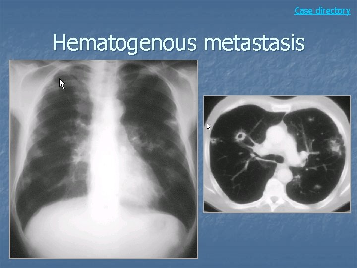
DDX cavitary lesion n n Infection: bacterial abscess Neoplasm: squamous cell, hematogenous metastasis Vasculitis: Wegener’s granulomatosis Granulomatosis: TB, sarcoidosis Septic pulmonary emboli
Case directory Hematogenous metastasis
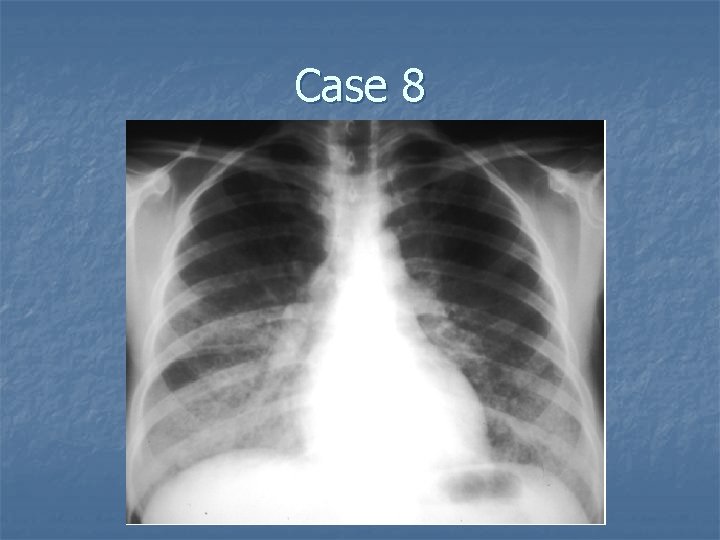
Case 8
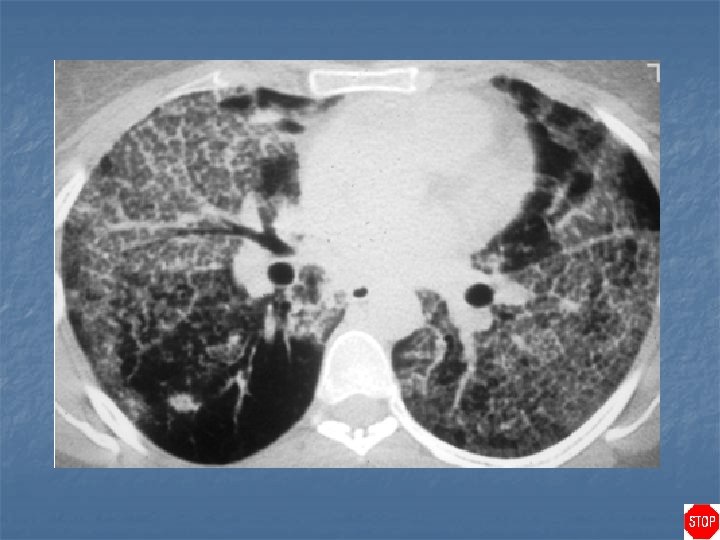
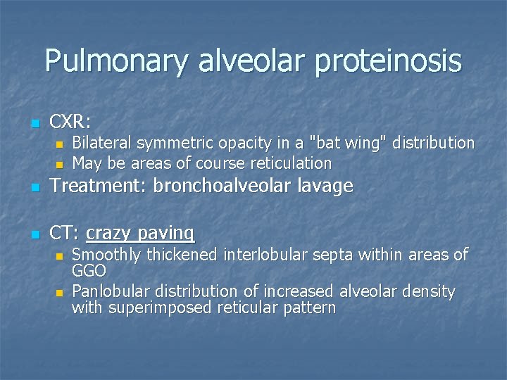
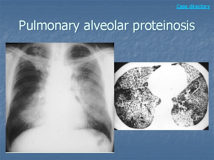
Pulmonary alveolar proteinosis n CXR: n n Bilateral symmetric opacity in a "bat wing" distribution May be areas of course reticulation n Treatment: bronchoalveolar lavage n CT: crazy paving n n Smoothly thickened interlobular septa within areas of GGO Panlobular distribution of increased alveolar density with superimposed reticular pattern
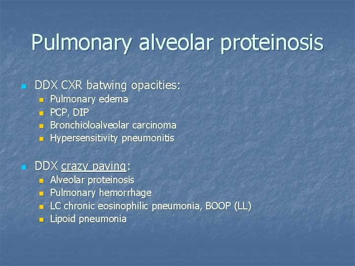
Pulmonary alveolar proteinosis n DDX CXR batwing opacities: n n n Pulmonary edema PCP, DIP Bronchioloalveolar carcinoma Hypersensitivity pneumonitis DDX crazy paving: n n Alveolar proteinosis Pulmonary hemorrhage LC chronic eosinophilic pneumonia, BOOP (LL) Lipoid pneumonia
Case directory Pulmonary alveolar proteinosis
Case 9
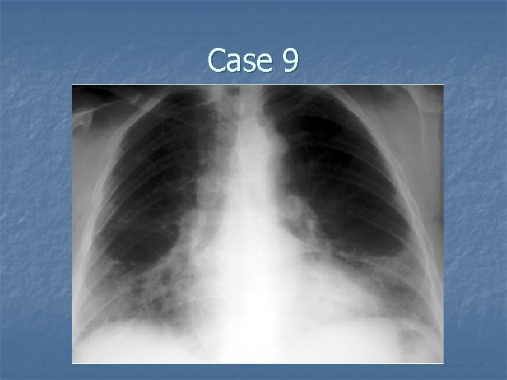
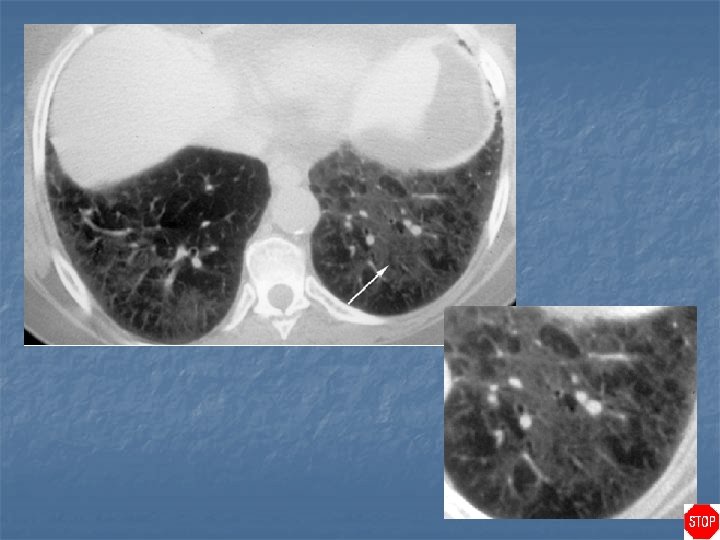
Case directory Desquamative interstitial pneumonia (DIP) n Case findings: n n CXR: MC LL GGO CT: patchy GGO in middle and lower lung n Alveolar macrophage pneumonia 90% are smokers Respond better to steroids than UIP Bilateral patchy GGO: LL and subpleural predominance n DDX: n n n Acute hypersensitivity pneumonitis (HP) RB, RB-ILD Alveolar hemorrhage
Case 10
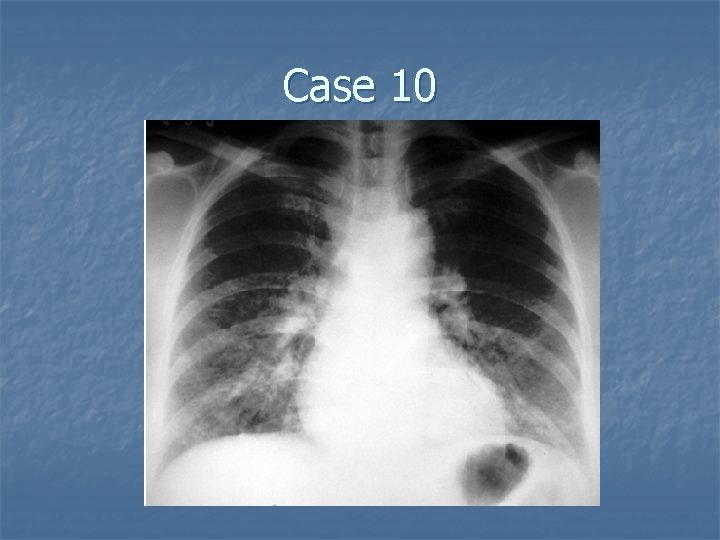
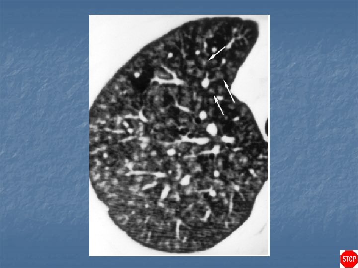
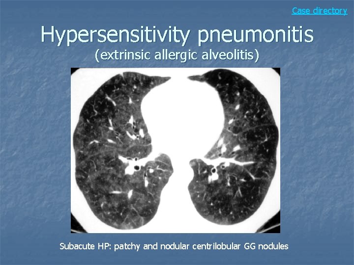
Hypersensitivity pneumonitis (extrinsic allergic alveolitis) n Case findings: n n Subacute: MC fine reticular or reticulonodular pattern involving both lungs, centrilobular GG nodules CT: numerous centrilobular, peribronchiolar, indistinct nodular opacities with peribronchiolar thickening GGO in a panlobular distribution Example: farmer’s lung, bird fancier’s lung
Hypersensitivity pneumonitis (extrinsic allergic alveolitis) n Subacute: n n n Weeks to months after exposure MC patchy or diffuse GGO Small ill-defined centrilobular GG nodular opacities Air trapping: small airway obstruction from bronchiolitis Head cheese sign: GGO and decreased lung attenuation reflecting air-trapping (mosaic perfusion) on inspiratory scan Chronic: n n Fibrosis MC in mid lung zones Sparing of the apex and bases
Case directory Hypersensitivity pneumonitis (extrinsic allergic alveolitis) Subacute HP: patchy and nodular centrilobular GG nodules
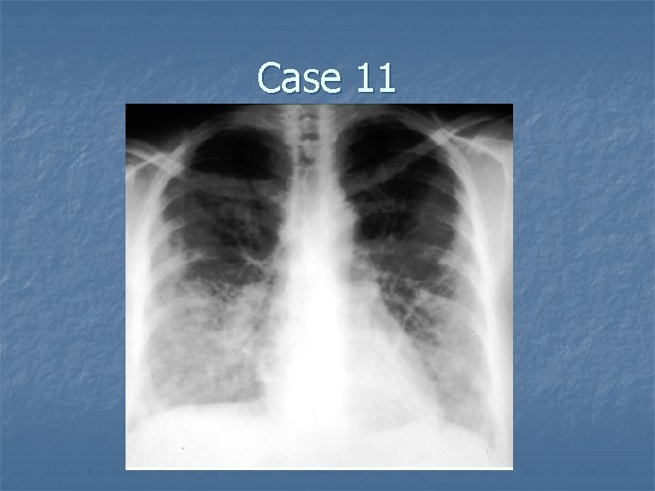
Case 11
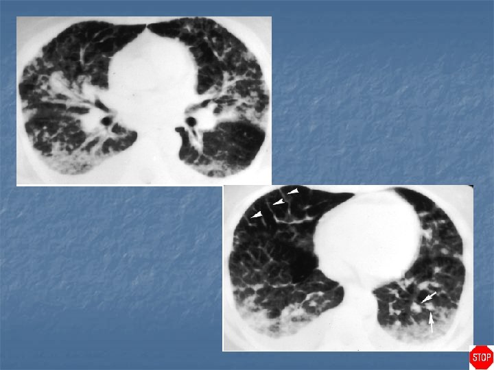
Acute eosinophilic pneumonia (Hamman-Rich syndrome) n CXR: n n n Acute progression of diffuse opacities involving both lungs Kerley's lines and pleural effusions are frequently present CT: n n Diffuse alveolar opacities with associated thickening of bronchovascular bundles and interlobular septa, simulating pulmonary edema Mediastinal lymphadenopathy and bilateral pleural effusions are common findings
Acute eosinophilic pneumonia (Hamman-Rich syndrome) n DDX: Pulmonary edema n BOOP n Lymphangitic carcinomatosis n Hypersensitivity pneumonitis n
Acute eosinophilic pneumonia (Hamman-Rich syndrome) n Diffuse alveolar damage (DAD): injury to the alveolar lining and endothelial cells chronic fibrotic phase follows n n n CXR: n n n Findings similar to ARDS/pulmonary edema Reticular opacities with Kerley B lines progress to interstitial infiltrates and airspace consolidations Extensive bilateral GGO, airspace consolidation, architectural distortion Diagnosis of exclusion: may be an idiopathic form of ARDS CT: n n Bilateral, patchy airspace opacification, spares CP angles With progression, lungs become diffusely consolidated Similar to ARDS with bilateral, patchy ground glass opacities, consolidation Traction bronchiectasis
Case directory Cryptogenic eosinophilic pneumonia n Umbrella term for several conditions: n Loffler’s syndrome: n n Acute eosinophilic pneumonia: n n Peripheral consolidation Prompt response to steroids Churg Strauss syndrome: n n Rapidly progressing illness of several days duration with fever, severe hypoxemia Diffuse lung consolidation mimicking ARDS Chronic eosinophilic pneumonia: n n Migrating pulmonary consolidation Allergic angiitis and granulomatosis Lung nodules, which unlike Wegener’s granulomatosis, do NOT cavitate Peripheral areas of consolidation similar to eosinophilic pneumonia Hypereosinophilic syndrome: n Idiopathic eosinophilic syndrome
Case 12
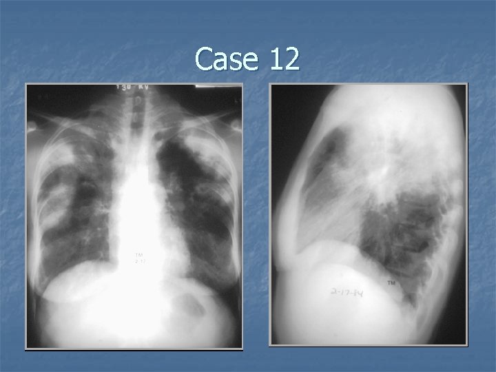
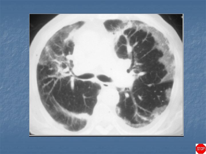
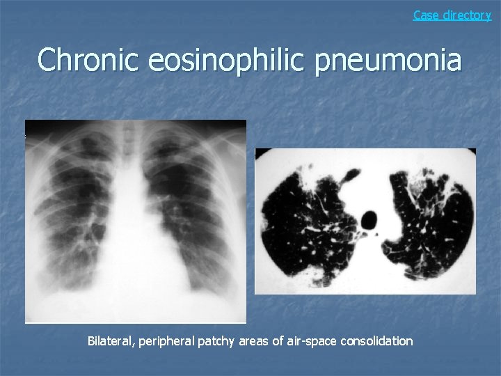
Chronic eosinophilic pneumonia n Case findings: n n CXR: photographic negative of pulmonary edema peripheral infiltrate (in the outer 2/3 of the lung) Peripheral infiltrates are MC in UL and occur bilaterally. About 20% patients present migratory infiltrates Mediastinal adenopathy in 50% DDX: n n Bronchiolitis obliterans with organizing pneumonia (BOOP) Sarcoidosis TB PCP
Case directory Chronic eosinophilic pneumonia Bilateral, peripheral patchy areas of air-space consolidation
Case 13
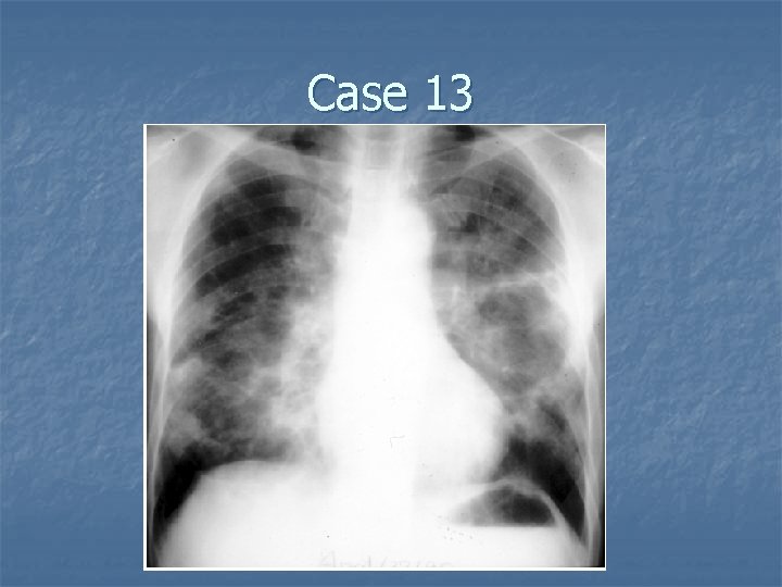
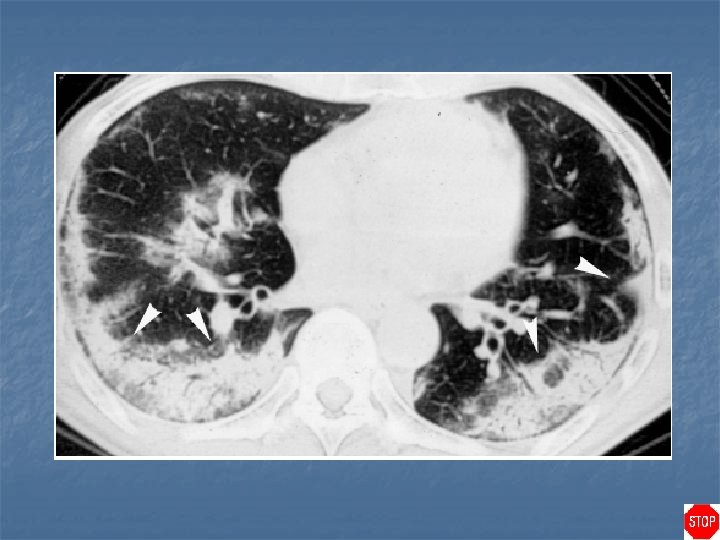
Case directory Bronchiolitis obliterans organizing pneumonia (BOOP) n Case findings: n n Patchy consolidation may be fleeting and migratory GGO is associated with the presence of alveolitis Consolidations are MC in subpleural regions DDX: n n Chronic eosinophilic pneumonia Pulmonary hemorrhage Multifocal infection Diffuse alveolar damage, acute eosinophilic pneumonia
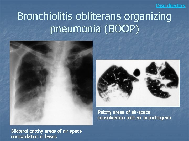
Bronchiolitis obliterans organizing pneumonia (BOOP) n n BOOP: cryptogenic organizing pneumonia MC idiopathic, respond well to steroids BOOP-like reaction seen with: pulmonary infection, drugs, CVD, Wegner’s, toxic fume inhalation Findings: n n Patchy consolidation or GGO, with a subpleural and/or peribronchial distribution Peripheral consolidation (DDX: chronic eosinophilic pneumonia) Irregular nodules of peribronchial consolidation Crazy paving (MC LL)
Case directory Bronchiolitis obliterans organizing pneumonia (BOOP) Patchy areas of air-space consolidation with air bronchogram Bilateral patchy areas of air-space consolidation in bases
Case 14
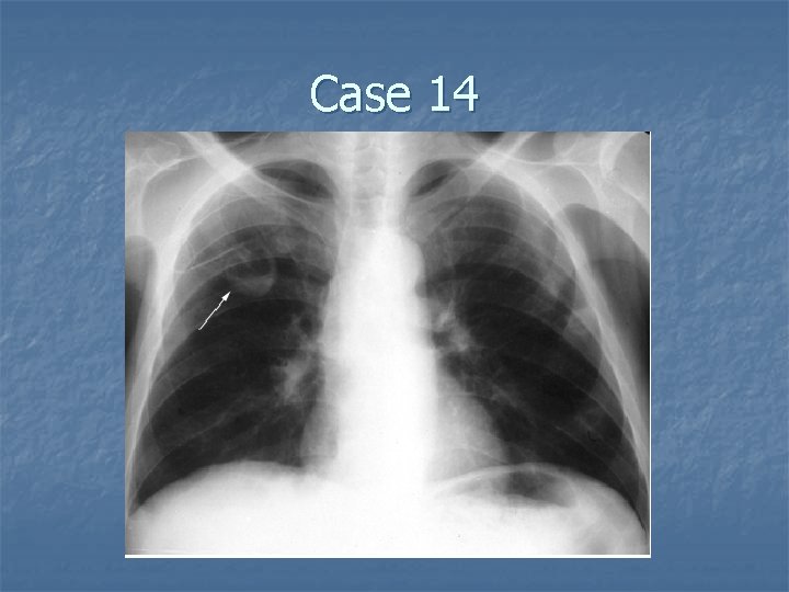
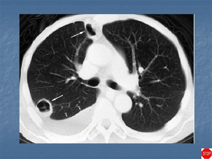
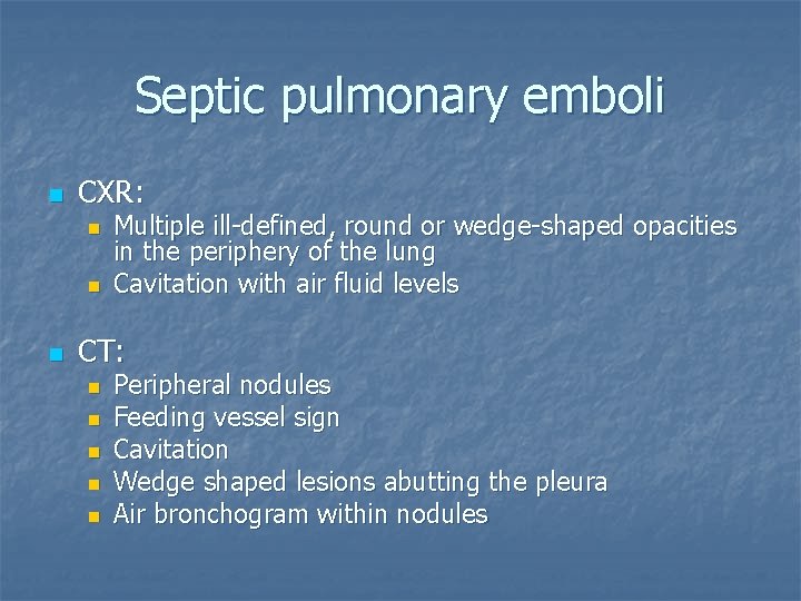
Septic pulmonary emboli n CXR: n n n Multiple ill-defined, round or wedge-shaped opacities in the periphery of the lung Cavitation with air fluid levels CT: n n n Peripheral nodules Feeding vessel sign Cavitation Wedge shaped lesions abutting the pleura Air bronchogram within nodules
Case directory DDX cavitary lesion n n Infection Neoplasm: squamous cell, hematogenous metastasis Vasculitis: Wegener’s granulomatosis Granulomatosis: TB, sarcoidosis Septic pulmonary emboli
Case 15
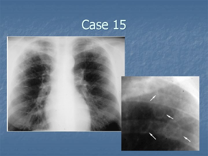
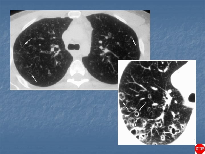
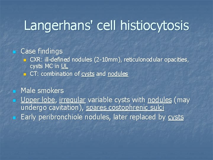
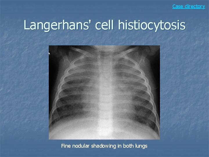
Langerhans' cell histiocytosis n Case findings n n n CXR: ill-defined nodules (2 -10 mm), reticulonodular opacities, cysts MC in UL CT: combination of cysts and nodules Male smokers Upper lobe, irregular variable cysts with nodules (may undergo cavitation), spares costophrenic sulci Early peribronchiole nodules, later replaced by cysts
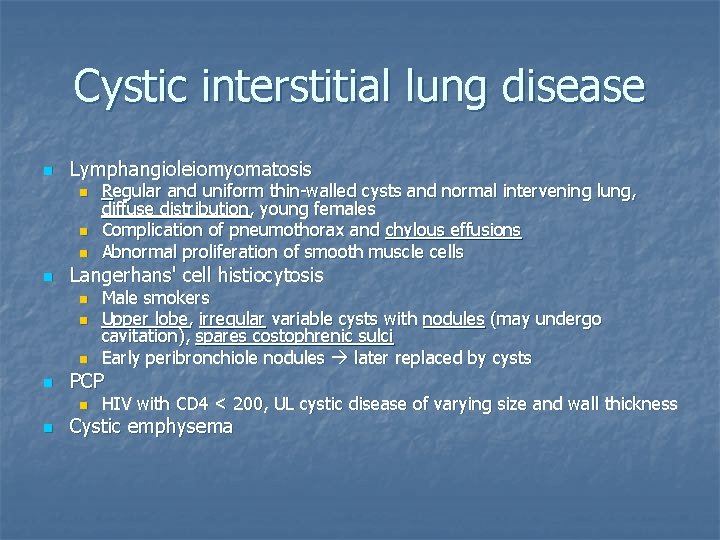
Cystic interstitial lung disease n Lymphangioleiomyomatosis n n Langerhans' cell histiocytosis n n Male smokers Upper lobe, irregular variable cysts with nodules (may undergo cavitation), spares costophrenic sulci Early peribronchiole nodules later replaced by cysts PCP n n Regular and uniform thin-walled cysts and normal intervening lung, diffuse distribution, young females Complication of pneumothorax and chylous effusions Abnormal proliferation of smooth muscle cells HIV with CD 4 < 200, UL cystic disease of varying size and wall thickness Cystic emphysema
Case directory Langerhans' cell histiocytosis Fine nodular shadowing in both lungs
Case 16
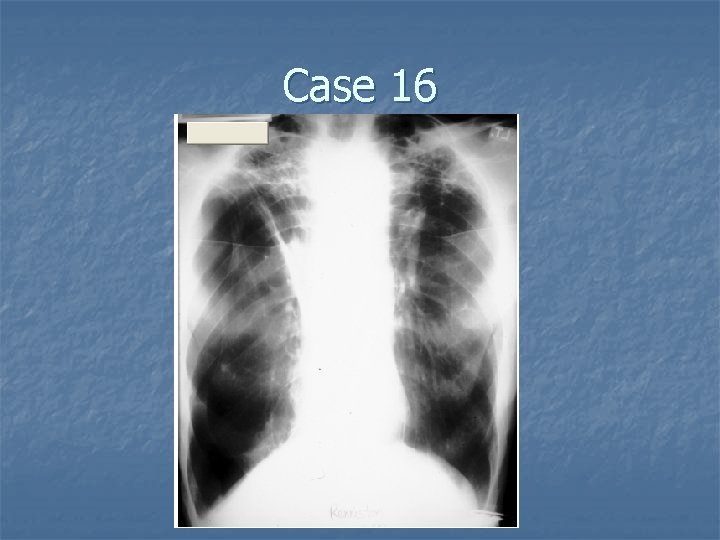
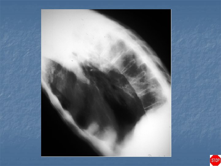
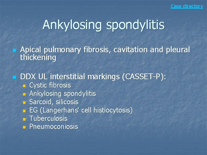
Case directory Ankylosing spondylitis n Apical pulmonary fibrosis, cavitation and pleural thickening n DDX UL interstitial markings (CASSET-P): n n n Cystic fibrosis Ankylosing spondylitis Sarcoid, silicosis EG (Langerhans' cell histiocytosis) Tuberculosis Pneumoconiosis
Case 17
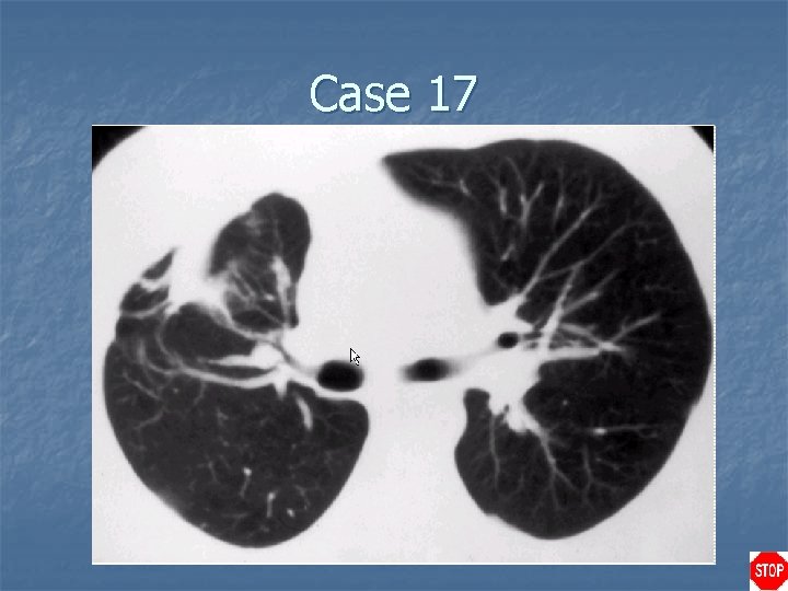
Rounded atelectasis n Case findings: Comet-tail sign: juxtapleural mass with associated pleural thickening and distorsion of adjacent vessels and bronchi producing a "comet tail" appearance as they lead towards the mass n Focal area of pulmonary collapse adjacent to thickened pleura n
Case directory Rounded atelectasis n Associated with: Asbestos-related pleural disease n Chronic pleural thickening n n High association between asbestos-related pleural disease and bronchogenic carcinoma
Case 18
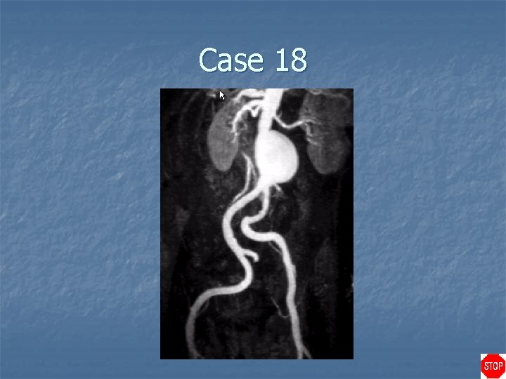
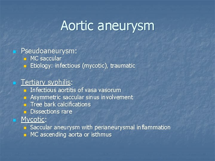
Aortic aneurysm n Pseudoaneurysm: n n n Tertiary syphilis: n n n MC saccular Etiology: infectious (mycotic), traumatic Infectious aortitis of vasa vasorum Asymmetric saccular sinus involvement Tree bark calcifications Dissections rare Mycotic: n n Saccular aneurysm with perianeurysmal inflammation MC ascending aorta or isthmus
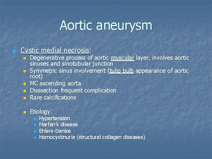
Aortic aneurysm n Cystic medial necrosis: n Degenerative process of aortic muscular layer, involves aortic sinuses and sinotubular junction Symmetric sinus involvement (tulip bulb appearance of aortic root) MC ascending aorta Disssection frequent complication Rare calcifications n Etiology: n n n n Hypertension Marfan’s disease Ehlers-Danlos Homocystinuria (structural collagen diseases)
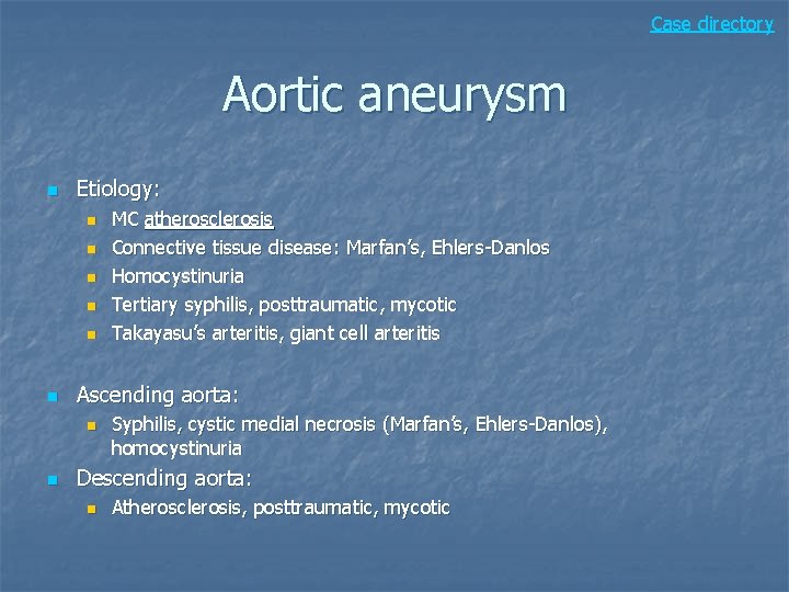
Case directory Aortic aneurysm n Etiology: n n n Ascending aorta: n n MC atherosclerosis Connective tissue disease: Marfan’s, Ehlers-Danlos Homocystinuria Tertiary syphilis, posttraumatic, mycotic Takayasu’s arteritis, giant cell arteritis Syphilis, cystic medial necrosis (Marfan’s, Ehlers-Danlos), homocystinuria Descending aorta: n Atherosclerosis, posttraumatic, mycotic
Case 19
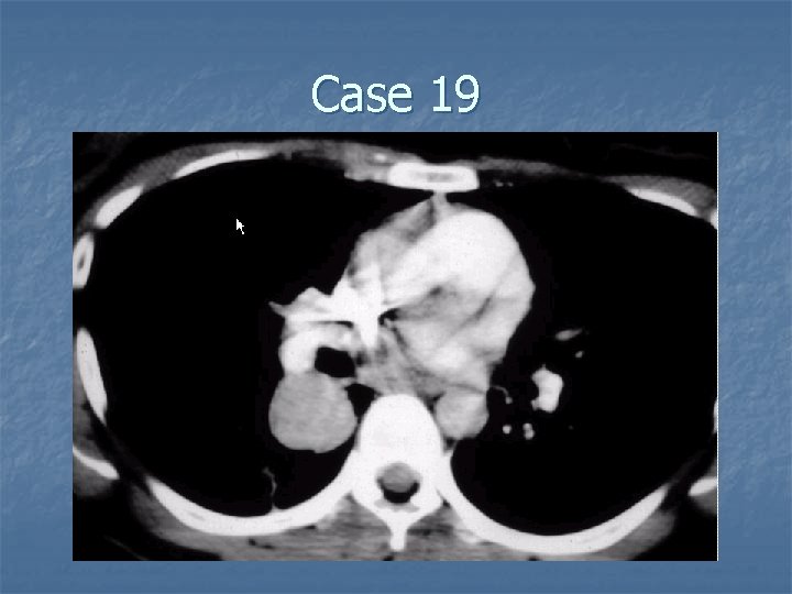
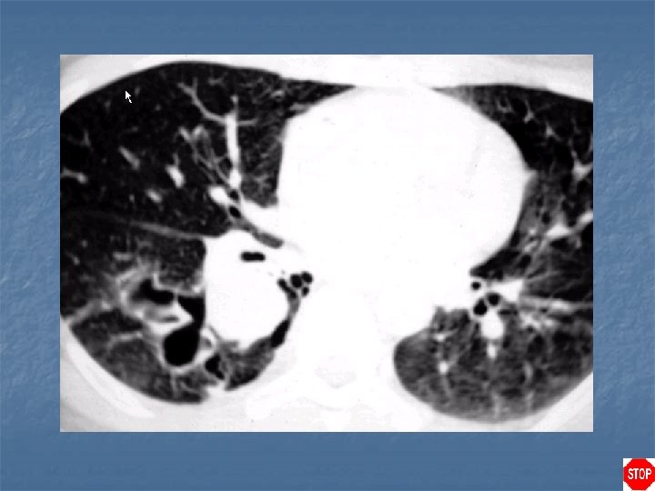
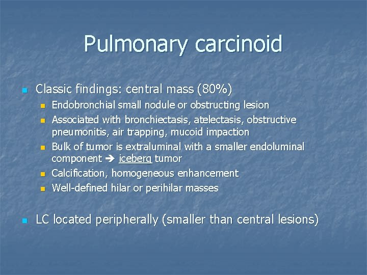
Pulmonary carcinoid n Classic findings: central mass (80%) n n n Endobronchial small nodule or obstructing lesion Associated with bronchiectasis, atelectasis, obstructive pneumonitis, air trapping, mucoid impaction Bulk of tumor is extraluminal with a smaller endoluminal component iceberg tumor Calcification, homogeneous enhancement Well-defined hilar or perihilar masses LC located peripherally (smaller than central lesions)
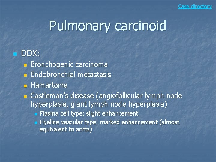
Case directory Pulmonary carcinoid n DDX: n n Bronchogenic carcinoma Endobronchial metastasis Hamartoma Castleman’s disease (angiofollicular lymph node hyperplasia, giant lymph node hyperplasia) n n Plasma cell type: slight enhancement Hyaline vascular type: marked enhancement (almost equivalent to aorta)
Case 20
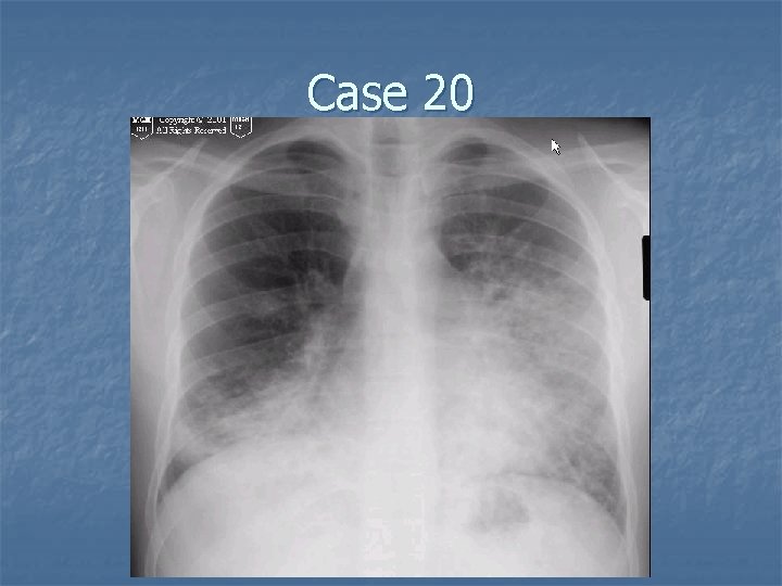
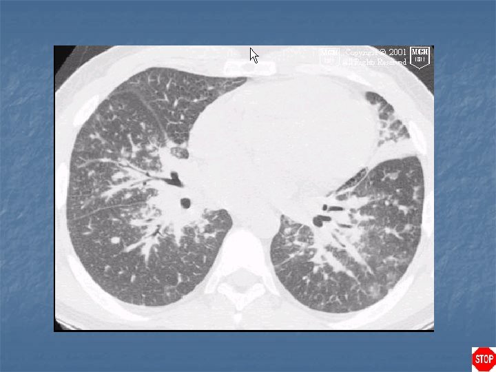
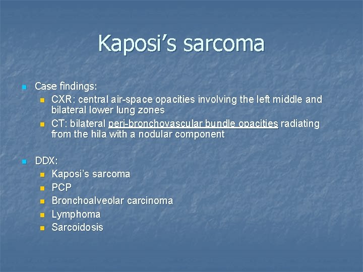
Kaposi’s sarcoma n n Case findings: n CXR: central air-space opacities involving the left middle and bilateral lower lung zones n CT: bilateral peri-bronchovascular bundle opacities radiating from the hila with a nodular component DDX: n Kaposi’s sarcoma n PCP n Bronchoalveolar carcinoma n Lymphoma n Sarcoidosis
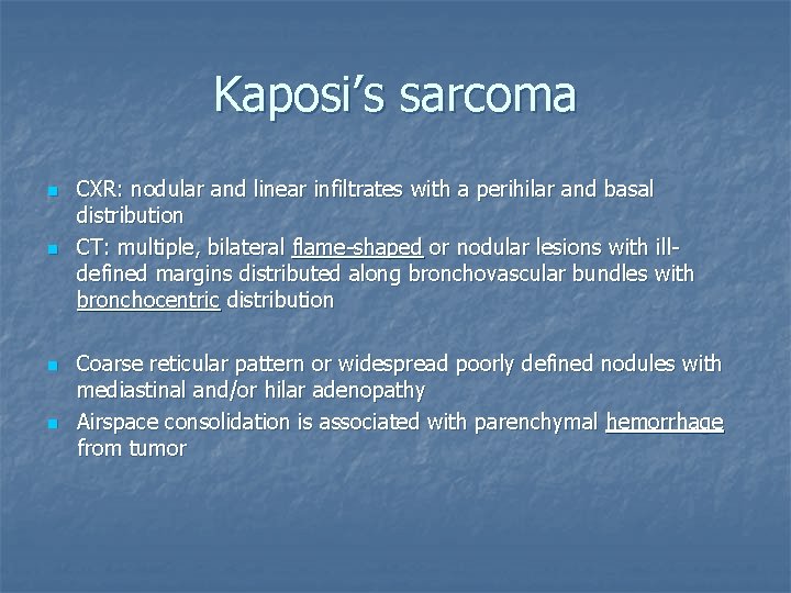
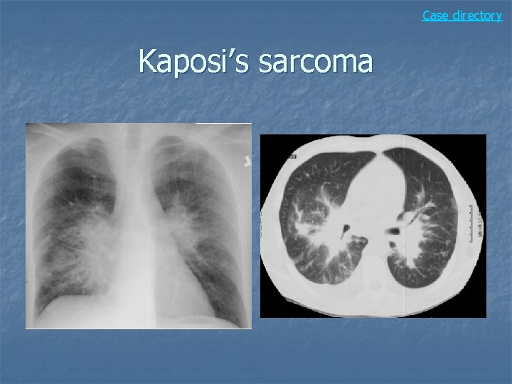
Kaposi’s sarcoma n n CXR: nodular and linear infiltrates with a perihilar and basal distribution CT: multiple, bilateral flame-shaped or nodular lesions with illdefined margins distributed along bronchovascular bundles with bronchocentric distribution Coarse reticular pattern or widespread poorly defined nodules with mediastinal and/or hilar adenopathy Airspace consolidation is associated with parenchymal hemorrhage from tumor
Case directory Kaposi’s sarcoma
Case 21
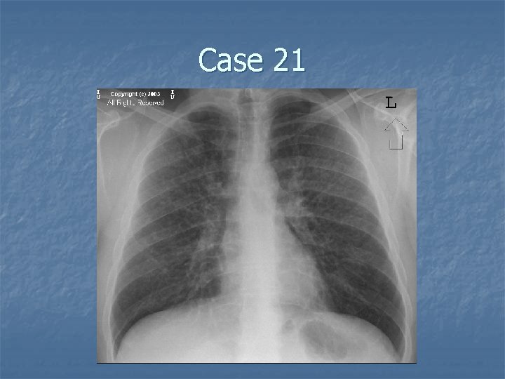
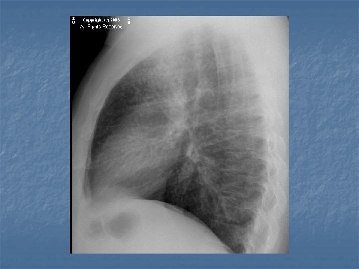
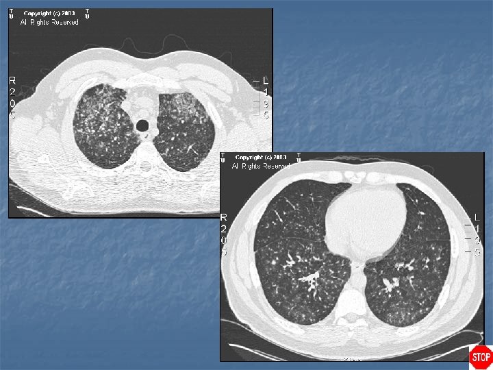
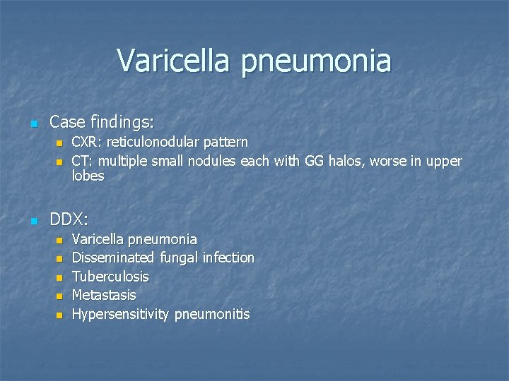
Varicella pneumonia n Case findings: n n n CXR: reticulonodular pattern CT: multiple small nodules each with GG halos, worse in upper lobes DDX: n n n Varicella pneumonia Disseminated fungal infection Tuberculosis Metastasis Hypersensitivity pneumonitis
Case directory Varicella pneumonia n n n Acute phase: multiple, bilateral 5 – 10 mm nodular opacities that coalesce into diffuse patchy air space disease Opacities usually resolve completely, however, in less than 2% of patients they involute and calcify to form innumerable small calcified nodules Pleural effusions are rare
Case 22
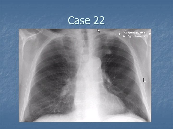
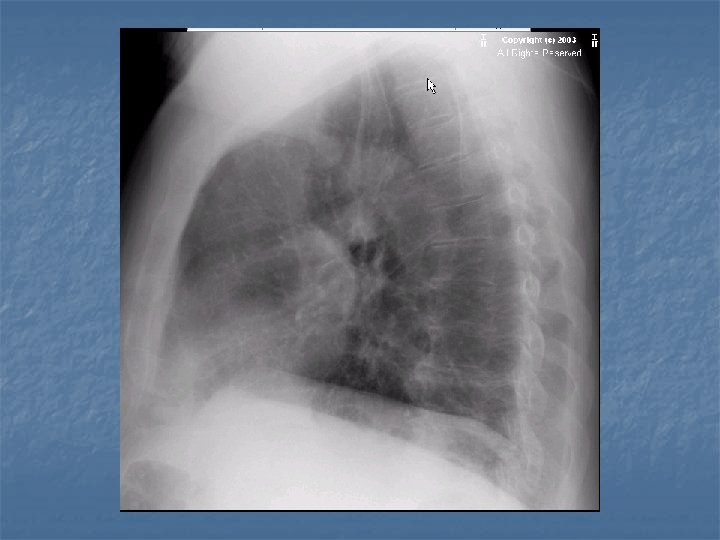
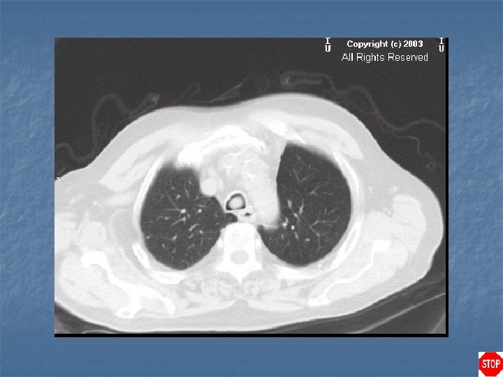
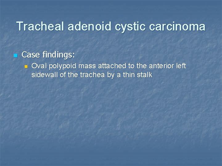
Tracheal adenoid cystic carcinoma n Case findings: n Oval polypoid mass attached to the anterior left sidewall of the trachea by a thin stalk
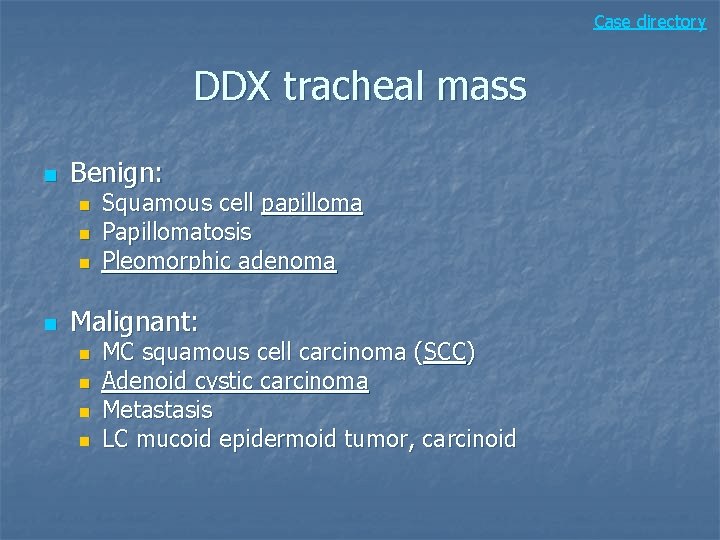
Case directory DDX tracheal mass n Benign: n n Squamous cell papilloma Papillomatosis Pleomorphic adenoma Malignant: n n MC squamous cell carcinoma (SCC) Adenoid cystic carcinoma Metastasis LC mucoid epidermoid tumor, carcinoid
Case 23
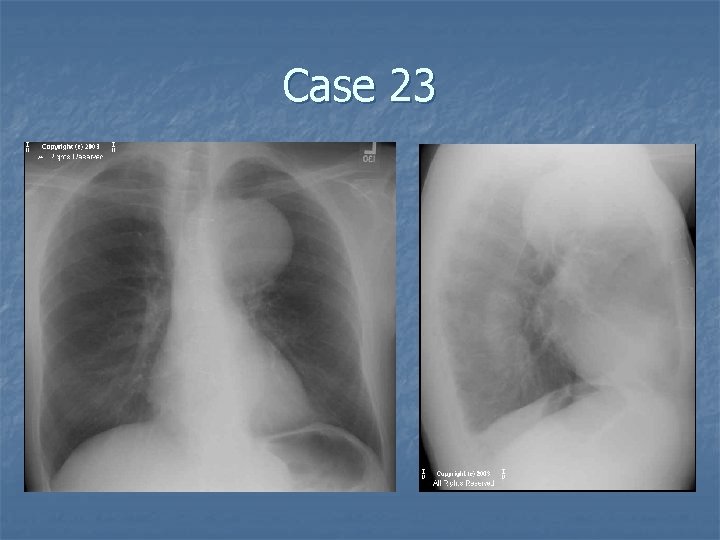
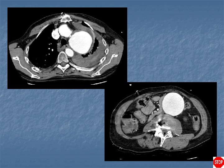
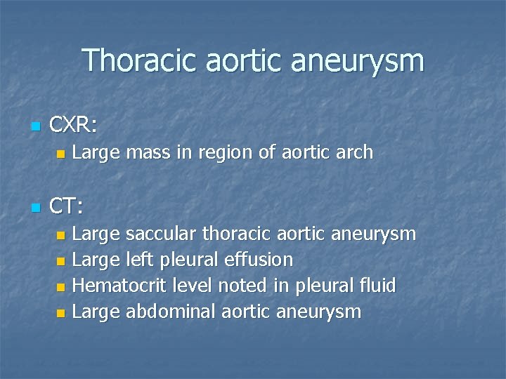
Thoracic aortic aneurysm n CXR: n n Large mass in region of aortic arch CT: Large saccular thoracic aortic aneurysm n Large left pleural effusion n Hematocrit level noted in pleural fluid n Large abdominal aortic aneurysm n
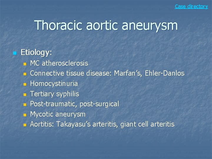
Case directory Thoracic aortic aneurysm n Etiology: n n n n MC atherosclerosis Connective tissue disease: Marfan’s, Ehler-Danlos Homocystinuria Tertiary syphilis Post-traumatic, post-surgical Mycotic aneurysm Aortitis: Takayasu’s arteritis, giant cell arteritis
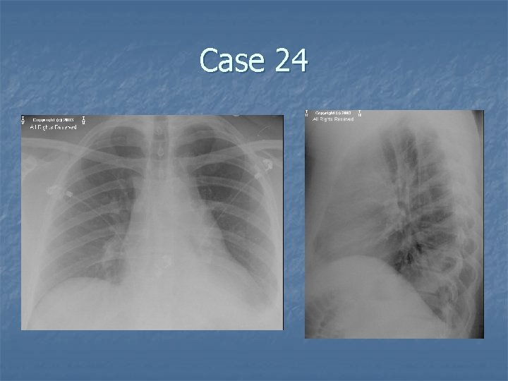
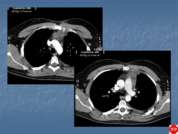
Case 24
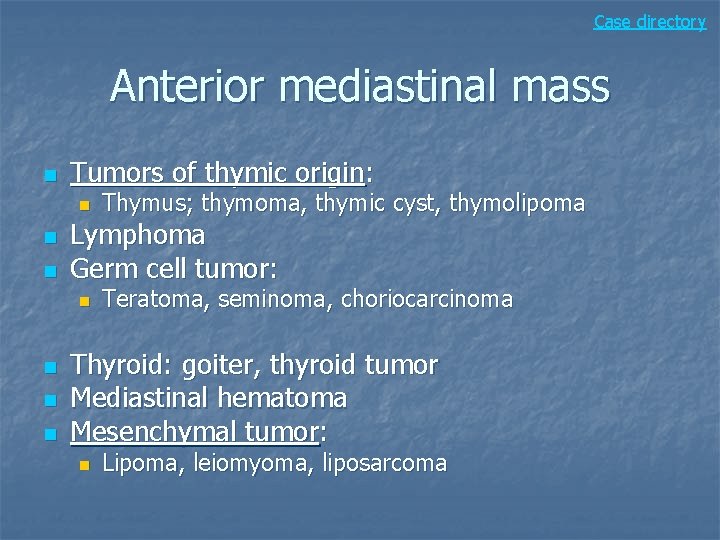
Case directory Anterior mediastinal mass n Tumors of thymic origin: n n n Lymphoma Germ cell tumor: n n Thymus; thymoma, thymic cyst, thymolipoma Teratoma, seminoma, choriocarcinoma Thyroid: goiter, thyroid tumor Mediastinal hematoma Mesenchymal tumor: n Lipoma, leiomyoma, liposarcoma
Case 25
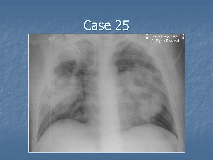
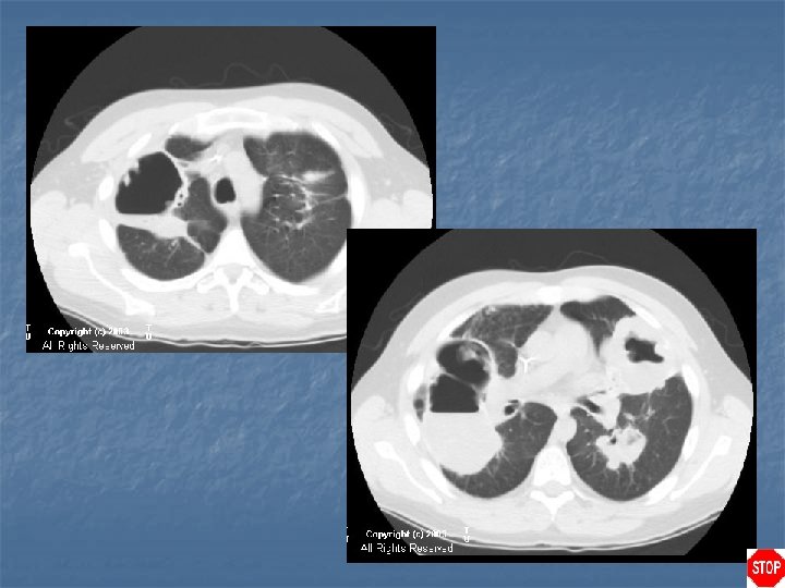
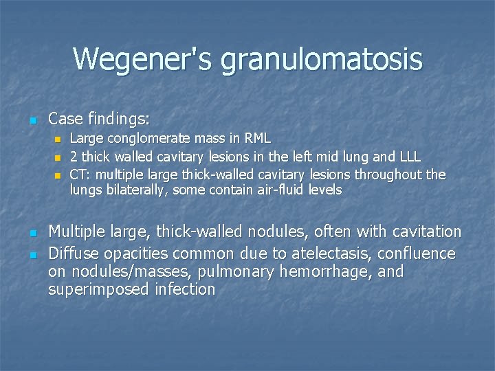
Wegener's granulomatosis n Case findings: n n n Large conglomerate mass in RML 2 thick walled cavitary lesions in the left mid lung and LLL CT: multiple large thick-walled cavitary lesions throughout the lungs bilaterally, some contain air-fluid levels Multiple large, thick-walled nodules, often with cavitation Diffuse opacities common due to atelectasis, confluence on nodules/masses, pulmonary hemorrhage, and superimposed infection
Case directory DDX multiple cavitary lesions n Metastasis: n n n n Newly treated or fast-growing carcinoma Wegener’s granulomatosis Septic emboli Multiple abscesses (e. g. , aspiration) Multifocal necrotizing pneumonia with infarction Fungal infection: MC in immunocompromised Rheumatoid nodules