The Cardiovascular System The Heart Heart pumps over
- Slides: 34
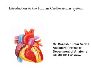
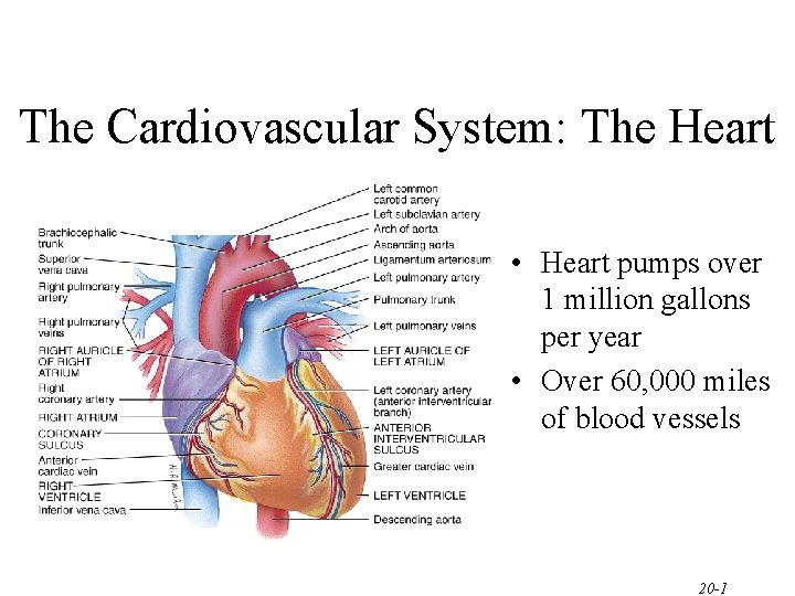
The Cardiovascular System: The Heart • Heart pumps over 1 million gallons per year • Over 60, 000 miles of blood vessels 20 -1
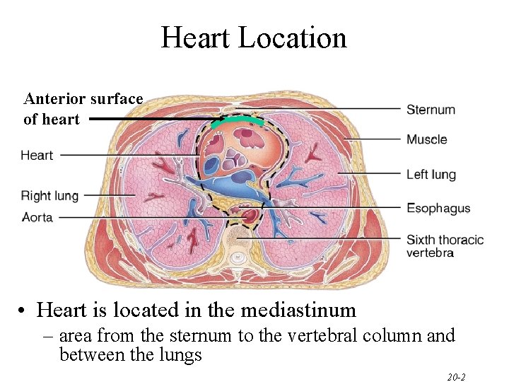
Heart Location Anterior surface of heart • Heart is located in the mediastinum – area from the sternum to the vertebral column and between the lungs 20 -2
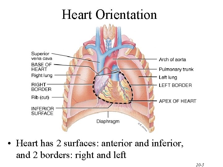
Heart Orientation • Heart has 2 surfaces: anterior and inferior, and 2 borders: right and left 20 -3
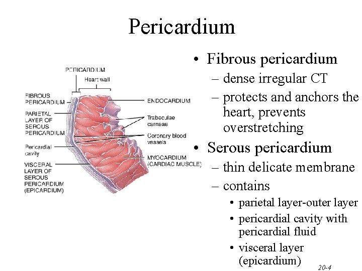
Pericardium • Fibrous pericardium – dense irregular CT – protects and anchors the heart, prevents overstretching • Serous pericardium – thin delicate membrane – contains • parietal layer-outer layer • pericardial cavity with pericardial fluid • visceral layer (epicardium) 20 -4
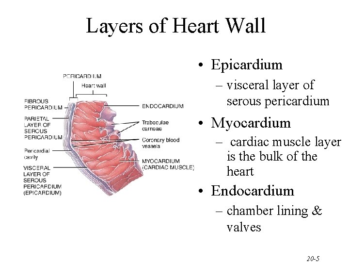
Layers of Heart Wall • Epicardium – visceral layer of serous pericardium • Myocardium – cardiac muscle layer is the bulk of the heart • Endocardium – chamber lining & valves 20 -5
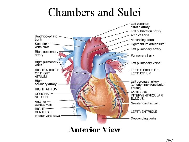
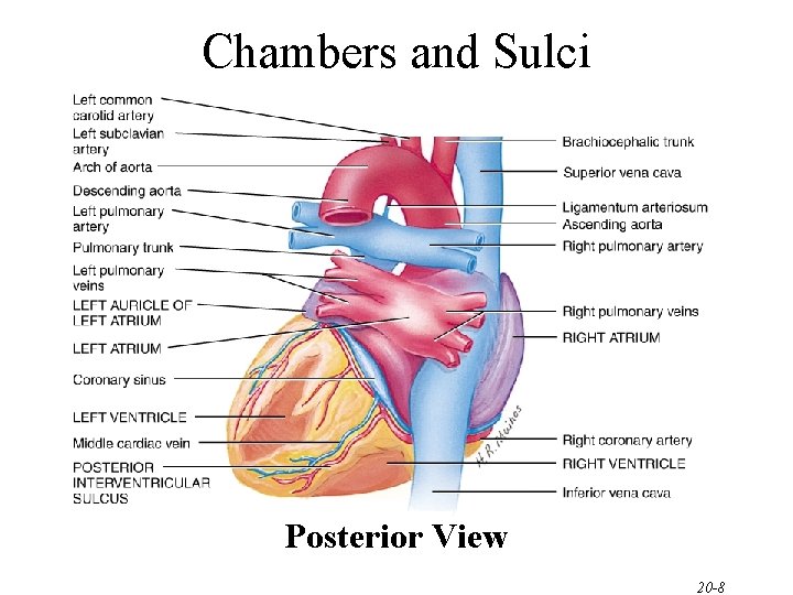
Chambers and Sulci of the Heart • Four chambers – 2 upper atria – 2 lower ventricles • Sulci - grooves on surface of heart containing coronary blood vessels and fat – coronary sulcus • encircles heart and marks the boundary between the atria and the ventricles – anterior interventricular sulcus • marks the boundary between the ventricles anteriorly – posterior interventricular sulcus • marks the boundary between the ventricles posteriorly 20 -6
Chambers and Sulci Anterior View 20 -7
Chambers and Sulci Posterior View 20 -8
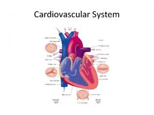
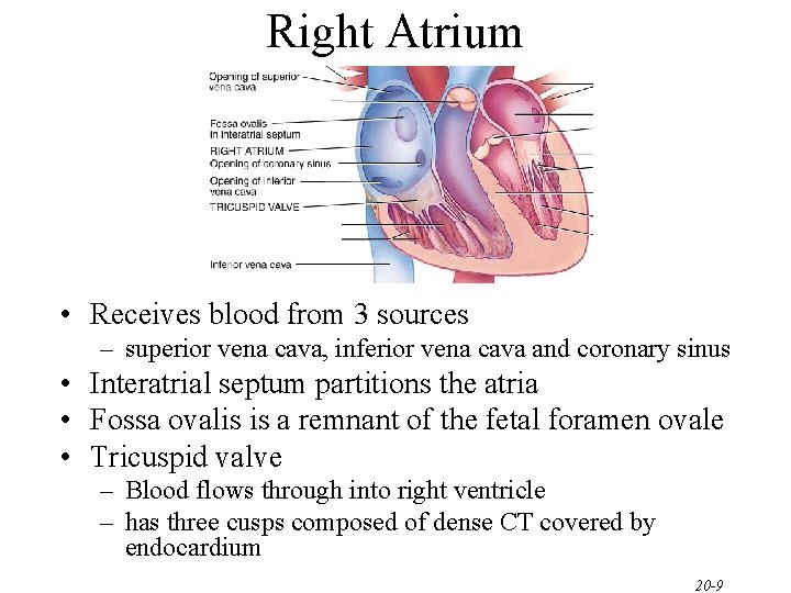
Right Atrium • Receives blood from 3 sources – superior vena cava, inferior vena cava and coronary sinus • Interatrial septum partitions the atria • Fossa ovalis is a remnant of the fetal foramen ovale • Tricuspid valve – Blood flows through into right ventricle – has three cusps composed of dense CT covered by endocardium 20 -9
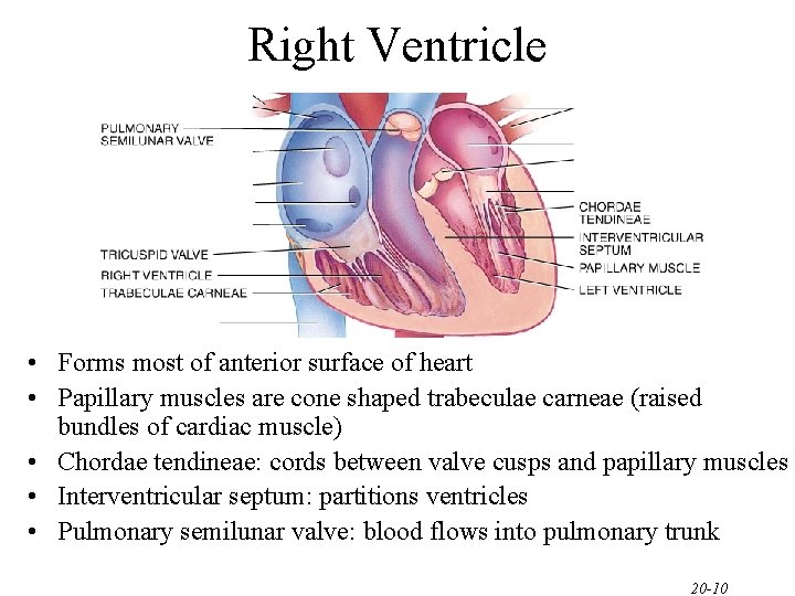
Right Ventricle • Forms most of anterior surface of heart • Papillary muscles are cone shaped trabeculae carneae (raised bundles of cardiac muscle) • Chordae tendineae: cords between valve cusps and papillary muscles • Interventricular septum: partitions ventricles • Pulmonary semilunar valve: blood flows into pulmonary trunk 20 -10
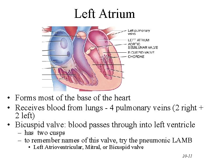
Left Atrium • Forms most of the base of the heart • Receives blood from lungs - 4 pulmonary veins (2 right + 2 left) • Bicuspid valve: blood passes through into left ventricle – has two cusps – to remember names of this valve, try the pneumonic LAMB • Left Atrioventricular, Mitral, or Bicuspid valve 20 -11
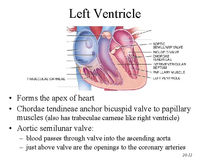
Left Ventricle • Forms the apex of heart • Chordae tendineae anchor bicuspid valve to papillary muscles (also has trabeculae carneae like right ventricle) • Aortic semilunar valve: – blood passes through valve into the ascending aorta – just above valve are the openings to the coronary arteries 20 -12
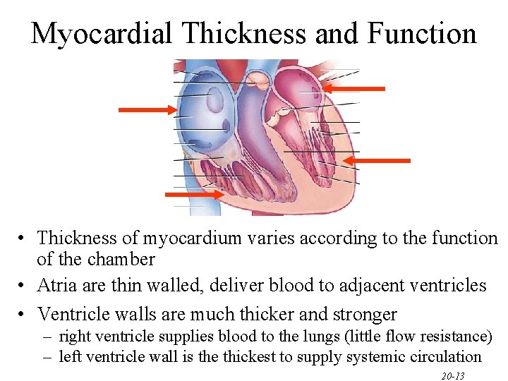
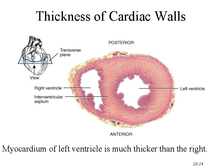
Myocardial Thickness and Function • Thickness of myocardium varies according to the function of the chamber • Atria are thin walled, deliver blood to adjacent ventricles • Ventricle walls are much thicker and stronger – right ventricle supplies blood to the lungs (little flow resistance) – left ventricle wall is the thickest to supply systemic circulation 20 -13
Thickness of Cardiac Walls Myocardium of left ventricle is much thicker than the right. 20 -14
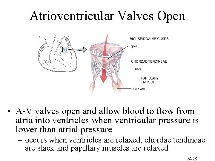
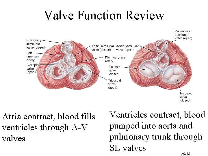
Atrioventricular Valves Open • A-V valves open and allow blood to flow from atria into ventricles when ventricular pressure is lower than atrial pressure – occurs when ventricles are relaxed, chordae tendineae are slack and papillary muscles are relaxed 20 -15
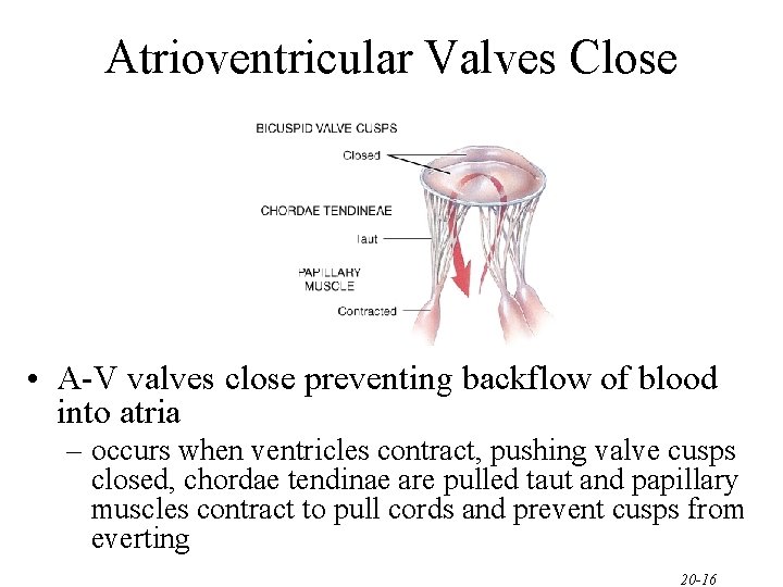
Atrioventricular Valves Close • A-V valves close preventing backflow of blood into atria – occurs when ventricles contract, pushing valve cusps closed, chordae tendinae are pulled taut and papillary muscles contract to pull cords and prevent cusps from everting 20 -16
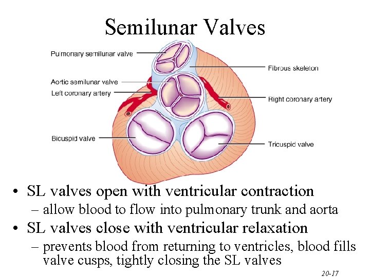
Semilunar Valves • SL valves open with ventricular contraction – allow blood to flow into pulmonary trunk and aorta • SL valves close with ventricular relaxation – prevents blood from returning to ventricles, blood fills valve cusps, tightly closing the SL valves 20 -17
Valve Function Review Atria contract, blood fills ventricles through A-V valves Ventricles contract, blood pumped into aorta and pulmonary trunk through SL valves 20 -18
One Cardiac Cycle • At 75 beats/min, one cycle requires 0. 8 sec. – systole (contraction) and diastole (relaxation) of both atria, plus the systole and diastole of both ventricles 20 -19
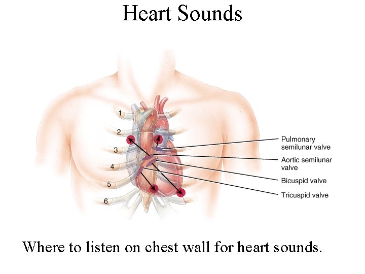
Auscultation • Stethoscope • Sounds of heartbeat are from turbulence in blood flow caused by valve closure – first heart sound (lubb) is created with the closing of the atrioventricular valves – second heart sound (dupp) is created with the closing of semilunar valves 20 -20
Heart Sounds Where to listen on chest wall for heart sounds.
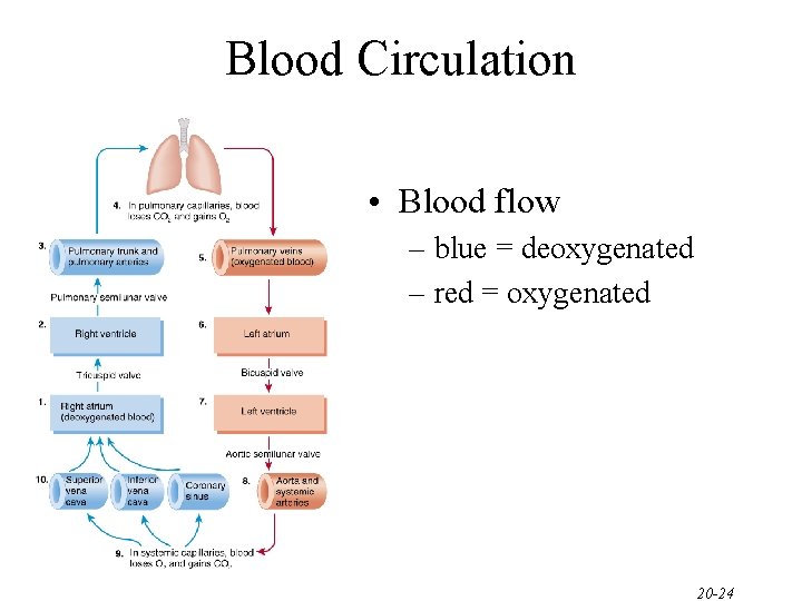
Blood Circulation • Two closed circuits, the systemic and pulmonic • Systemic circulation – left side of heart pumps blood through body – left ventricle pumps oxygenated blood into aorta – aorta branches into many arteries that travel to organs – arteries branch into many arterioles in tissue – arterioles branch into thin-walled capillaries for exchange of gases and nutrients – deoxygenated blood begins its return in venules – venules merge into veins and return to right atrium 20 -22
Blood Circulation (cont. ) • Pulmonary circulation – right side of heart pumps deoxygenated blood to lungs – right ventricle pumps blood to pulmonary trunk – pulmonary trunk branches into pulmonary arteries – pulmonary arteries carry blood to lungs for exchange of gases – oxygenated blood returns to heart in pulmonary veins 20 -23
Blood Circulation • Blood flow – blue = deoxygenated – red = oxygenated 20 -24
Coronary Circulation • Coronary circulation is blood supply to the heart • Heart as a very active muscle needs lots of O 2 • When the heart relaxes high pressure of blood in aorta pushes blood into coronary vessels • Many anastomoses – connections between arteries supplying blood to the same region, provide alternate routes if one artery becomes occluded 20 -25
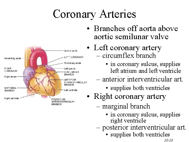
Coronary Arteries • Branches off aorta above aortic semilunar valve • Left coronary artery – circumflex branch • in coronary sulcus, supplies left atrium and left ventricle – anterior interventricular art. • supplies both ventricles • Right coronary artery – marginal branch • in coronary sulcus, supplies right ventricle – posterior interventricular art. • supplies both ventricles 20 -26
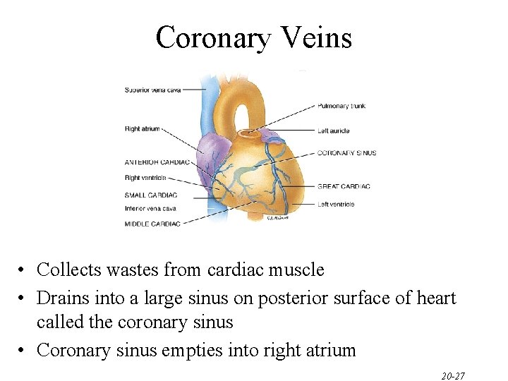
Coronary Veins • Collects wastes from cardiac muscle • Drains into a large sinus on posterior surface of heart called the coronary sinus • Coronary sinus empties into right atrium 20 -27
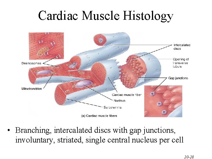
Cardiac Muscle Histology • Branching, intercalated discs with gap junctions, involuntary, striated, single central nucleus per cell 20 -28
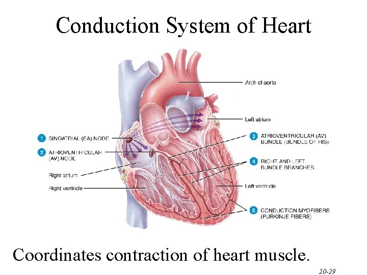
Conduction System of Heart Coordinates contraction of heart muscle. 20 -29
Conduction System of Heart • Autorhythmic Cells – Cells fire spontaneously, act as pacemaker and form conduction system for the heart • SA node – cluster of cells in wall of Rt. Atria – begins heart activity that spreads to both atria – excitation spreads to AV node • AV node – in atrial septum, transmits signal to bundle of His • AV bundle of His – the connection between atria and ventricles – divides into bundle branches & purkinje fibers, large diameter fibers that conduct signals quickly 20 -30
Rhythm of Conduction System • SA node fires spontaneously 90 -100 times per minute • AV node fires at 40 -50 times per minute • If both nodes are suppressed fibers in ventricles by themselves fire only 20 -40 times per minute • Artificial pacemaker needed if pace is too slow • Extra beats forming at other sites are called ectopic pacemakers – caffeine & nicotine increase activity 20 -31
Congestive Heart Failure • Causes of CHF – coronary artery disease, hypertension, MI, valve disorders, congenital defects • Left side heart failure – less effective pump so more blood remains in ventricle – heart is overstretched & even more blood remains – blood backs up into lungs as pulmonary edema – suffocation & lack of oxygen to the tissues • Right side failure – fluid builds up in tissues as peripheral edema 20 -32
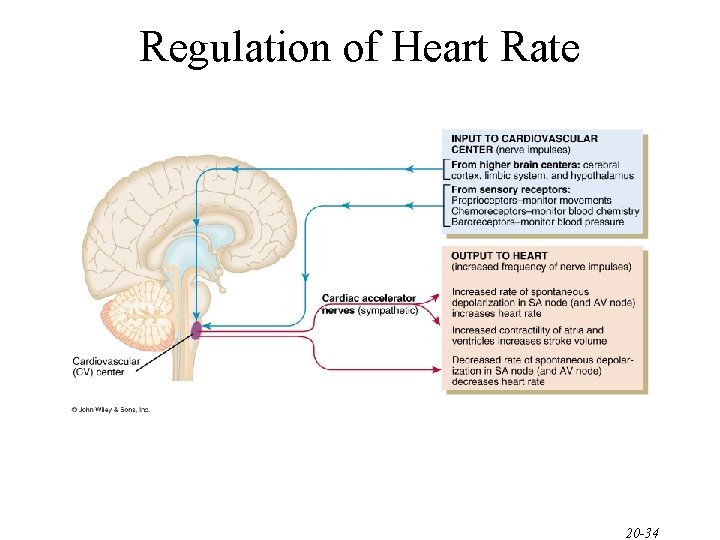
Regulation of Heart Rate • Nervous control from the cardiovascular center in the medulla – Sympathetic impulses increase heart rate and force of contraction – parasympathetic impulses decrease heart rate. – Baroreceptors (pressure receptors) detect change in BP and send info to the cardiovascular center • located in the arch of the aorta and carotid arteries • Heart rate is also affected by hormones – epinephrine, norepinephrine, thyroid hormones – ions (Na+, K+, Ca 2+) – age, gender, physical fitness, and temperature 20 -33
Regulation of Heart Rate 20 -34