Circulatory System Heart and Blood Vessels Crash Course

Circulatory System Heart and Blood Vessels

Crash Course • https: //www. youtube. com/watch? v=9 fxm 85 F y 4 s. Q&index=27&list=PL 3 EED 4 C 1 D 684 D 3 ADF

• The circulatory system is the system that transports materials around the body to and from the cells.

• Question: Why do humans need a circulatory system whereas bacteria and simple organisms do not? • Answer: Because a complex organism such as a human has many cells that are far from the outside environment where nutrients would come from. The system brings the materials to the cells that would not normally receive them.

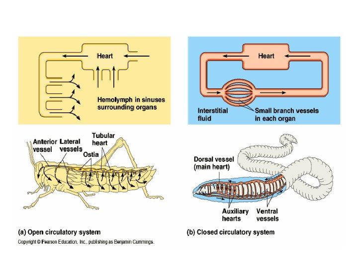
• Humans have a closed circulatory system: This means that the blood is always contained in tubes and vessels.

Parts of the Circulatory System • The human circulatory system is composed of the following: 1. Blood Vessels 2. Heart 3. Blood
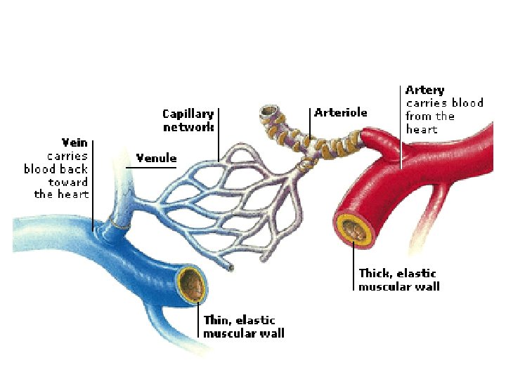
 Arteries Structure: • Thick, elastic • Smallest arteries are called arterioles")
Blood Vessels a) Arteries Structure: • Thick, elastic • Smallest arteries are called arterioles • DO NOT CONTAIN VALVES Function: Transport blood AWAY from the heart to the organs and tissues of the body.
 Veins Structure: • Thin and slightly elastic • Smallest veins are")
Blood Vessels b) Veins Structure: • Thin and slightly elastic • Smallest veins are called venules • Contain VALVES – to help blood flow back to the heart against the force of gravity. Lower pressure than arteries • Function: To RETURN blood from the body tissues to the heart.
 Capillaries Structure: • Microscopic blood vessels that connect arterioles and venules")
Blood Vessels c) Capillaries Structure: • Microscopic blood vessels that connect arterioles and venules • Thin walled and narrow • Blood cells pass through them in single file • Function: Allows material and gas exchange between the body cells and the blood.

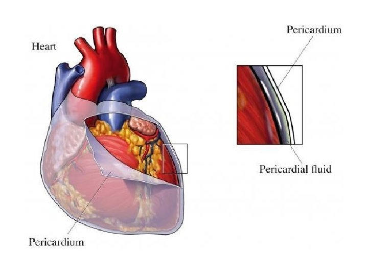
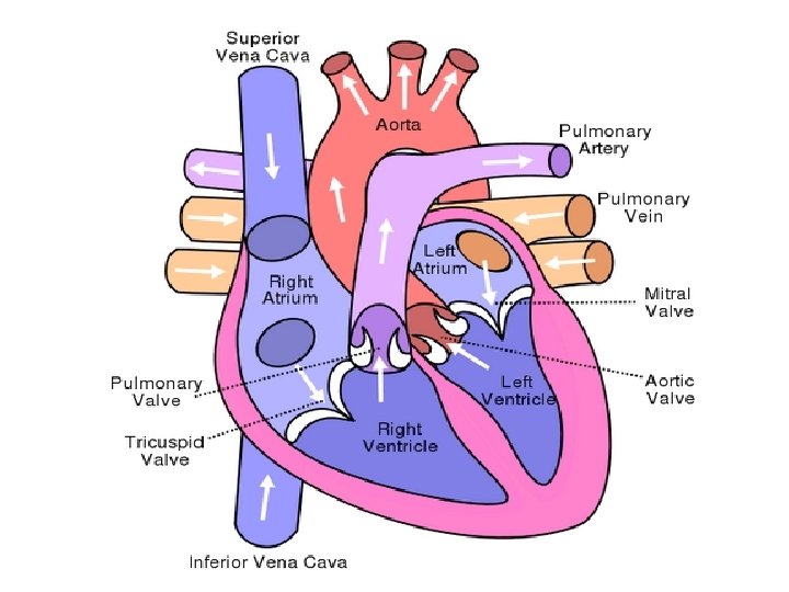
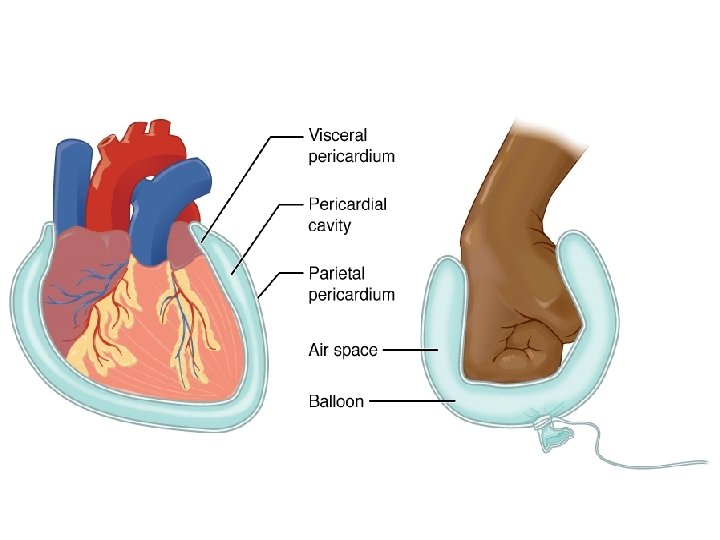
The Heart Structure: • A four chambered muscular organ located in the chest cavity of a human. • Made of cardiac muscle. • It is covered by a pericardium that protects it. • Pericardium: A tough membrane that surrounds the heart.

• Function: Pump blood around the body supplying the cells with nutrients and removing wastes (CO 2) from the cells.


Video Time • http: //studyjams. scholastic. com/studyjams/ja ms/science/human-body/circulatorysystem. htm

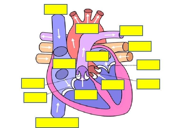
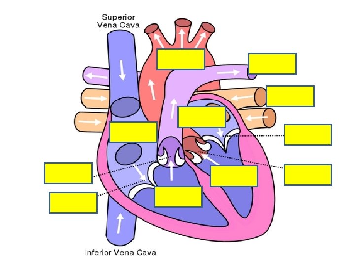
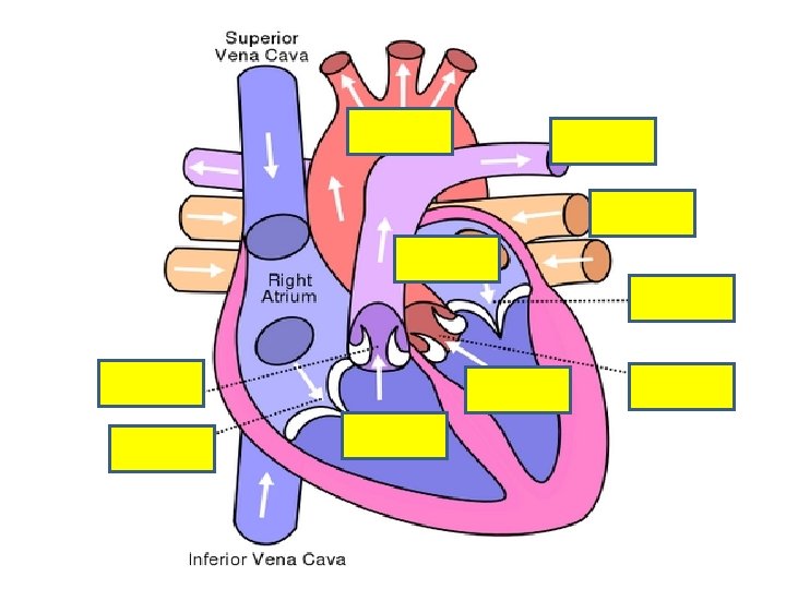
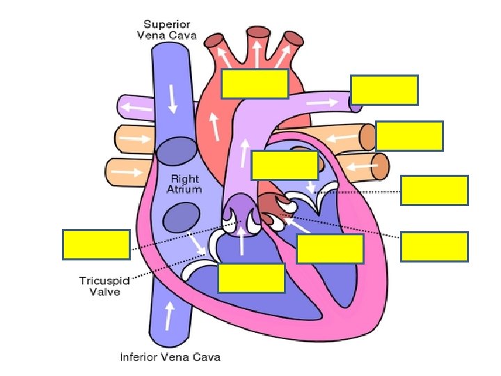
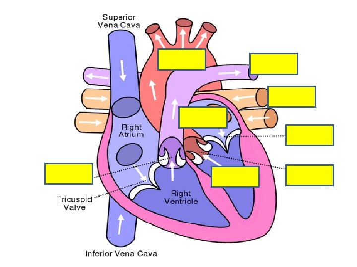
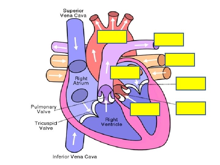
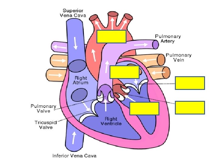
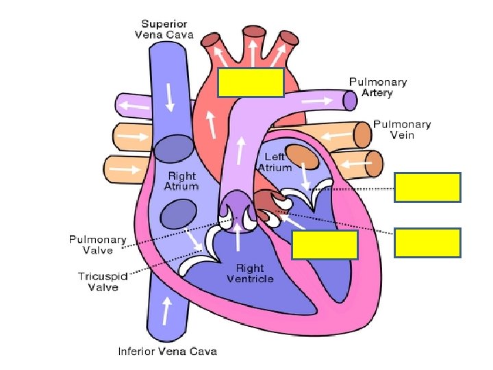
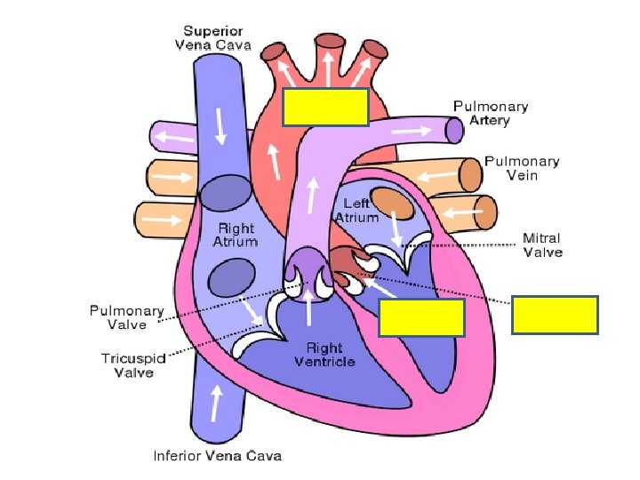
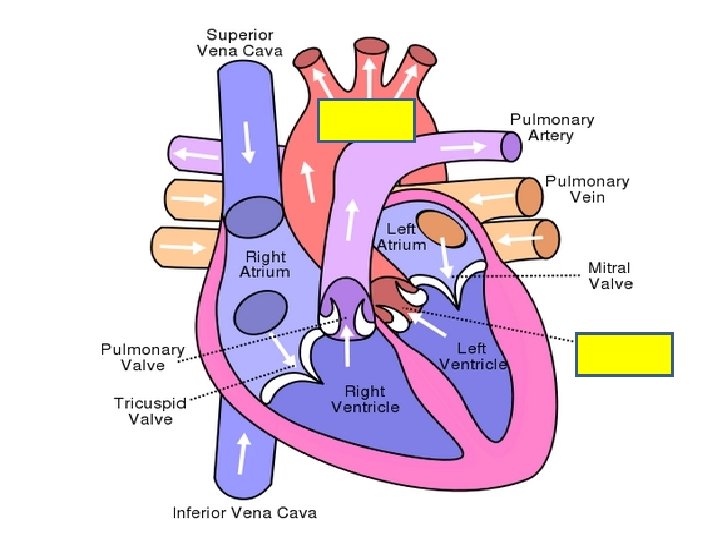
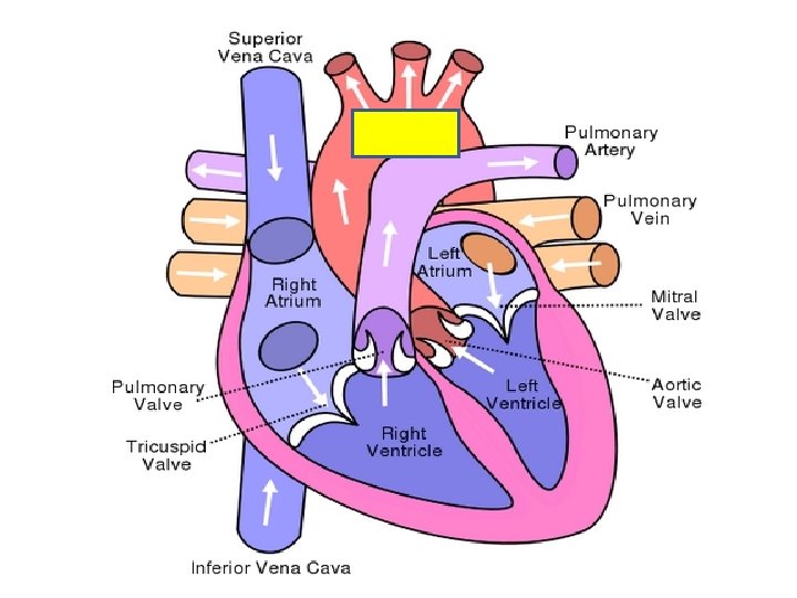
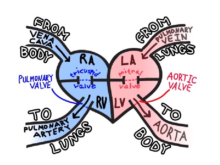
Blood Flow through the Heart • Deoxygenated blood from the body enters the inferior right atrium _______via the _____and superior vena cava ___________. • Here the blood is passed through the right ventricle tricuspid valve _______to the ________.

• The right ventricle contracts and forces blood semilunar up through the ______ valves and out pulmonary through the left and right _____ arteries. lungs • This brings blood to the ____ to be oxygenated.

• Oxygenated blood from the lungs returns to pulmonary the heart via the left and right ______ left atrium veins to the ______. left ventricle • The blood is passed to the _______ bicuspid through the _____ valve. • The left ventricle contracts and pushes blood through the semilunar valves and out through aorta the _______ to the body.

The Heartbeat Cycle • A single beat of the heart consists of contractions of the atria followed by contractions on the ventricles and then a period of relaxation of all four chambers of the heart. • Contractions are called systoles, and relaxations are called diastoles.

Video Time • https: //www. youtube. com/watch? v=j. LTdgrhp DCg

The “Lub. Dub” sound of the Heartbeat • The “Lub. Dub” sound of the heartbeat is caused by the closing of the heart’s valves. • Lub Sound -- caused by the closing of the A-V valves (tricuspid, bicuspid). • Dub Sound -- caused by the closing of the semilunar valves.

Heart Sounds • http: //depts. washington. edu/physdx/heart/d emo. html

Crash Course • https: //www. youtube. com/watch? v=X 9 ZZ 6 tcx Ar. I

CONTROL OF THE HEARTBEAT • The heart is caused to beat regularly by a sinoatrial node structure called the ________ (S - A PACEMAKER node) or the _______.

How it Happens • An electrical impulse from the brain is received by the S-A node (pacemaker) in the right atrium. The impulse spreads quickly over both atria causing atria muscles to contract. The impulse reaches the A-V node (atrioventricular node) in the right ventricle.

• The pacemaker controls the heartbeat for a human for their entire life. • The impulse is transmitted by the AV node down a nerve bundle (bundle of His). • The impulse branches to both ventricles causing contraction

Q. What happens if the pacemaker gives out? • A. The person’s heart will stop beating because the atria and ventricles are not receiving electrical impulses causing them to contract.

• A person whose pacemaker gives out can get artificial an ______ one inserted into their chest.
 at which the heart")
CONTROL OF THE HEART RATE • The heart rate (speed) at which the heart beats is controlled by two nerves. Cardioaccelerator nerve • ___________ in the Medulla Oblongata: Nerve in the brain that causes the heart to speed up when needed. Vagus nerve • ______: Nerve in the brain that causes the heart to slow down when needed
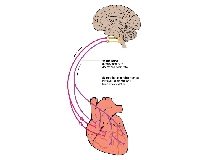

• The medulla sends a message to the SA node to cause an impulse to be sent to the AV node causing the heart to contract more or less in an attempt to set the heart rate.

BLOOD PRESSURE • Blood Pressure: A measure of the pressure blood exerts on the walls of blood vessels.

Q. How is blood pressure measured? • A. Blood pressure is measured using a blood pressure cuff or Sphygmomanometer.
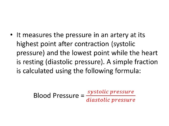

• For example: A person with a pressure 120/80 means that the person has a pressure of 120 while the heart is contracting and 80 when the heart is relaxing. • P. S. Normal blood pressure is different for each person but is usually around 120/80. (high is over 140/90)

Divisions of Circulation

There are two types of circulation that happen in the human organism. 1. Pulmonary circulation 2. Systemic circulation

Pulmonary Circulation

Systemic Circulation

Coronary Circulation

Hepatic-portal Circulation

Renal Circulation
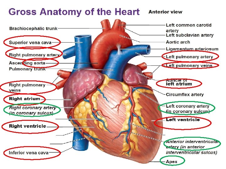

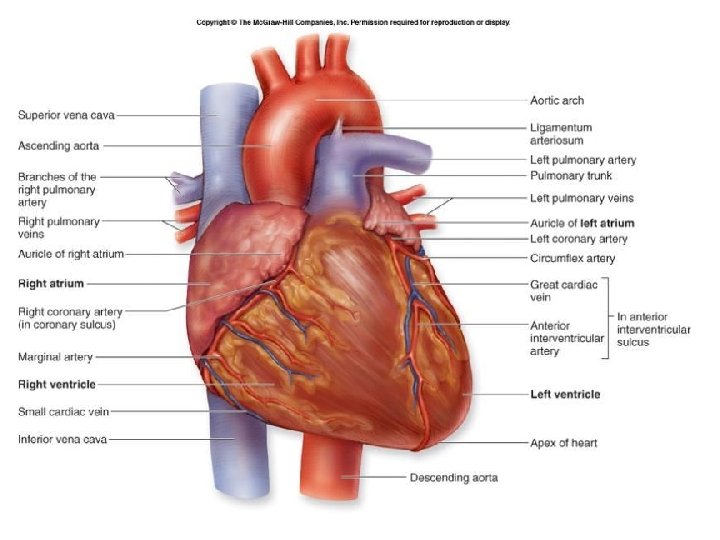
Coronary Artery • Supplies blood to nourish the heart muscles
. The atria")
Coronary Sulcus • Encircles the heart like a crown (corona = crown). The atria are found above and the ventricles are found below this blood vessel.

What happens when there is a blockage within the coronary artery? • The blood flow to the heart muscles will be blocked, leading to a heart attack.
 position of the heart. This vessel")
Anterior Interventricular Sulcus • Marks the front (ventral) position of the heart. This vessel follows the septum that separates the right and left ventricles.

Apex • The pointed end of the heart. Only the left ventricle forms the heart apex.

Pericardium • The double-walled sac around the heart. • Function: – Protects and anchors the heart – Prevents overfilling of the heart with blood – Allows for the heart to work in a relatively frictionfree environment

Myocardium • Cardiac muscle layer forming the bulk of the heart

Why do you think there is a thicker layer of myocardium in the ventricles than compared to the atria?

Why do you think there is a thicker layer of myocardium in the left ventricle than compared to the right ventricle?
- Slides: 69