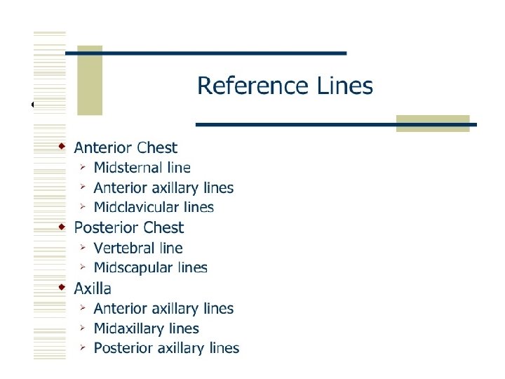
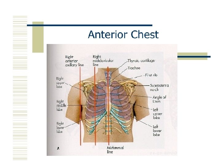
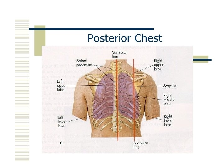
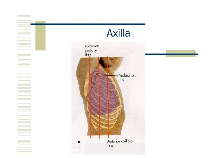
Assessment of the respiratory system TOPOGRAPHICAL LANDMARKS History


 Shape of chest – elliptical, symmetrical b) Symmetry of chest – measurement")
 Shape of chest: • Barrel chest – over inflation of lungs –")


 Breathing patterns i) Rate • Eupnea – Normal – 12 -20 /")
 Depth • Hyperpnea – h depth • Hyperventilation – h depth &")

 Rhythm • Apnea – Not breathing • Cheyne-stokes – Varying depth apnea")

 Tracheal deviation: Normally: centrally aligned Abnormal: shifted to one side • Pleural")
 Intercostal spaces: • Bulging intercostal spaces – Obstruction – Emphysema • Marked")
 Identify the areas of tenderness b) Assessment of Respiratory Excursion c) Elicit")


 • Pneumonia • Pulmonary embolism • Pleurisy")


- Slides: 57
Assessment of the respiratory system
TOPOGRAPHICAL LANDMARKS
History • • • Physical problems Functional problems Life style Smoking Past medical history Family Hx Personal and social hx Allergens / environment Anxiety
The basic steps of the examination • • Inspection Palpation Percussion Auscultation
INSPECTION a) Shape of chest – elliptical, symmetrical b) Symmetry of chest – measurement of chest expansion c) Breathing pattern d) Position of trachea e) Intercoastal spaces
Inspection a) Shape of chest: • Barrel chest – over inflation of lungs – anterior-posterior diameter increased.
Inspection • Funnel chest – Depression of the lower portion of the sternum – Complications • Heart damage • i Cardiac output
Inspection • Pigeon chest – Sternum protrudes outward – anterior-posterior diameter • h
Inspection • Scoliosis – Increased Lateral curvature of thoracic spine – Complications • Lung & heart damage • Back problems • Body image
Inspection • Kyphosis • Hunchback – Abnormal increased curvature of the thoracic spine
Inspection • Lordosis • Sway-back – Abnormal, anteriorly increased curvature of the lumbar spine
Inspection: c) Breathing patterns i) Rate • Eupnea – Normal – 12 -20 / min • Tachypnea – h rate – Pnuemonia, pulm edema, acidosis, septicemia, pain • Bradypnea – i rate
Inspection ii) Depth • Hyperpnea – h depth • Hyperventilation – h depth & rate • Hypoventilation – i depth & rate
Inspection • Kussmaul's – h rate & depth – Assoc. with sever acidosis • Apneustic – Prolonged gasping
Inspection iii) Rhythm • Apnea – Not breathing • Cheyne-stokes – Varying depth apnea
Inspection • Biot’s – h rate & depth w/ abrupt pauses
Inspection: d) Tracheal deviation: Normally: centrally aligned Abnormal: shifted to one side • Pleural effusion • Tension pneumothorax • Atelectasis
Inspection e) Intercostal spaces: • Bulging intercostal spaces – Obstruction – Emphysema • Marked retraction of intercostal spaces – Blockage
Palpation a) Identify the areas of tenderness b) Assessment of Respiratory Excursion c) Elicit tactile fremitus
Assessment of Respiratory Excursion. posterior • Place your thumbs about the level of and parallel to the 10 th rib, your hands grasping the lateral rib cage. • As you position your hands, slide them medially in order to raise loose skin folds between your thumbs and the patient's spine. • Feel for range of symmetry of respiratory movement.
Assessment of Respiratory Excursion. Anterior • Place your thumbs along each costal margin with your hands along the lateral rib cage. • As you position your hands, slide them medially a bit to raise a between the thumbs. • Ask the patient to inhale deeply. • Watch for your thumbs to separate as the thorax expands. • Feel for the range and symmetry of respiratory movement
Elicit tactile fremitus • Use the ball of the hand (the palm of the hand at the base of the fingers), palpate and compare like areas of the lungs. . Do not let your fingers touch the patient's chest. • Have the patient repeat a sound that will make full and rich sounds such as "ninety-nine" or "one-oneone. " Symmetrically move your hand over the patient's chest. • You should feel vibrations of equal intensity on either side of the patient's chest.
Percussion Rational • To determine if underlying tissue is filled with air or solid material Procedure • Pt sitting • Tap starting at shoulder • compare right to left
Percussion: results • Resonance – drum like – Normal • Hyper-resonance – Too much air – Emphysema • Flatness / dull – – Fluid or solid Pleural effusion Pneumonia Tumor
Percussion
Percussion
Auscultation of the Lungs Ø Sitting is the optimal position Ø Anterior and posterior walls Ø Expose the chest and ask the patient to breath deeply through open mouth Ø Compare the pitch, intensity and quality of breathing sounds of one side to the other side Ø Perform symmetrically Ø Prevent the patient from fall Ø Prevent the patient from dizziness secondary to hyperventilation Ø Rechecking can be done after deep inspiration
Auscultation Process of listening to sounds within the body specifically breath sounds during examination of the lungs. Breath sounds occur because of movement of air in the airways during inspiration and expiration. Purpose • Asses air flow through bronchial tree Procedure • Diaphragm of stethoscope • Superior inferior • Compare rt to lft
Auscultation: Results Normal • Vesicular – Lung field – Soft and low • Bronchial – Trachea & bronchi – Hollow • Bronchovesicular – – Mixed Between scapulae Side of sternum 1 st & 2 nd intercostal space
Auscultation: Results Adventitious • Crackles (short interrupted sounds due to opening of previously closed airways) • On start of inspiration: problem in large airways • On mid of inspiration: medium airways are closed • On end of inspiration: smaller airways or lung tissues
Auscultation: Results • Wheezes – Sonorous wheezes • Deep low pitched • Snoring • Caused by air narrowed passages • h secretions • Pleural effusion – Sibilant Wheezes • High pitched • Whistle-like • Caused by air narrowed passages • constriction – Asthma
Auscultation: Results • Pleural friction rub – inflammation of pleural membranes, pleural surfaces rub together – Produced due to friction between two surfaces – Grating, creaking sound – Best heard • Anterior, Lower, lateral area
Auscultation: Results • Stridor – Crowing – Partial obstruction of the larynx or trachea
Dyspnea • Significance – Common with cardiac & resp. disease – Sudden onset – healthy person • Pneumothorax – Sudden onset ill, post-op or injury • Pulmonary emboli
Dyspnea • Orthopnea – Sit up to breath • COPD • CHF
Cough is a forced expulsive maneuver against an initially closed glottis, causing a characteristic sound. – Acute: lasting less than 3 weeks – Chronic: more than 8 weeks
Cough Onset Duration Nature Sputum Severity Associated sypmtoms
Sputum Production Definition • Matter discharged from resp. tract that contains mucus and pus, blood, fibrin, or bacteria
Sputum Production Significance • Purulent – Thick, yellow/green – Bacteria • Rusty • Thin, mucous – Viral
Sputum Production • Pink-tinged – Lung CA – TB • Pink tinged, profuse, – Pulmonary edema • Malodorous – Lung abscess
Chest pain Definition • Cardiac or pulmonary
Chest pain Significance • CA (late stage) • Pneumonia • Pulmonary embolism • Pleurisy
Chest pain • Pleurisy – Inflammation of pleura – Sharp with breath – i breath sounds
Clubbed fingers Definition • Sponginess of the nail bed • Loss of the nail bed angle • Finger tip is round and bulbous
Clubbed Finger Significance • chronic hypoxia
Hemoptysis Definition • Expectoration of blood from the respiratory tract
Hemoptysis Significance • Pulm or cardiac • Common causes – – – Pulm infection CA of lungs Pulm. Emboli Pulm. Infarction TB
Hemoptysis • Hemoptysis – Definition? • Coughed up blood – From? • Pulm hemorrhage – Description • Pink, red, mixed with sputum – Blood p. H • Alkaline blood • Hematemesis – Definition? • Vomited blood – From? • Stomach / GI – Description • “Coffee ground” – Blood ph • Acidic blood
Cyanosis Definition • Bluish coloring of skin
Cyanosis Significance • Very late indicator of hypoxia • Appears when O 2 sats < 85% • NOT a reliable sign of hypoxia – Esp. with anemia
Cyanosis • O 2 sat definition – % of hemoglobin carrying oxygen compared to total # of hemoglobin
Cyanosis • Normal Breath 100 O 2 molecules 98 cross into blood Blood: 100 Hgb • O 2 SATS – 98% – No cyanosis
Cyanosis • Hypoxia Breath 100 O 2 molecules 75 cross into blood Blood: 100 Hgb • O 2 SATS – 75% – Cyanosis