Chapter 13 Part 1 The Respiratory System Organs


 · Pharynx - Muscular passage from nasal cavity to larynx · About")
 · Three regions of the pharynx: · Nasopharynx – superior region behind")

 are also found in")

 · Functions of the Larynx: 1. Routes air and food into")


 · Pair of folds ·")
 · Connects larynx with bronchi · About 4 inches long · Lined")

 · The trachea is fairly rigid because its walls are reinforced with")


 pleura covers the lung surface · Parietal")


 – Has gas (air) flowing past on")


")
- Slides: 52
Chapter 13 – Part 1 The Respiratory System
Organs of the Respiratory system · Nose · Pharynx · Larynx · Trachea · Bronchi · Lungs – alveoli
Function of the Respiratory System · Oversees gas exchanges between the blood and external environment · Exchange of gasses takes place within the lungs in the alveoli · Passageways to the lungs purify, warm, and humidify the incoming air
The Nose · The only externally visible part of the respiratory system · Air enters the nose through the external nares (nostrils) · The interior of the nose consists of a nasal cavity divided by a nasal septum
Figure 13. 2
Anatomy of the Nasal Cavity · Olfactory receptors for the sense of smell are located in the mucosa on the slitlike superior part of the nasal cavity · The rest of the cavity is lined with respiratory mucosa · Warm the air · Moistens the air · Traps incoming foreign particles (cleanse)
Anatomy of the Nasal Cavity · The ciliated cells of the nasal mucosa create a gentle current that moves contaminated mucous posteriorly towards the throat (pharynx) · It is then swallowed and digested by stomach juices. · When it is extremely cold, these cilia become sluggish, allowing mucus to accumulate in the nasal cavity and to dribble outward through the nostrils. · This is why you have a “runny” nose on a cold day.
Nosebleeds · The respiratory mucosa rests on a rich network of thin-walled veins (warms the air as it flows by). · Because of the superficial location of these blood vessels, nosebleeds are common and often profuse.
Anatomy of the Nasal Cavity · The lateral walls of the nasal cavity have three projections or lobes called conchae · Increases surface area · Increases air turbulence within the nasal cavity · Helps to deflect inhaled particles onto the mucus-coated surfaces, where they are trapped and prevented from entering the lungs.
Anatomy of the Nasal Cavity · The nasal cavity is separated from the oral cavity by the palate · Anterior hard palate (bone) · Posterior soft palate (muscle)
Cleft Palate · Cleft palate – The bones forming the palate fail to fuse medially · Genetic defect · Results in breathing difficulties and problems with oral cavity functions (chewing and speaking)
Paranasal Sinuses · The nasal cavity is surrounded by a ring of paranasal sinuses. · Are located in the: · Frontal bone · Sphenoid bone · Ethmoid bone · Maxillary bone
Paranasal Sinuses · Function of the sinuses 1. Lighten the skull 2. Act as resonance chambers for speech 3. Produce mucus that drains into the nasal cavity • The suctioning effect created by nose blowing helps to drain the sinuses. • The nasolacrimal ducts, which drain tears from the eyes, also empty into the nasal cavities
Sinusitis · Sinusitis – sinus inflammation · Difficult to treat · Can cause marked changes in voice quality · When the passageways connecting the sinuses to the nasal cavity are blocked with mucus or infectious matter, the air in the sinus cavities is absorbed · The result is a partial vacuum and a sinus headache
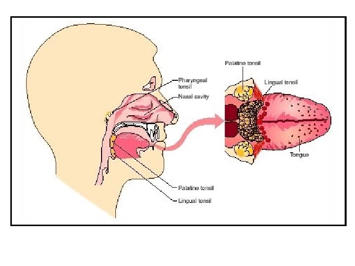
Pharynx (Throat) · Pharynx - Muscular passage from nasal cavity to larynx · About 5 inches long · Commonly called the throat · Serves as a common passageway for food and air · Is continuous with the nasal cavity anteriorly via the internal nares
Pharynx (Throat) · Three regions of the pharynx: · Nasopharynx – superior region behind nasal cavity · Oropharynx – middle region behind mouth · Laryngopharynx – inferior region attached to larynx · The oropharynx and laryngopharynx are common passageways for air and food · Air then passes through the larynx, while food is directed into the esophagus posteriorly
Structures of the Pharynx · The auditory tubes, which drain the middle ear, open into the nasopharynx · Since the mucosae of these two regions are continous, ear infections may follow a sore throat or other types of pharyngeal infections
Structures of the Pharynx · Tonsils (clusters of lymphatic tissue) are also found in the pharynx · Their job is to trap and remove any bacteria or other foreign pathogens entering the throat · Pharyngeal tonsil – located high in the nasopharynx · Palatine tonsils – located in the oropharynx at the end of the soft palate · Lingual tonsils – located at the base of the tongue
Tonsillitis · Tonsillitis – Inflammation and swelling of the pharyngeal tonsil · Can occur during a bacterial infection · It obstructs the nasopharnyx and forces the person to breathe through the mouth · In mouth breathing, air is not properly moistened, warmed, or filtered before entering the lungs · Years ago, the belief was that the tonsils were more trouble than they were worth and they were routinely removed. · Now, this is no longer necessary because of the large use of antibiotics
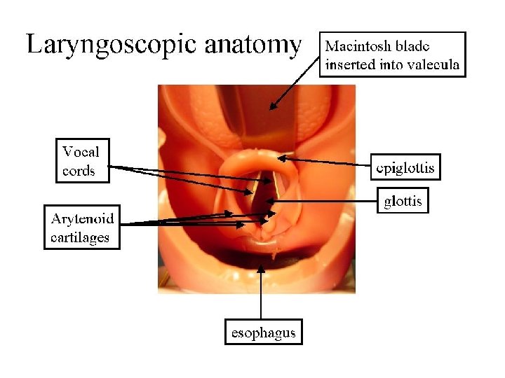
Larynx (Voice Box) · Functions of the Larynx: 1. Routes air and food into proper channels 2. Plays a role in speech (voice production) 3. Acts as an airway · Made of eight rigid hyaline cartilages and a spoon-shaped flap of elastic cartilage (epiglottis)
Structures of the Larynx · Thyroid cartilage · Largest hyaline cartilage · Shield-shaped · Protrudes anteriorly · Commonly called the Adam’s apple
Structures of the Larynx · Epiglottis · Protects the superior opening of the larynx · Routes food to the esophagus and air toward the trachea · The epiglottis moves positions when swallowing · When we are not swallowing: the epiglottis does not restrict the passage of air into the lower respiratory passages · When we are swallowing: the larynx is pulled upward and the epiglottis tips, forming a lid over the opening of the larynx; this routes food into the esophagus
Structures of the Larynx · Palpate your larynx by placing your hand midway on the anterior surface of your neck. Swallow. Can you feel the larynx rising as you swallow?
Cough Reflex · If anything other than air enters the larynx, a cough reflex is triggered to expel the substance and to prevent it from continuing into the lungs. · Because this protective reflex does not work when we are unconscious, it is never a good idea to try to give fluids to an unconscious person when attempting to revive him or her.
Structures of the Larynx · Vocal cords (vocal folds) · Pair of folds · Vibrate with expelled air to create sound · Allows us to speak · Glottis – the slitlike passageway between the vocal cords
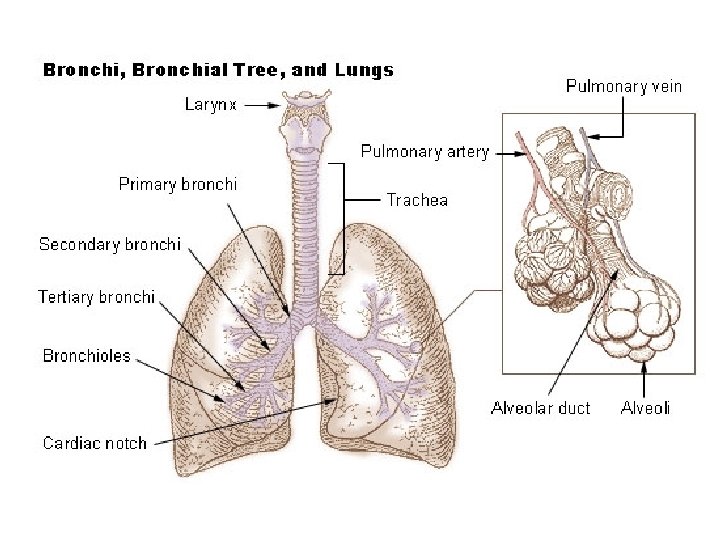
Trachea (Windpipe) · Connects larynx with bronchi · About 4 inches long · Lined with ciliated mucosa · Beat continuously in the opposite direction of incoming air · Propels mucus loaded with dust and other debris away from lungs to the throat, where it can be swallowed or spat out
The Trachea and Smoking · Smoking inhibits ciliary activity and ultimately destroys the cilia · Without these cilia, coughing is the only means of preventing mucus from accumulating in the lungs
Trachea (Windpipe) · The trachea is fairly rigid because its walls are reinforced with C-shaped hyaline cartilage · These rings form two purposes: 1. Support the trachea and keep it open in spite of the pressure changes that occur during breathing 2. Allows it to expand anteriorly when we swallow a large piece of food
Heimlich Maneuver · Heimlich maneuver – a procedure in which the air in a person’s own lungs is used to “pop out, ” or expel, an obstructing piece of food · Because the trachea is the only way air can get into the lungs, tracheal obstruction is lifethreatening · Many people have suffocated after choking on a piece of food that suddenly closed off the trachea · Has saved many people from choking to death
Primary Bronchi · The right and left primary bronchi is formed by the division of the trachea · Enters the lung at the hilus (medial depression) · Right bronchus is wider, shorter, and straighter than left · Consequently it is the more common site for an inhaled foreign object to become lodged · Bronchi subdivide into smaller and smaller branches · By the time air enters the bronchi, it is warmed, cleansed of most impurities, and well humidified
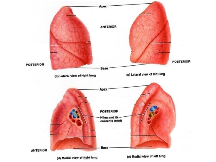
Lungs · The paired lungs are fairly large organs · Occupy most of the thoracic cavity · Apex is near the clavicle (narrow, superior portion) · The broad base rests on the diaphragm (inferior portion) · The bronchi enters the lung at the hilus (medial depression)
Lungs · Each lung is divided into lobes by fissures · Left Lung Has two lobes · Right Lung Has three lobes
Coverings of the Lungs · Pulmonary (visceral) pleura covers the lung surface · Parietal pleura lines the walls of the thoracic cavity · Pleural fluid fills the area between layers of pleura to allow gliding during breathing movements · Can slide easily from side to side across one another, but resists being pulled apart.
Lungs
Coverings of the Lungs · Pleurisy – Inflammation of the pleura · Can be caused by the decreased secretion of pleural fluid · The pleural surfaces become dry and rough · Results in friction and stabbing pain with each breath
Respiratory Tree Divisions · This branching and rebranching within the lungs is often referred to as the bronchial or respiratory tree: 1. Primary bronchi 2. Secondary bronchi 3. Tertiary bronchi 4. Bronchioles 5. Terminal bronchioles
Bronchioles · Bronchioles - Smallest branches of the bronchi · All but the smallest branches have reinforcing cartilage
Bronchioles · Terminal bronchioles end in alveoli, or air sacs.
Respiratory Zone · The respiratory zone is the only site of gas exchange · Includes the following structures: · Respiratory bronchioles · Alveolar ducts · Alveolar sacs · Alveoli · All other respiratory passages are conducting zone structures · Serve as conduits to and from the respiratory zone.
Alveoli · There are millions of the clustered alveoli, which resemble bunches of grapes. · They make up the bulk of the lungs · Consequently, the lungs are mostly air space · In spite of their relatively large size, the lungs weigh only about 2 ½ pounds, and they are soft and spongy
Alveoli · Structure of alveoli · Alveolar duct · Alveolar sac · Alveoli · Gas exchange takes place within the alveoli in the respiratory membrane
Alveoli · Structure of alveoli · Alveolar duct · Alveolar sac · Alveoli · Gas exchange takes place within the alveoli in the respiratory membrane
Respiratory Membrane · Respiratory Membrane (Air-Blood Barrier) – Has gas (air) flowing past on one side and blood flowing past on the other · Made up of the alveolar and capillary walls and their fused basement membranes.
Respiratory Membrane · The walls of the alveoli are composed largely of a single, thin layer of squamous epithelial cells · The thinness of their walls is hard to imagine, but a sheet of tissue paper is much thicker · The external surfaces of the alveoli are covered with a “cobweb” of pulmonary capillaries
Respiratory Membrane · Alveolar pores connect neighboring air sacs and provide alternate routes for air to reach alveoli · In case feeder bronchioles have been clogged by mucus or otherwise blocked
Gas Exchange · Gas exchanges occur by simple diffusion through the respiratory membrane · Oxygen enters the blood · Carbon dioxide enters the alveoli · The total gas exchange surface provided by the alveolar walls is 40 times greater than the surface of your skin
Gas Exchange · The final line of defense for the respiratory system is in the alveoli · Macrophages wander in and out of the alveoli picking up bacteria, carbon particles, and other debris · Surfactant coats the gas-exposed alveolar surfaces · Lowers the surface tension of the film of water lining each alveolar sac so that the alveoli do not collapse between each breathe
Respiratory Membrane (Air-Blood Barrier)