Respiratory System Respiratory system structure Upper respiratory system


 • Function: – Filtering")


")


- Slides: 30
Respiratory System
Respiratory system structure • Upper respiratory system – – – nose Pharynx larynx Upper part Functions: 1. Filtration 2. Warming 3. Moistering 4. Sound produce • Lower respiratory system ( from the larynx down ) – – – trachea bronchi Lungs Functions = gas exchange
Figure 23. 1
The Nose • Otorhinolaryngology (Oto= Ear, Rhino= Nose, Larynx= Throat) • Function: – Filtering Air – Odors Detection – Amplifies The Voice – Moisture Addition – Warming Air
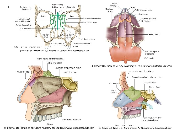
External nose 1. Cartilage 2. Nasal bones
Nasal chamber surface Superior surface Ethmoeid Sphenoied Lateral surface 3 choncae ( superior - middle - inferior ) 3 Meatuses ( superior - middle - inferior ) Meatuses function : produce air turbulence warm and humidify incoming air trap particles Inferior surface (floor of nasal cavity ) Hard palate + Soft palate Medial surface ( septum ) the perpendicular plate of the ethmoid bone (superior) the vomer (inferior) septial cartilage (anterior)
Nostrils Coana nasal vestibule contain of nasal hairs first particle filtration system
Para nasal sinuses 1. Maxillary sinus 2. Ethmoied sinus 3. Frontal sinus 4. Sphenoid sinus
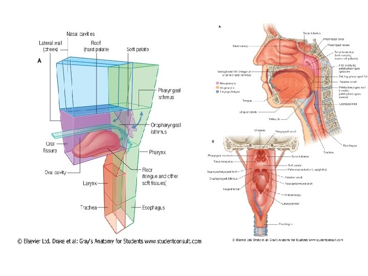
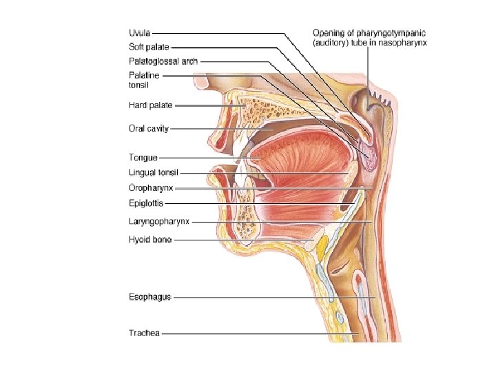
The Pharynx A chamber shared by digestive and respiratory systems Extends from internal coana to entrances to larynx and esophagus
The Nasopharynx • Superior portion of the pharynx • Contains : 1. 2. 3. pharyngeal tonsils C 1 openings to left and right auditory tubes
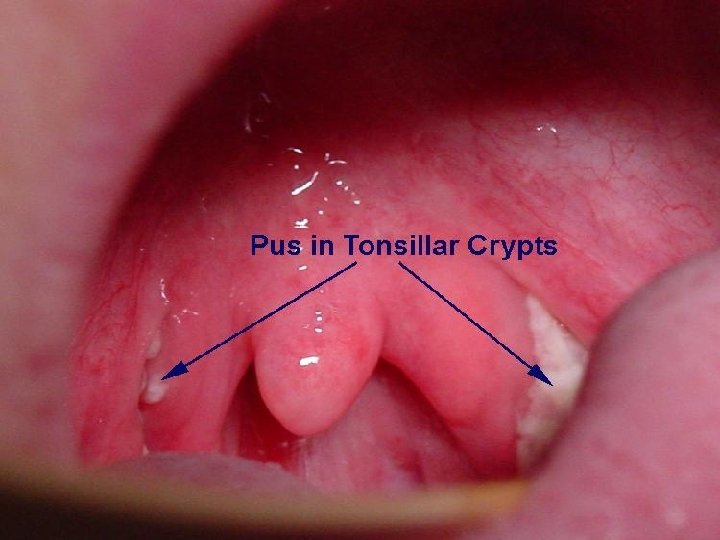
The Oropharynx • Middle portion of the pharynx • C 2 • Communicates with oral cavity • Contain of palatal tonsils
The Laryngopharynx • Inferior portion of the pharynx • C 3 , C 4 , C 5 , C 6 • Extends to esophagus
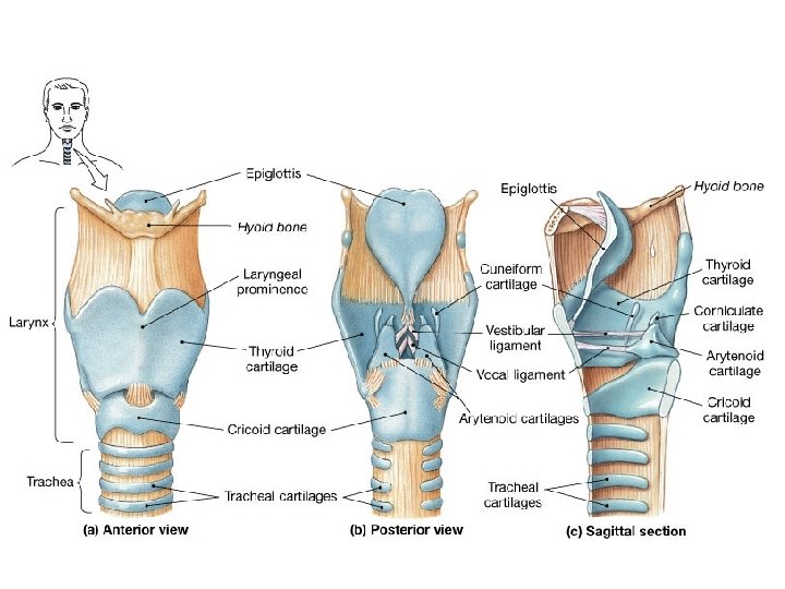
Larynx • Function : air way • C 3 ~ C 6 • 4 cm • Glottis & sound production
Cartilages of the Larynx • 9 cartilage : • unpaired : 1. thyroid 2. epiglotic 3. cricoid • Pair : 1. arytenoid 2. corniculate 3. cuneiform
The Glottis Two pairs of folds : 1. Vestibular folds or false vocal cords ( upper ) 2. Vocal folds or true vocal cords ( lower )
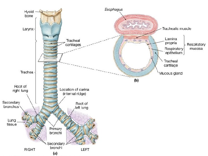
Trachea • Anterior to esophagus • From the cricoid cartilage C 6 • At T 5 it divides into right and left bronchi (CARINA ) • Tracheal rings- composed of, in the shape of a “c” • 15– 20 tracheal cartilages ( hyaline cartilage ) : – The “c” opens posterior to allow the esophagus to expand while swallowing – Protect airway – Trachealis muscle
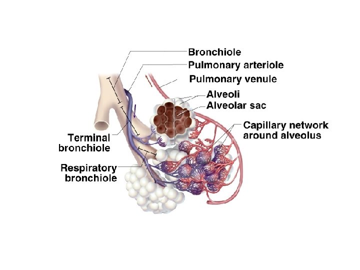
Bronchial division 1. R & L bronchus 2. Lobar bronchus ( 3 in R --- 2 in L ) 3. Segmental bronchus 4. Bronchiole 5. Terminal Bronchiole 6. Respiratory Bronchiole 7. Alveolar duct 8. Alveolar sac
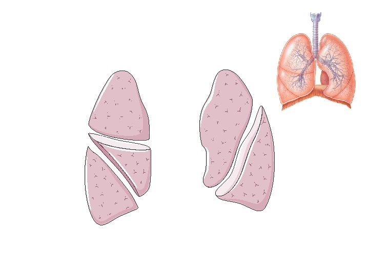
Lungs • Cone shaped organs • Lie in thoracic cavity • Separated by the mediastinum • surrounded by the pleural membrane • The pleural membrane has two layers: – Parietal pleura – Visceral pleura • Base- inferior portion • Apex- superior portion, narrow • Costal surface • Mediastinal -- contains 0 f hilus
• Right lung: – is wider – is displaced upward by liver – Has 3 lobes: separated by horizontal and oblique fissures – Superior – Middle – inferior • Left lung: – is longer – cardiac notch – Has 2 lobes: are separated by an oblique fissure 1. Superior 10 segments per lung 2. inferior Each segment contains lobules
Lobes (1)
Hillum
Radiographic Landmarks