
Good Morning CHEST RADIOGRAPHS More Than 50 Shades






- Slides: 68
Good Morning!
CHEST RADIOGRAPHS: More Than 50 Shades of Grey* Donna Magid, MD, M. Ed * More like 1024…
Lecture objectives • Introduction and basic approach to the chest radiograph (Ur unit of imaging) • Correlation between anatomy and radiology • Extrapolation from image to patient • Just a hint of future clinical relevance … which will be revisited and expanded across this Horizontal Strand
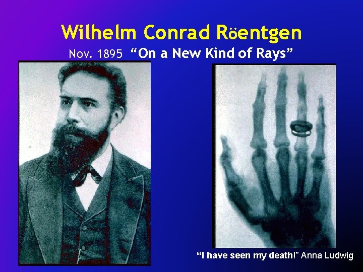
Wilhelm Conrad Röentgen Nov. 1895 “On a New Kind of Rays” “I have seen my death!” Anna Ludwig
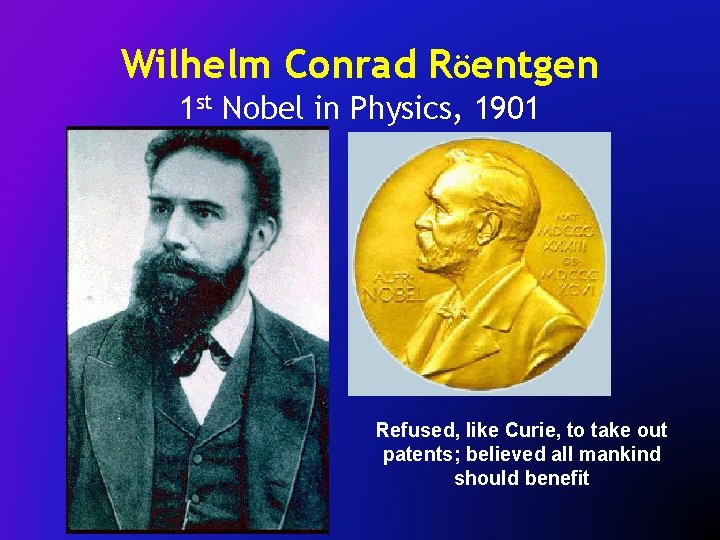
Wilhelm Conrad Röentgen 1 st Nobel in Physics, 1901 Refused, like Curie, to take out patents; believed all mankind should benefit
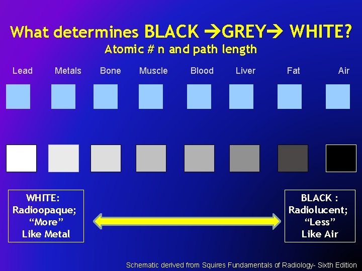
What determines BLACK GREY WHITE? Atomic # n and path length Lead Metals WHITE: Radioopaque; “More” Like Metal Bone Muscle Blood Liver Fat Air BLACK : Radiolucent; “Less” Like Air Schematic derived from Squires Fundamentals of Radiology- Sixth Edition
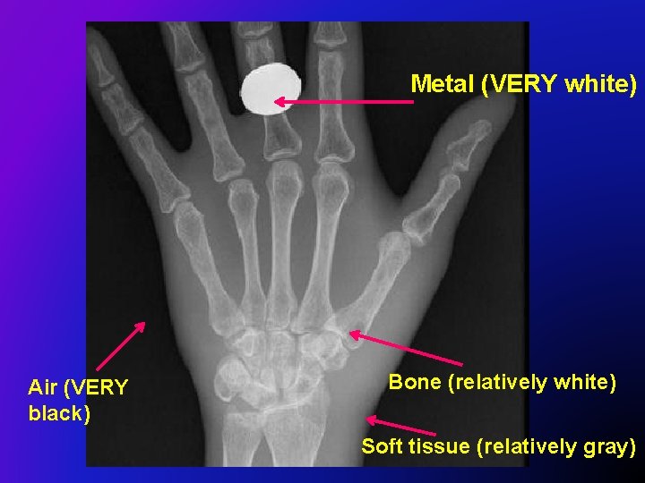
Metal (VERY white) Air (VERY black) Bone (relatively white) Soft tissue (relatively gray)
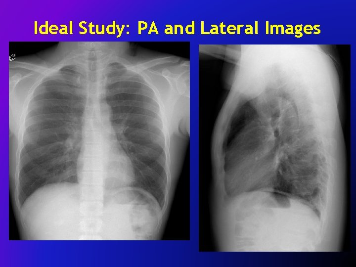
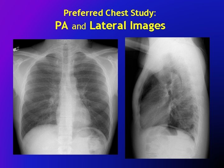
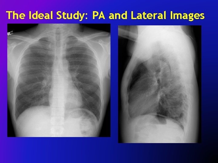
Ideal Study: PA and Lateral Images
“ONE View is NO View”
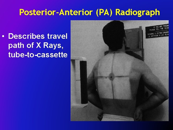
AP vs PA Chest xray’s path, (source cassette) A P P A
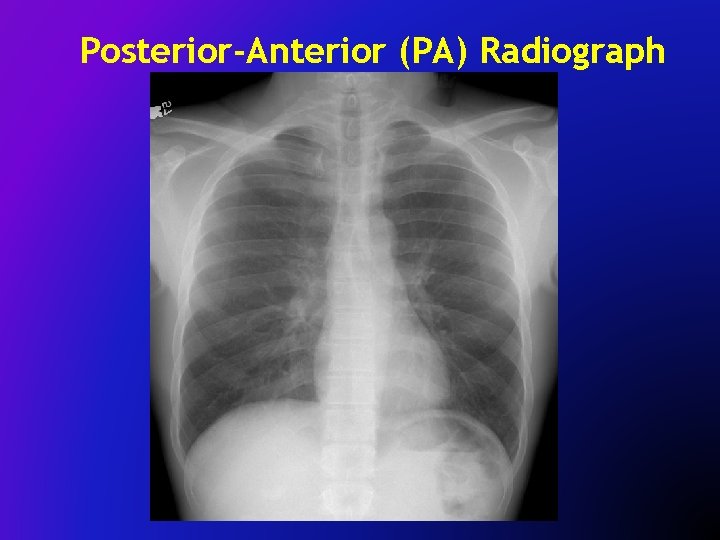
Posterior-Anterior (PA) Radiograph • Describes travel path of X Rays, tube-to-cassette
Posterior-Anterior (PA) Radiograph
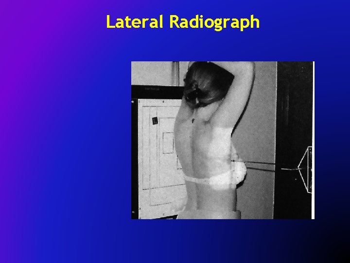
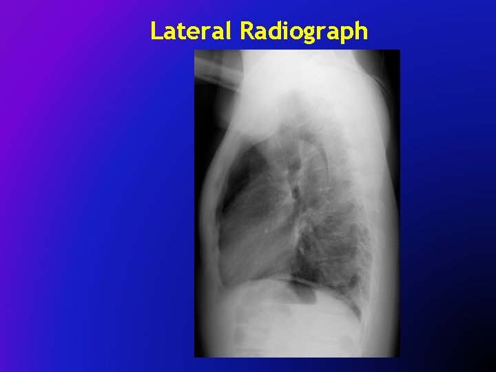
Lateral Radiograph
Lateral Radiograph
Preferred Chest Study: PA and Lateral Images
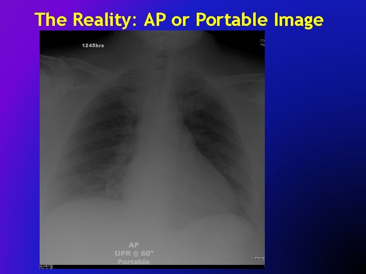
The Reality: AP or Portable Image
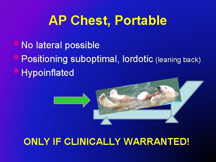
AP Chest, Portable § No lateral possible § Positioning suboptimal, lordotic (leaning back) § Hypoinflated ONLY IF CLINICALLY WARRANTED!
The Reality: AP or Portable Image
The Ideal Study: PA and Lateral Images
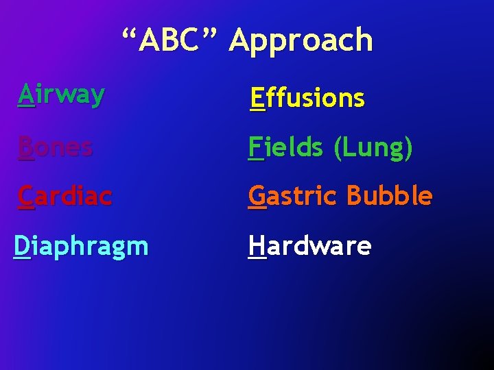
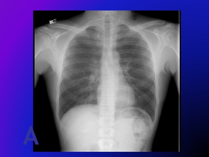
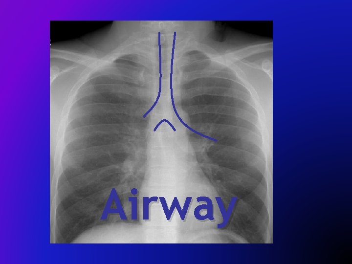
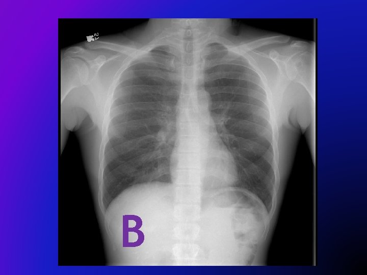
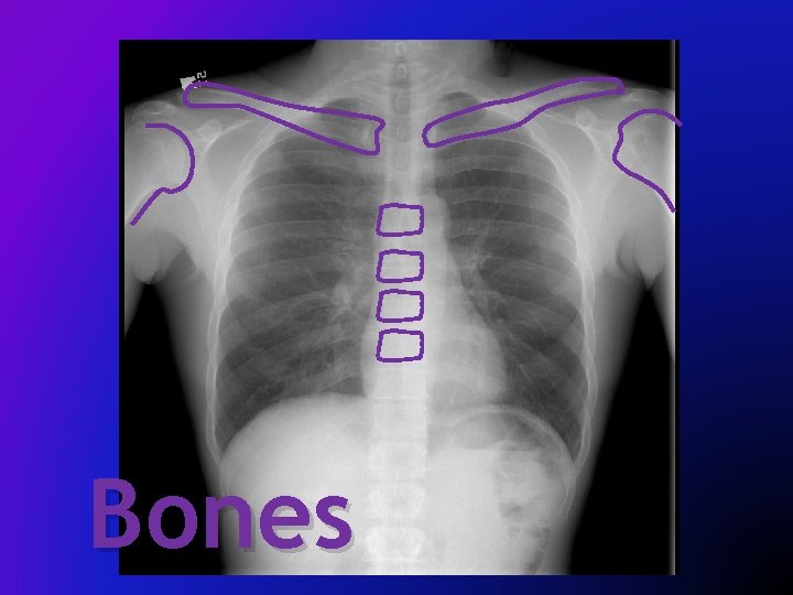
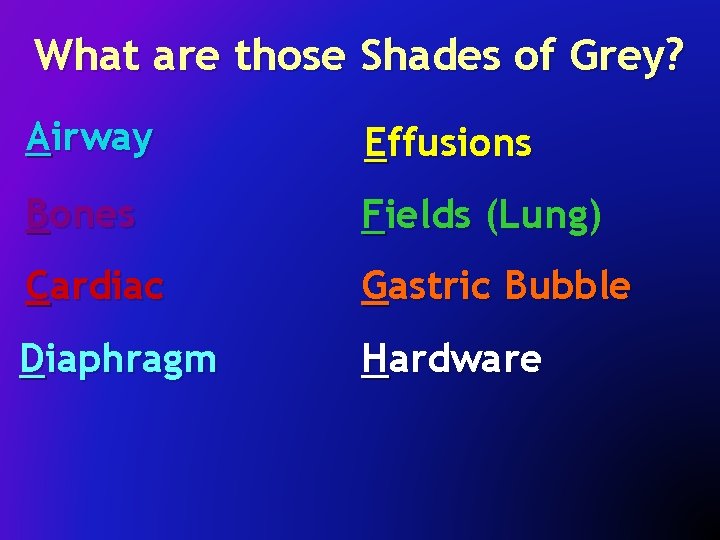
“ABC” Approach Airway Effusions Bones Fields (Lung) Cardiac Gastric Bubble Diaphragm Hardware
A
Airway
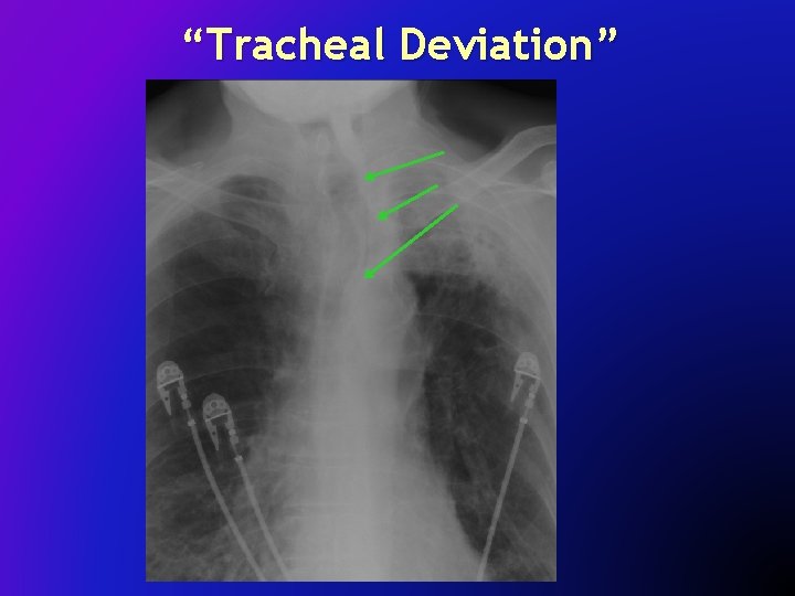
“Tracheal Deviation”
B
Bones
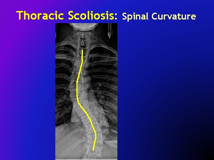
Thoracic Scoliosis: Spinal Curvature
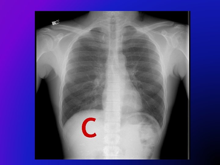
C
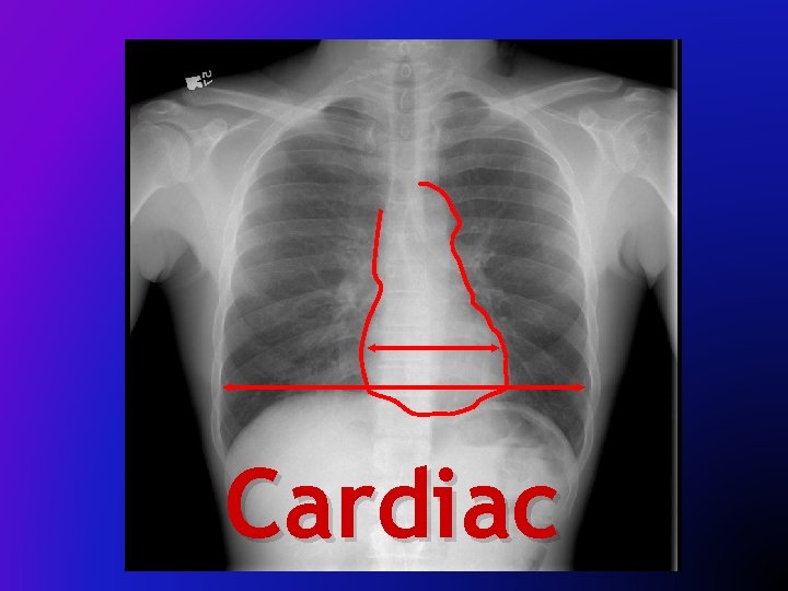
Cardiac
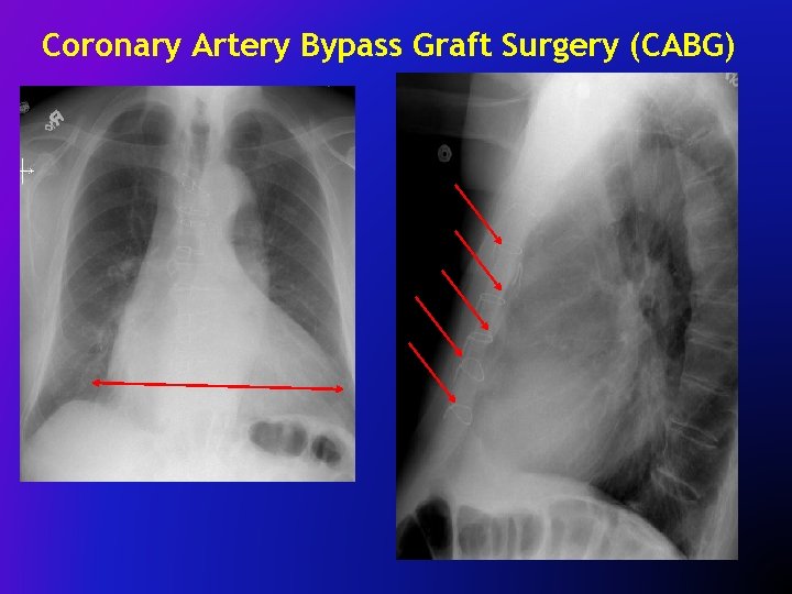
Coronary Artery Bypass Graft Surgery (CABG)
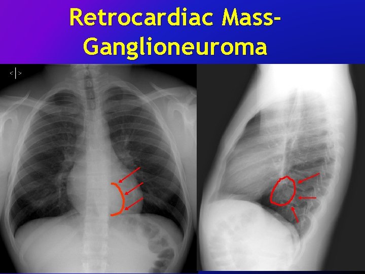
Retrocardiac Mass. Ganglioneuroma
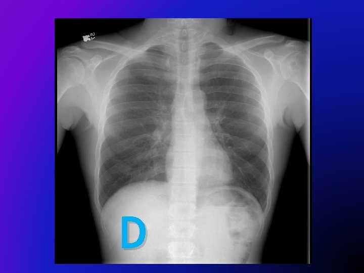
D
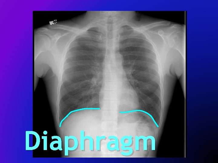
Diaphragm
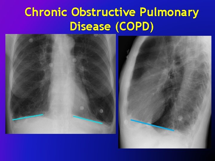
Chronic Obstructive Pulmonary Disease (COPD)
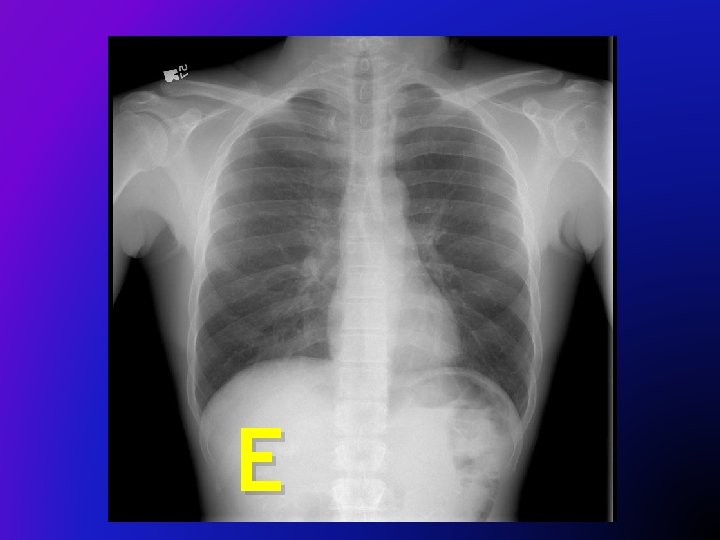
E
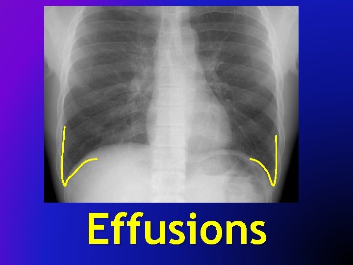
Effusions
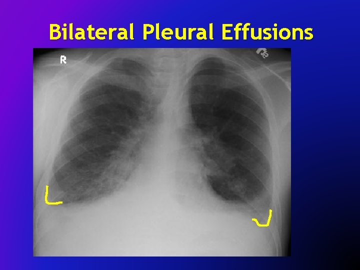
Bilateral Pleural Effusions
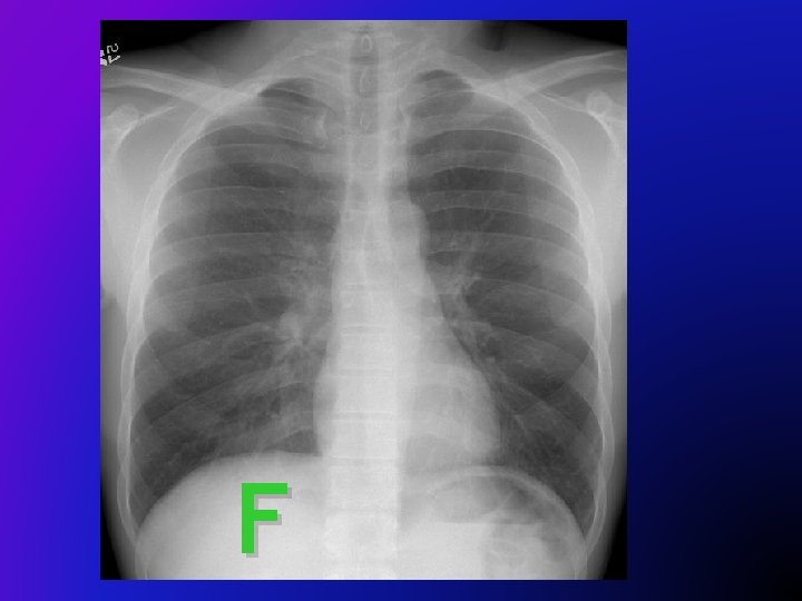
F
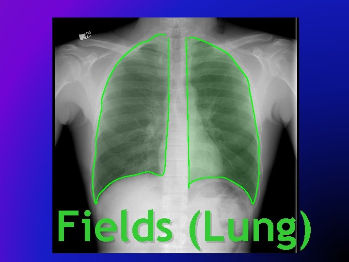
Fields (Lung)
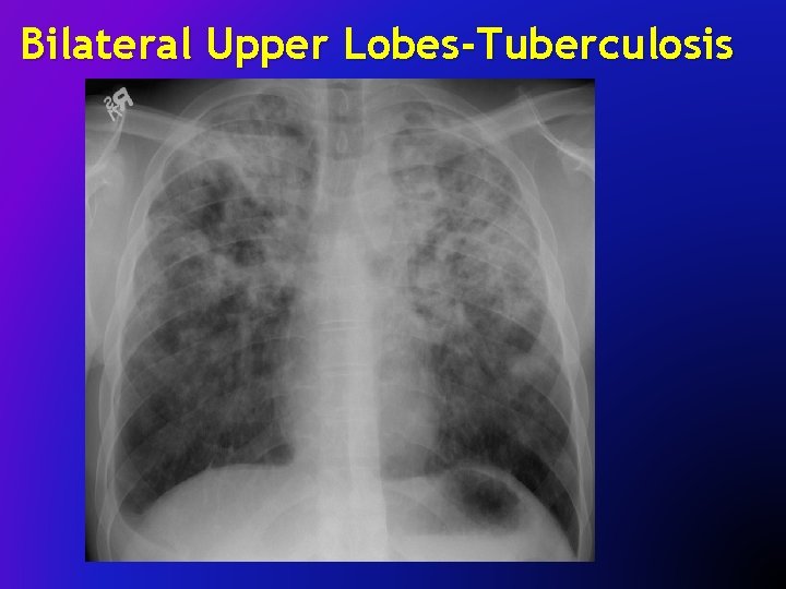
Bilateral Upper Lobes-Tuberculosis
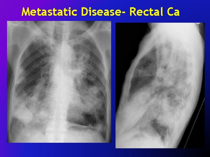
Metastatic Disease- Rectal Ca
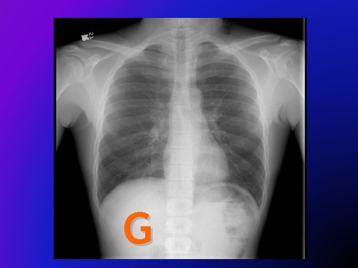
G
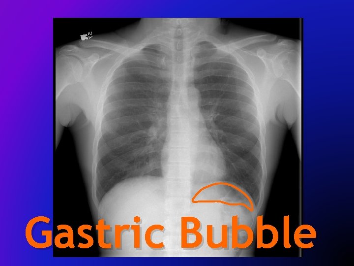
Gastric Bubble
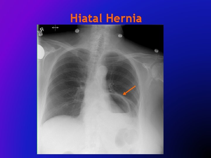
Hiatal Hernia
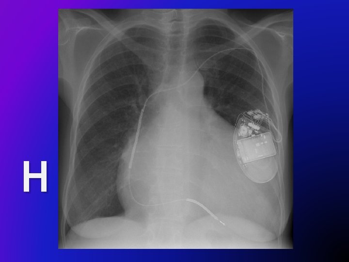
H
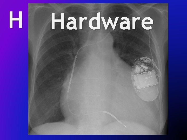
H Hardware
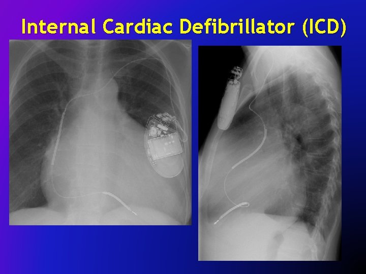
Internal Cardiac Defibrillator (ICD)
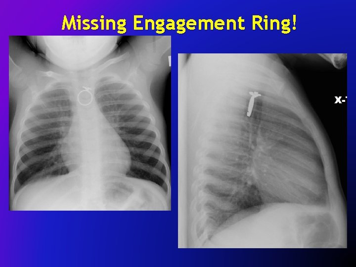
Missing Engagement Ring!
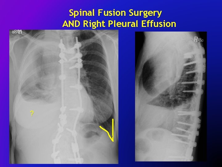
Spinal Fusion Surgery AND Right Pleural Effusion Effusio ?
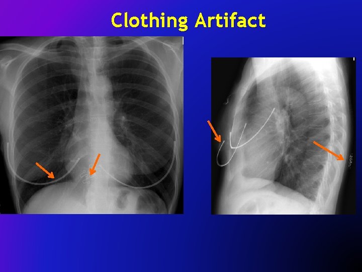
Clothing Artifact
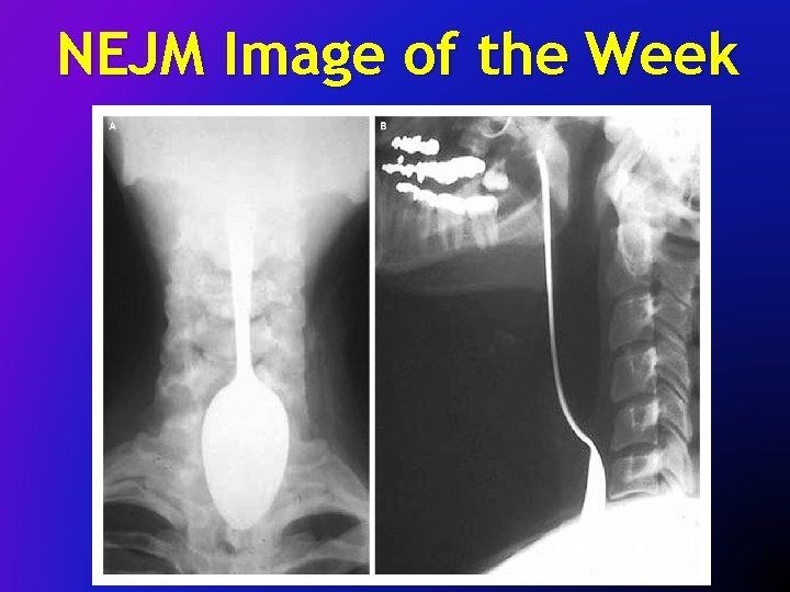
NEJM Image of the Week
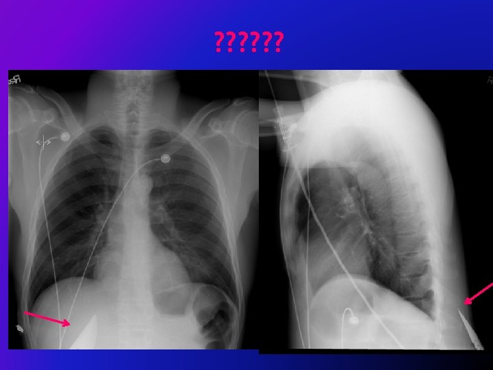
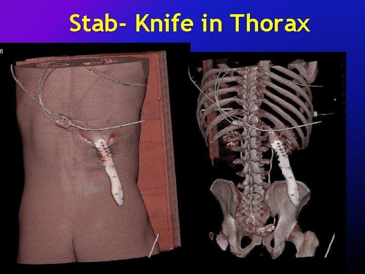
Stab- Knife in Thorax
What are those Shades of Grey? Airway Effusions Bones Fields (Lung) Cardiac Gastric Bubble Diaphragm Hardware
What else can radiologists do in the THORAX? ?
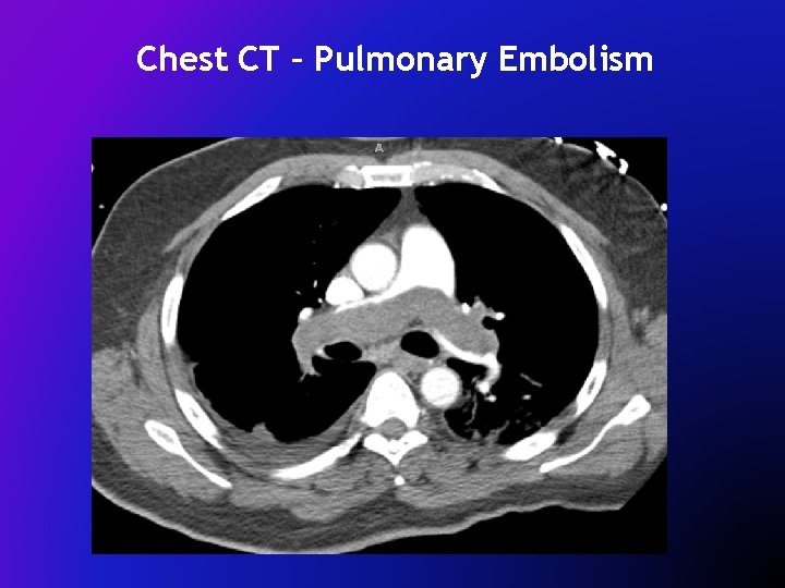
Chest CT – Pulmonary Embolism
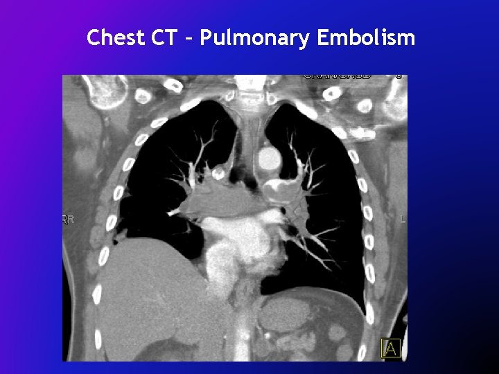
Chest CT – Pulmonary Embolism
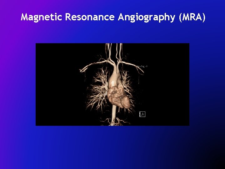
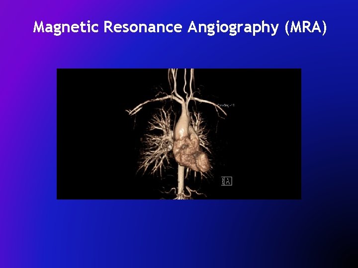
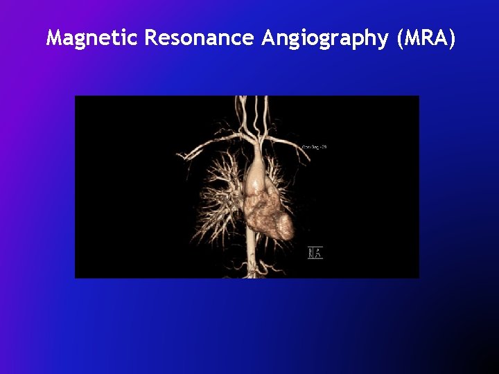
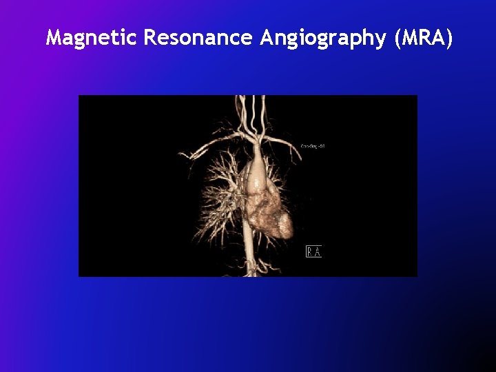
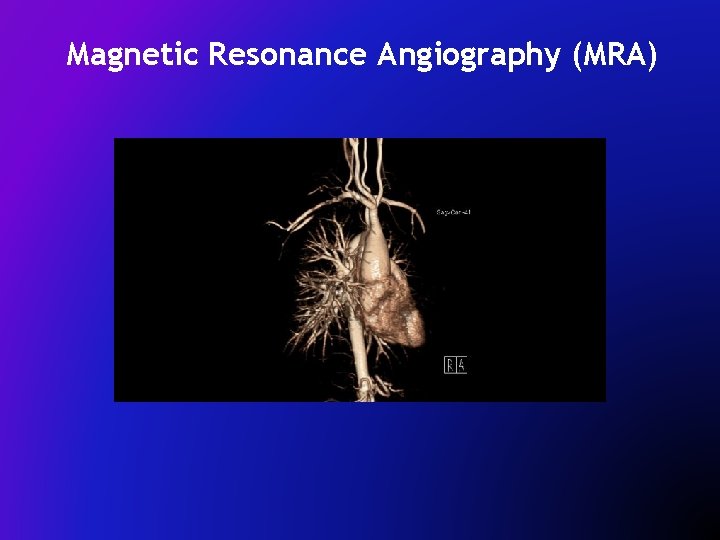
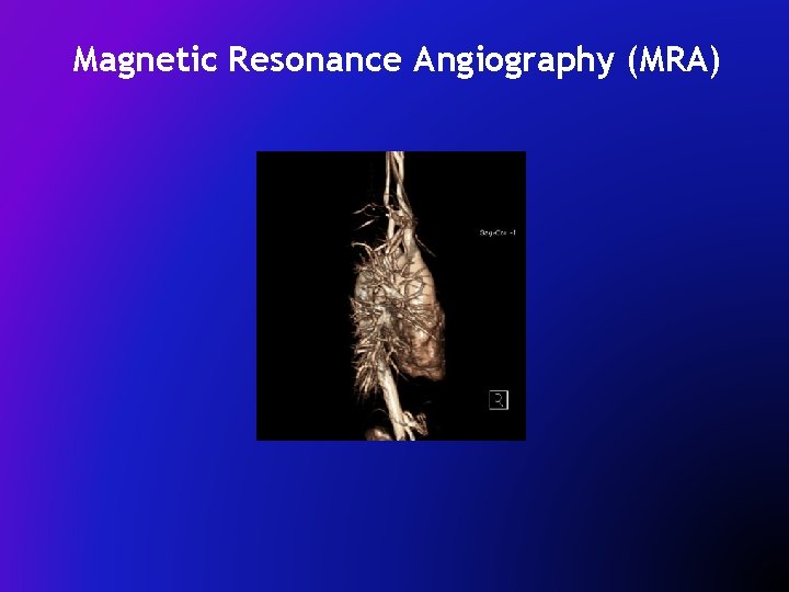
Magnetic Resonance Angiography (MRA)
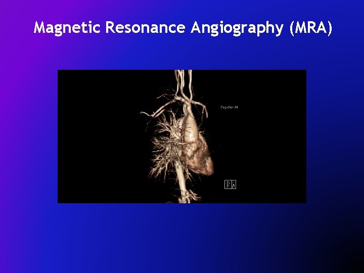
Magnetic Resonance Angiography (MRA)
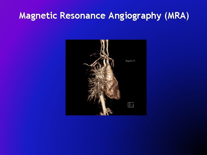
Magnetic Resonance Angiography (MRA)
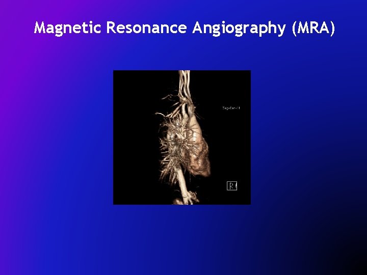
Magnetic Resonance Angiography (MRA)
Magnetic Resonance Angiography (MRA)
Magnetic Resonance Angiography (MRA)
Magnetic Resonance Angiography (MRA)
Magnetic Resonance Angiography (MRA)
Magnetic Resonance Angiography (MRA)
Take Home Points § ABC’s = starting point to organize approach to frontal chest images § Start to understand the “black and whites” of radiographic imaging § “One View Is No View”: Ideal Chest Study is PA/lateral images
Welcome to Hopkins! dmagid@jhmi. edu www. Teamrads. com