
XRays Michael R Jackson Neonatal Chest Radiography Diligent



- Slides: 64
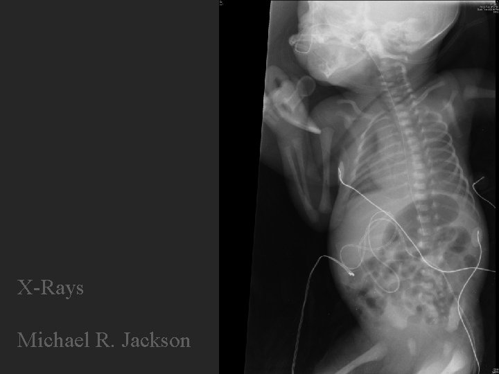
X-Rays Michael R. Jackson
Neonatal Chest Radiography Diligent Study Michael R. Jackson RRT-NPS CPFT Brigham & Women’s Hospital, Boston, MA
Neonatal Chest Radiography Diligent Study Michael R. Jackson RRT-NPS CPFT Brigham & Women’s Hospital, Boston, MA
Neonatal Chest Radiography Diligent Study Michael R. Jackson RRT-NPS CPFT Brigham & Women’s Hospital, Boston, MA
Neonatl Chest Radiography Diligent Study Michael R. Jackson RRT-NPS CPFT Brigham & Women’s Hospital, Boston, MA
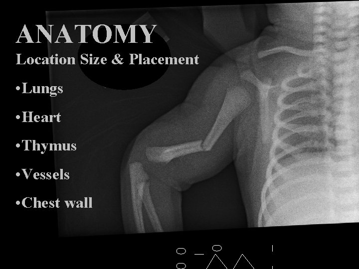
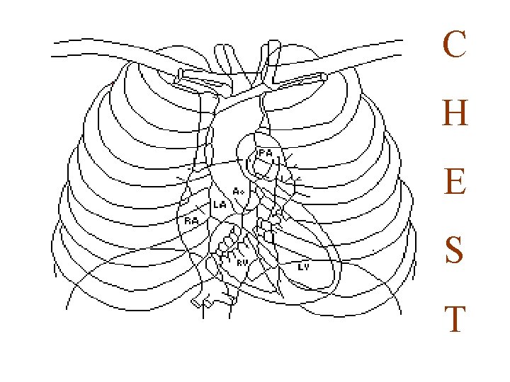
ANATOMY Location Size & Placement • Lungs • Heart • Thymus • Vessels • Chest wall
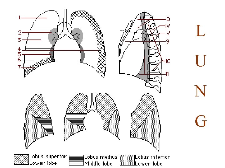
L U N G
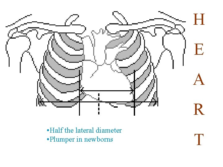
H E A R • Half the lateral diameter • Plumper in newborns T
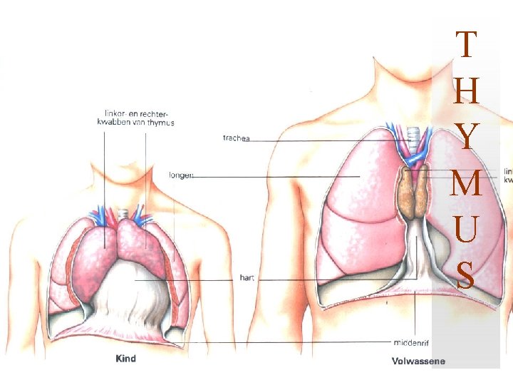
T H Y M U S
C H E S T
Method
• Quality Improvement in Radiography in a Neonatal Intensive Care Unit Linda Loovere, MD, FRCPC, Elaine M Boyle, MB, Ch. B, MSc, Susan Blatz, RN, Ph. D, Marion Bowslaugh, M. R. T. (R), ACR, Marilyn Kereliuk, M. R. T. (R), Bosco Paes, MBBS, FRCPC E D Dougeni. Dose and image quality optimization in neonatal radiography. Br J of Radiol. 2007; 80: 807 -815. Europen Commission. European guidelines on Quality Criteria for Diagnostic Radiographic Images in Pediatrics. EUR 1996; 16261: 1 -71. Key factors in the optimization of paediatric X-ray practice J V Cook, MRCP, FRCR 1, J C Kyriou, BSc, MSc 2, A Pettet, DCR, DMU 2, M C Fitzgerald, BSc, MSc 2, K Shah, DCR, DMU 1 and S M Pablot, FRCR 1 (n=3000)
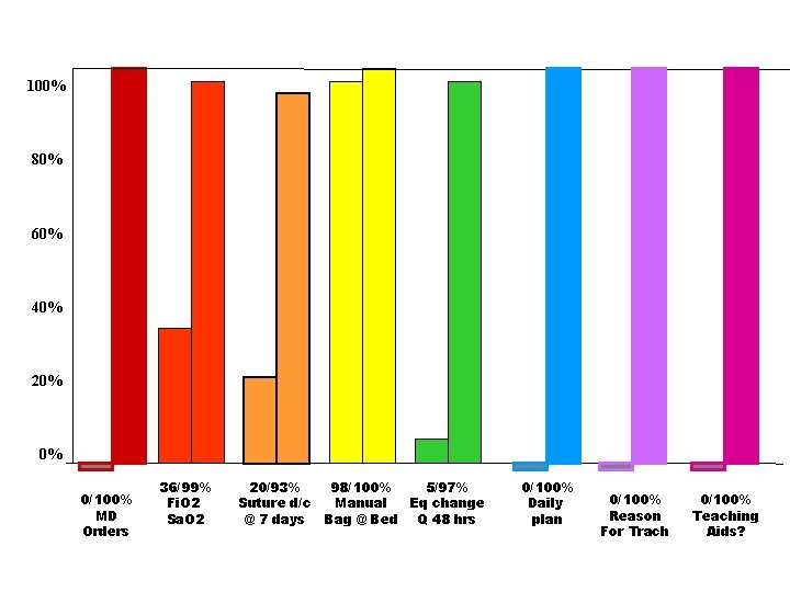
100% 80% 60% 40% 20% 0% 0/100% MD Orders 36/99% Fi. O 2 Sa. O 2 20/93% 98/100% 5/97% Suture d/c Manual Eq change @ 7 days Bag @ Bed Q 48 hrs 0/100% Daily plan 0/100% Reason For Trach 0/100% Teaching Aids?
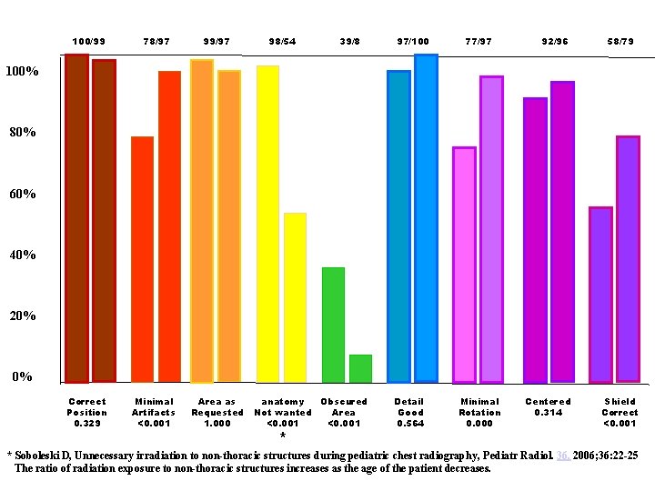
100/99 78/97 99/97 Correct Position 0. 329 Minimal Artifacts <0. 001 Area as Requested 1. 000 98/54 39/8 97/100 77/97 92/96 58/79 100% 80% 60% 40% 20% 0% anatomy Obscured Not wanted Area <0. 001 * Detail Good 0. 564 Minimal Rotation 0. 000 Centered 0. 314 Shield Correct <0. 001 * Soboleski D, Unnecessary irradiation to non-thoracic structures during pediatric chest radiography, Pediatr Radiol. 36, 2006; 36: 22 -25 The ratio of radiation exposure to non-thoracic structures increases as the age of the patient decreases.
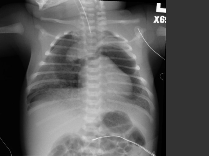
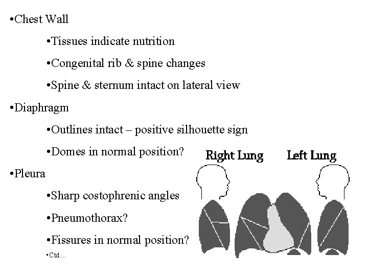
• Chest Wall • Tissues indicate nutrition • Congenital rib & spine changes • Spine & sternum intact on lateral view • Diaphragm • Outlines intact – positive silhouette sign • Domes in normal position? • Pleura • Sharp costophrenic angles • Pneumothorax? • Fissures in normal position? • Ctd…
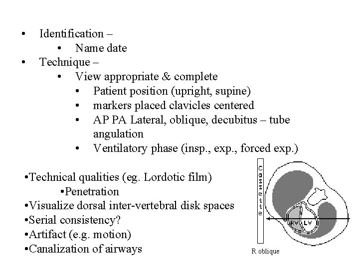
• • Identification – • Name date Technique – • View appropriate & complete • Patient position (upright, supine) • markers placed clavicles centered • AP PA Lateral, oblique, decubitus – tube angulation • Ventilatory phase (insp. , exp. , forced exp. ) • Technical qualities (eg. Lordotic film) • Penetration • Visualize dorsal inter-vertebral disk spaces • Serial consistency? • Artifact (e. g. motion) • Canalization of airways R oblique
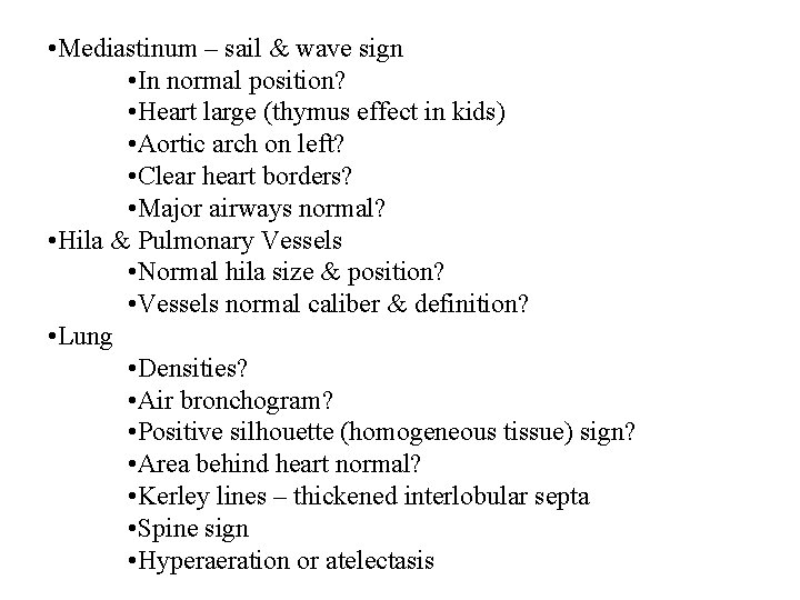
• Mediastinum – sail & wave sign • In normal position? • Heart large (thymus effect in kids) • Aortic arch on left? • Clear heart borders? • Major airways normal? • Hila & Pulmonary Vessels • Normal hila size & position? • Vessels normal caliber & definition? • Lung • Densities? • Air bronchogram? • Positive silhouette (homogeneous tissue) sign? • Area behind heart normal? • Kerley lines – thickened interlobular septa • Spine sign • Hyperaeration or atelectasis
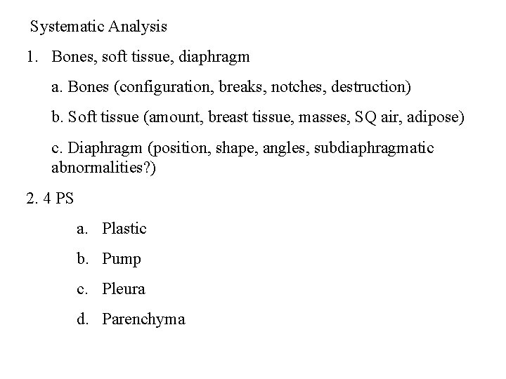
Systematic Analysis 1. Bones, soft tissue, diaphragm a. Bones (configuration, breaks, notches, destruction) b. Soft tissue (amount, breast tissue, masses, SQ air, adipose) c. Diaphragm (position, shape, angles, subdiaphragmatic abnormalities? ) 2. 4 PS a. Plastic b. Pump c. Pleura d. Parenchyma
• Illusions • Skin folds • Rotation from desired posture
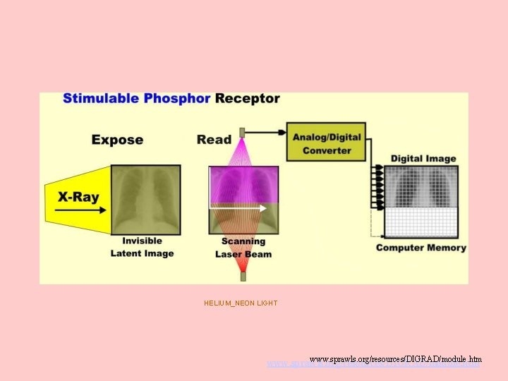
HELIUM_NEON LIGHT www. sprawls. org/resources/DIGRAD/module. htm
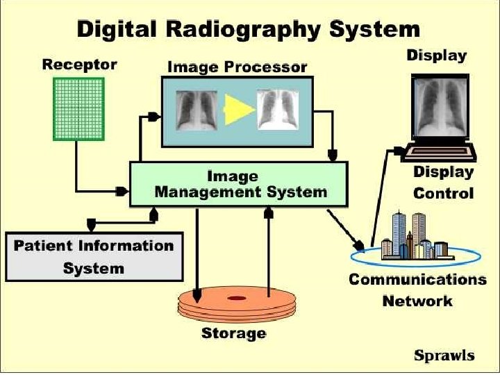
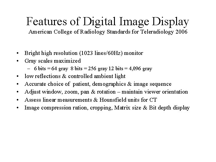
Features of Digital Image Display American College of Radiology Standards for Teleradiology 2006 • Bright high resolution (1023 lines/60 Hz) monitor • Gray scales maximized – 6 bits = 64 gray 8 bits = 256 gray 12 bits = 4, 096 gray • • • low reflections & controlled ambient light Accurate choice of patient, demographics & image sequence Adjust window, zoom, pan & rotation – maintain viewer orientation Assess linear measurements & Hounsfield units for CT Image compression ration, cropping, Matrix size & Bit depth display
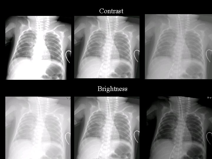
Contrast Brightness
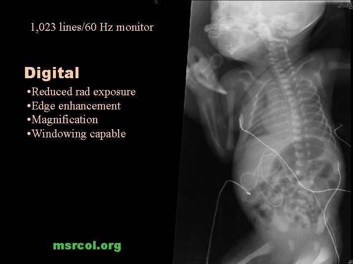
1, 023 lines/60 Hz monitor Digital • Reduced rad exposure • Edge enhancement • Magnification • Windowing capable msrcol. org
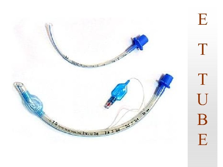
E T T U B E
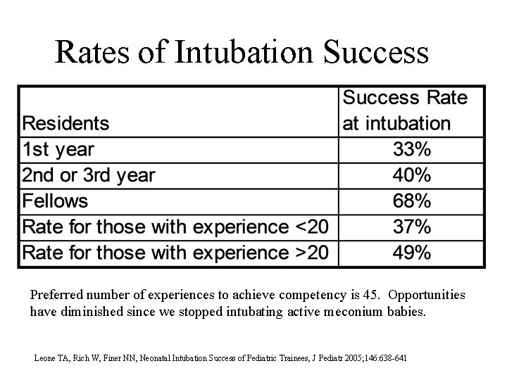
Rates of Intubation Success Preferred number of experiences to achieve competency is 45. Opportunities have diminished since we stopped intubating active meconium babies. Leone TA, Rich W, Finer NN, Neonatal Intubation Success of Pediatric Trainees, J Pediatr 2005; 146: 638 -641
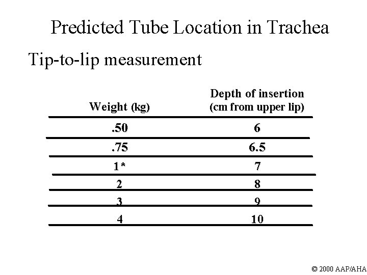
Predicted Tube Location in Trachea Tip-to-lip measurement Depth of insertion Weight (kg) (cm from upper lip) . 50. 75 6 6. 5 1* 2 3 4 7 8 9 10 © 2000 AAP/AHA
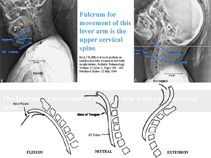
8. 3 5 Epiglottis Vocal Cord Thy. Cartilage Fulcrum for movement of this lever arm is the upper cervical spine. . Rost, J R, Effect of neck position on endotracheal tube location in low birth weight infants, Pediatric Pulmonology Volume 27, Issue 3, Pages 199 - 202 Published Online: 21 May 1999 10 Vocal Cord 5. 5 Epiglottis Thy. Cartilage The fulcrum for movement of this lever arm is the upper cervical spine. FLEXION NEUTRAL EXTENSION
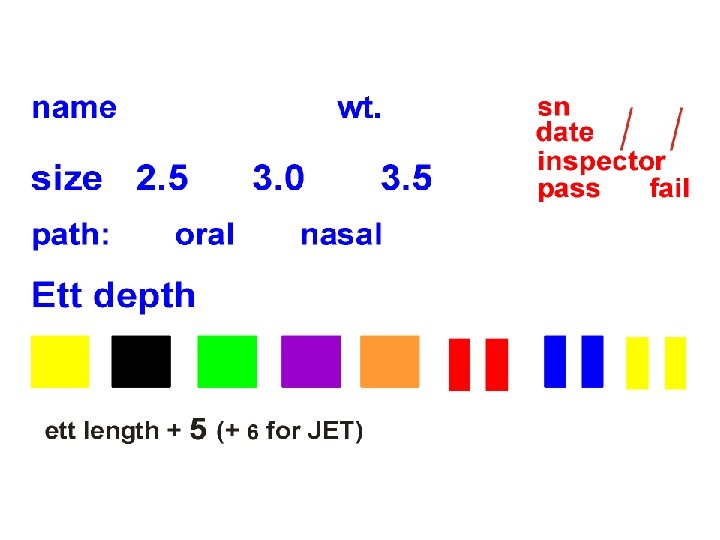
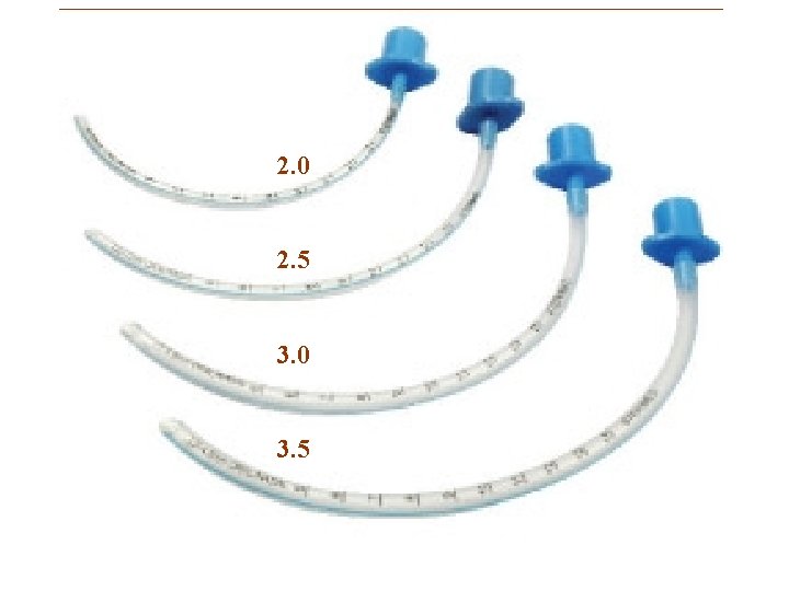
2. 0 2. 5 3. 0 3. 5
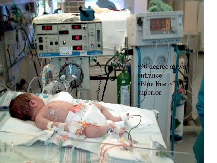
• 90 degree airway entrance • Blue line of ETT superior
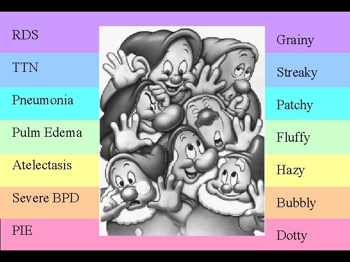
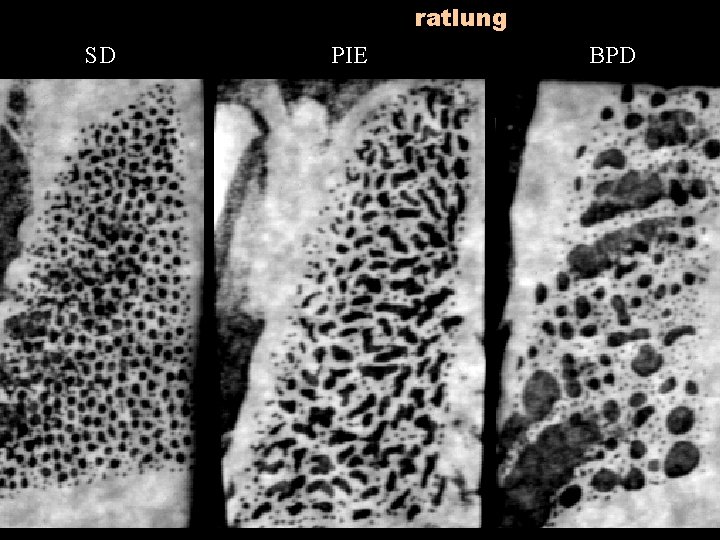
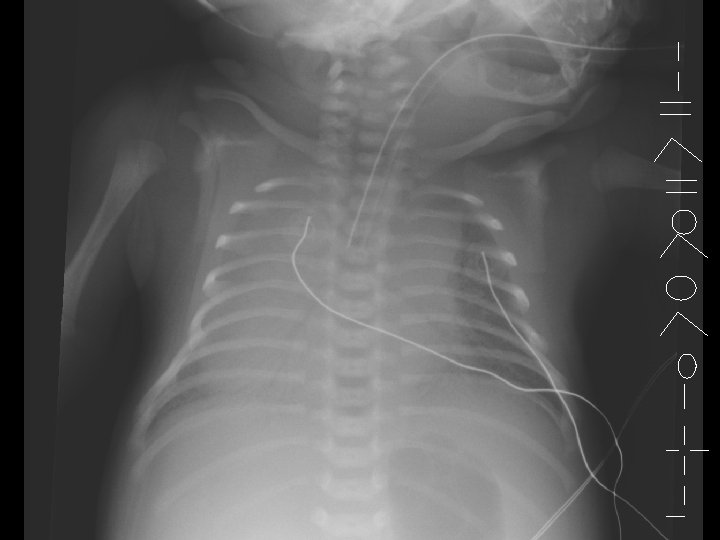
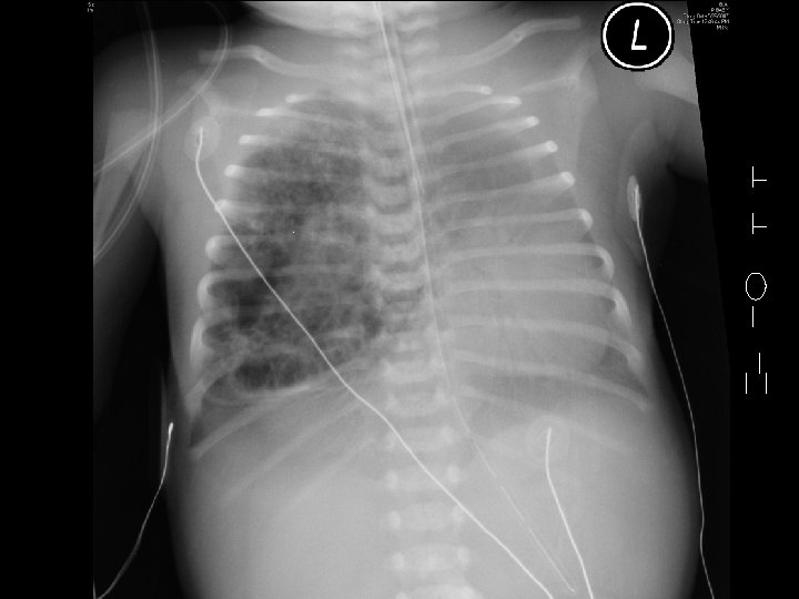
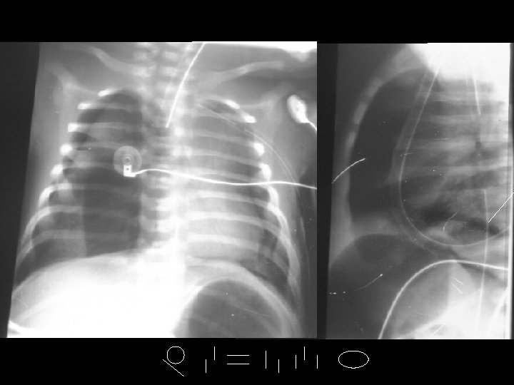
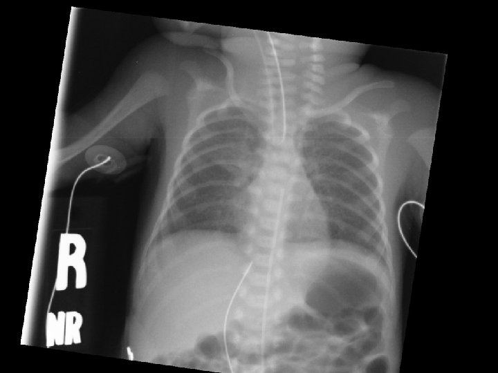
RDS Grainy TTN Streaky Pneumonia Patchy Pulm Edema Fluffy Atelectasis Hazy Severe BPD Bubbly PIE Dotty
ratlung SD PIE BPD
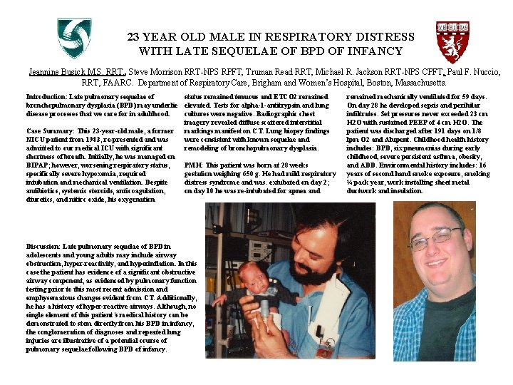
23 YEAR OLD MALE IN RESPIRATORY DISTRESS WITH LATE SEQUELAE OF BPD OF INFANCY Jeannine Busick M. S. RRT. , Steve Morrison RRT-NPS RPFT, Truman Read RRT, Michael R. Jackson RRT-NPS CPFT, Paul F. Nuccio, RRT, FAARC. Department of Respiratory Care, Brigham and Women’s Hospital, Boston, Massachusetts. Introduction: Late pulmonary sequelae of status remained tenuous and ETCO 2 remained bronchopulmonary dysplasia (BPD) may underlie elevated. Tests for alpha-1 -antitrypsin and lung disease processes that we care for in adulthood. cultures were negative. Radiographic chest imagery revealed diffuse scattered interstitial Case Summary: This 23 -year-old male, a former markings manifest on CT. Lung biopsy findings NICU patient from 1983, re-presented and was were consistent with known sequelae and admitted to our medical ICU with significant remodeling of bronchopulmonary dysplasia. shortness of breath. Initially, he was managed on BIPAP; however, worsening respiratory status, PMH: This patient was born at 28 weeks specifically severe hypoxemia, required gestation weighing 650 g. He had mild respiratory intubation and mechanical ventilation. Despite distress syndrome and was. extubated on day 2; antibiotics, systemic steroids, anticoagulation, on day 10 he was re-intubated for apnea and diuretics, and nitirc oxide, his oxygenation Discussion: Late pulmonary sequelae of BPD in adolescents and young adults may include airway obstruction, hyper-reactivity, and hyperinflation. In this case the patient has evidence of a significant obstructive airway component, as evidenced by pulmonary function testing prior to this most recent admission and emphysematous changes evident from CT. Additionally, he has a history of hyper-reactive airways. Although, no single element of this patient’s medical history can be demonstrated to stem directly from his BPD in infancy, the conglomeration of diagnoses and repeated lung injuries are illustrative of a potential course of pulmonary sequelae following BPD of infancy. remained mechanically ventilated for 59 days. On day 28 he developed sepsis and perihilar infiltrates. Set pressures never exceeded 23 cm H 2 O with sustained PEEP of 4 cm H 2 O. The patient was discharged after 191 days on 1/8 lpm O 2 and Alupent. Childhood health history includes: BPD, six pneumonias during early childhood, severe persistent asthma, obesity, and ADD. Environmental history includes: 16 years of second hand smoke exposure, smoking ¼ pack year, work installing sheet metal ductwork and insulation.