ILO CLASSIFICATION OF STANDARD CHEST RADIOGRAPHS OF PNEUMOCONIOSES


 Rounded b)")


 b. Costophrenic")


 Ø Remaining")


 looking like pleural plaques")
 on the")
 and face-on plaques (blue arrows)")


- Slides: 52
ILO CLASSIFICATION OF STANDARD CHEST RADIOGRAPHS OF PNEUMOCONIOSES Presenter : Dr Jyotsna Rayannavar Chairperson : Dr K R Pravinchandra
INTRODUCTION • 1930 First International Conference of Experts on Pneumoconioses in Johannesberg radiological appearances and impairment of lung functions. • 1958 Geneva classification – radiological appearances. • Periodically revised 1968, 1971, 1980 & 2000.
SCOPE OF CLASSIFICATION • Provides a means for describing and recording systematically the radiographic abnormalities in the chest provoked by inhalation of dusts. • Used to describe radiographic abnormalities that occur in any type of pneumoconioses. • Designed for classifying the appearances seen only on postero-anterior chest radiographs.
OBJECT OF CLASSIFICATION • To codify the radiographic abnormalities of the pneumoconioses in a simple, systematic and reproducible manner. • Neither defines pathological entities nor takes into account working capacity
USES OF CLASSIFICATION • Internationally for : Ø Epidemiological research Ø Screening and surveillance of those in dusty occupations Ø Clinical purposes • May lead to better international comparability of data regarding pneumoconioses.
CLASSIFICATION • ILO classification includes printed guidelines and 2 sets of standard radiographs. a. Complete set – 22 radiographs b. Quad set – 14 radiographs. • The reader compares the subject chest radiograph with those of the standard set
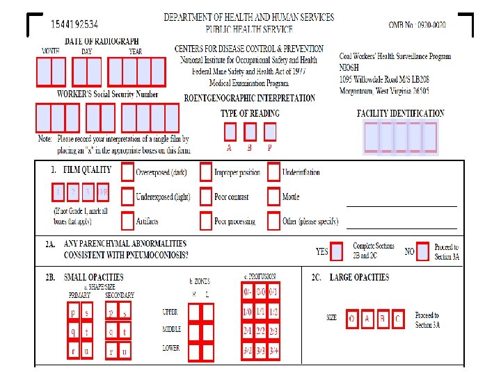
• To classify the chest radiograph of a dust exposed individual, 4 main points are to be considered. 1. Technical quality 2. Parenchymal abnormalities 3. Pleural abnormalities 4. Symbols or other abnormalities
TECHNICAL QUALITY Four grades : 1, 2, 3 & 4 Grade 1 : Good Grade 2 : Acceptable, with no technical defect Grade 3 : Acceptable, with some technical defect but still adequate for classification • Grade 4 : Unacceptable • •
PARENCHYMAL ABNORMALITIES • Include : Ø Small opacities Ø Large opacities
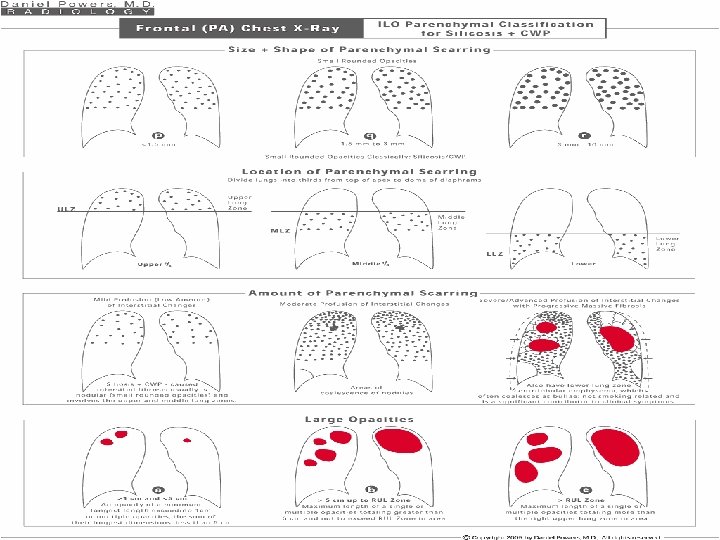
• SMALL OPACITIES : a. Profusion b. Affected zones of the lungs c. Shape d. Size
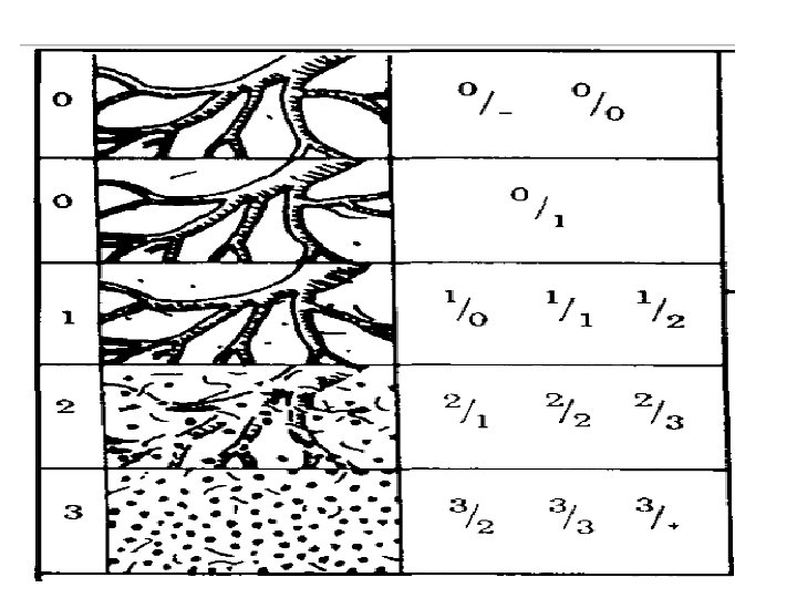
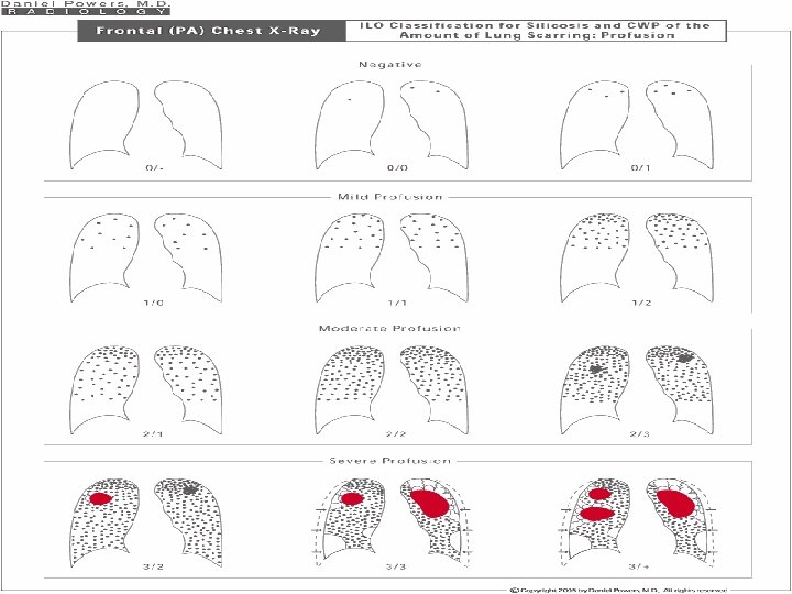
• PROFUSION : ü 4 categories : 0, 1, 2, 3 ü 12 subcategories : 0/-, 0/0, 0/1 1/0, 1/1, 1/2 2/1, 2/2, 2/3 3/2, 3/3, 3/+ ü Cat 0 refers to absence of small opacities & cat 3 to highest profusion. ü Compared with standard radiographs 0/0, 1/1, 2/2 & 3/3.
• AFFECTED ZONES : Ø Lung field is divided into 3 zones by horizontal lines drawn at 1/3 rd & 2/3 rd of vertical distance between lung apices & the domes of diaphragm. Ø Upper / middle / lower Ø Small opacities profusion is determined in affected lung zone.
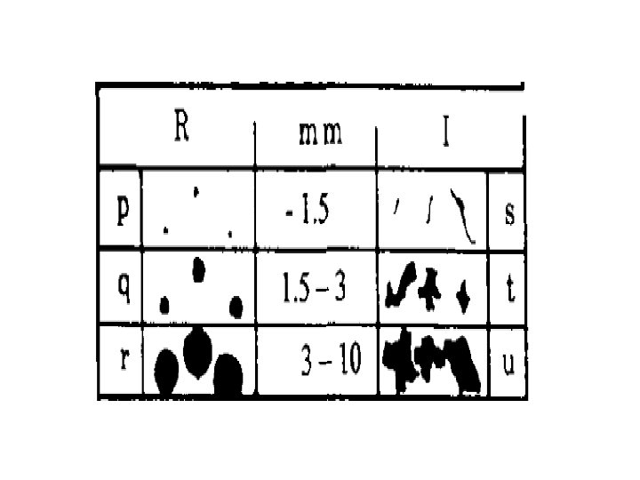
• SHAPE AND SIZE : Ø 2 shapes recognized : a) Rounded b) Irregular Ø 3 sizes determined in each shape. Ø Small rounded opacities : p, q, r Ø Small irregular opacities : s, t, u
Contd…. • • • p : diameters upto 1. 5 mm q : diameters 1. 5 – 3 mm r : diameters 3 - 10 mm s : widths upto 1. 5 mm t : widths 1. 5 – 3 mm u : widths 3 – 10 mm
• LARGE OPACITIES : ü Opacity having the longest dimension exceeding 10 mm ü 3 categories : A, B, C ü Category A : one large opacity having the longest dimension up to 50 mm or several large opacities with the sum of their longest dimensions not exceeding 50 mm.
Contd…. ü Category B : one large opacity having the longest dimension exceeding 50 mm but not exceeding the equivalent area of right upper zone ü Category C : one large opacity which exceeds the equivalent area of the right upper zone
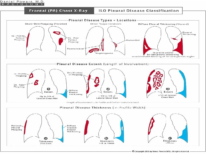
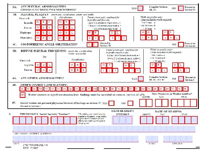
PLEURAL ABNORMALITIES • Comprise of : a. Pleural plaques (localized pleural thickening) b. Costophrenic angle obliteration c. Diffuse pleural thickening
• PLEURAL PLAQUES : Ø Localized pleural thickening of parietal pleura Ø Seen on diaphragm, on chest wall & at other sites Ø If present on chest wall, recorded as in-profile or face-on Ø Site, calcification and extent are to be recorded
Contd…. . Ø Extent is recorded only for plaques along the chest wall Ø Extent is defined in terms of the total length of involvement from with respect to projection of the lateral chest wall (apex to costophrenic angle) Ø 1 - one quarter of projection, 2 - b/w one quarter and one-half of projection, 3 - more than one-half.
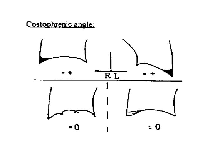
• COSTOPHRENIC ANGLE OBLITERATION : ü Present / absent • DIFFUSE PLEURAL THICKENING : Ø Recorded only in the presence of, and in continuity with, an obliterated costophrenic angle
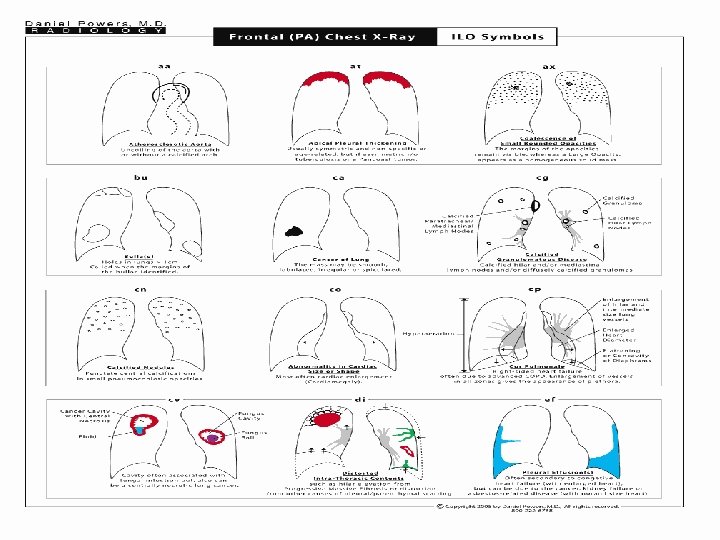
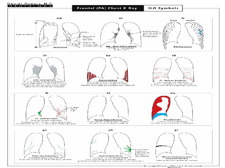
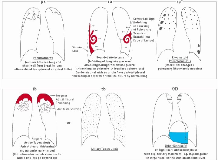
OTHER ABNORMALITIES/ SYMBOLS • Relevant as they describe additional features related to dust exposure and other aetiologies • Each definition of symbols assumes an introductory qualifying word or phrase such as “changes indicative of”, or “opacities suggestive of”, or “suspect”.
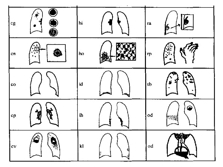
Contd…. • Symbols are : ü aa, at, ax, bu, ca, cg, cn, co, cp, cv, di, ü ef, em, es, fr, hi, ho, id, ih, kl, me, ü pa, pb, pi, px, ra, rp, tb, od
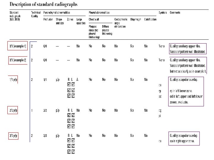
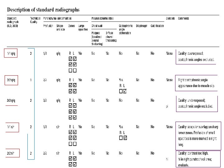
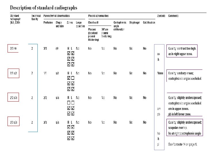
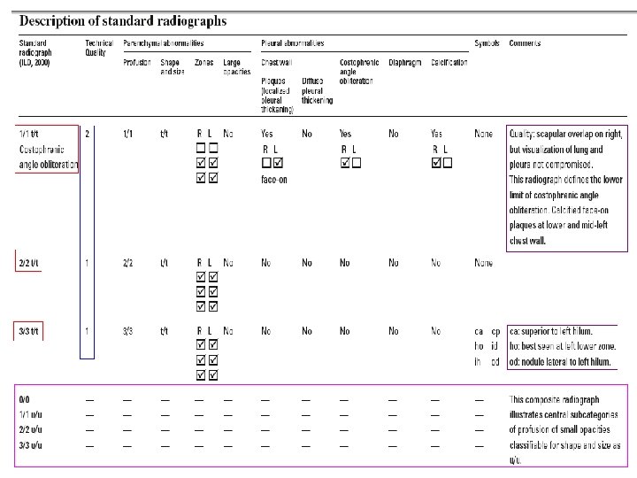
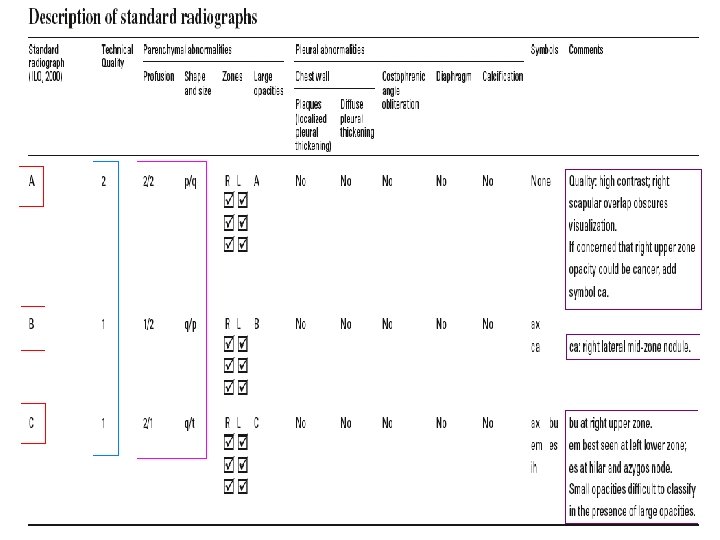
DESCRIPTION OF STANDARD RADIOGRAPHS • THE COMPLETE SET : Ø 22 standard radiographs Ø 2 : illustrate category 00 profusion of small opacities Ø 15: define small opacity profusion category (1/1, 2/2, 3/3) and some of the shapes & sizes of these opacities (p, q, r, s & t)
Contd…. . Ø 3 : large opacities are shown (A, B, C) Ø Remaining 2 radiographs are composite reproductions of sections from full-size chest radiographs. ü One depicts increasing profusion of irregular small ‘u’ sized opacities ü Other one illustrates various pleural abnormalities
• THE QUAD SET : Ø Includes 9 of the most commonly used standard radiographs from the complete set Ø Remaining 5 are composite reproductions of quadrant sections from other radiographs in the complete.
Normal chest radiograph showing normal anatomical features. Note the presence of opacities that represent lymph nodes and blood vessels.
Chest radiograph of poor quality showing overlapping scapula (white arrows) looking like pleural plaques on both right and left lung fields.
Silicotic chest radiograph showing a large opacity of category B (black arrows) on the right upper zone. Numerous rounded opacities (q type) are present in all zones of the lung field.
Chest radiograph showing calcified diaphragmatic pleural plaques (white arrows) and face-on plaques (blue arrows) bilaterally. In this film, pleural plaques can also be seen in other site (mediastinal region bilaterally).
Chest radiograph of asbestos-exposed worker showing the presence of small irregular opacities (of “t” type primarily and “s” type secondarily), predominantly on lower and middle lung zones. The right upper lobe is also involved. The profusion is 2/3. In addition, diffuse pleural thickening (DPT) is evident on the right and in-profile plaque on the left; also note the abnormal cardiac size. The film quality is of grade 2 (scapula overlap).
REFERENCES • Hunter’s Diseases of Occupations , 10 th edition • Guidelines for the use of the ILO international classification of radiographs of Pneumoconioses. Revised edition 2011, Occupational Safety and Health Series 22. • ILO Encyclopedia of Occupational Health and Safety. 4 th Edition Geneva, volume 2.
Thank you