Digital Radiography Chapter 26 Digital radiographs l l

. Filmless imaging using")


 Instant viewing of")


- Slides: 15
Digital Radiography Chapter 26
Digital radiographs l l l Different from analog images (traditional radiographs). Filmless imaging using pixels. Instant images viewed on computer monitor. Potential to improve quality of oral health care, while reducing radiation exposure. Images made with digital sensors and phosphor plates. No processing or darkroom needed.
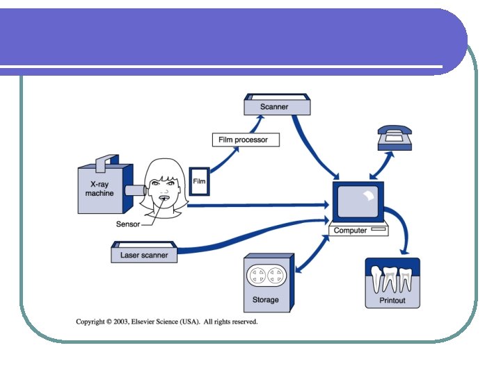
l Digital panoramic and cephalometric systems also available. l Indirect digital imaging – converting radiographs to digital images with a scanner; placing radiographs on viewbox and taking picture with digital camera; not good quality image.
l Direct digital imaging – use sensor; placed and exposed just like film; sensor transmits image to computer and then viewed on monitor. l Sensors available in different sizes (1, 2) l Large range of gray scale which helps in diagnosis.
The size of the electronic sensor is compared with sizes 0, 1, and 2 of traditional intraoral film.
Uses similar to film radiography: l Detect, confirm dental diseases and lesions. l Detect, evaluate trauma, growths and development. l Provide info during procedures (endo and surgery).
Equipment: Dental x-ray machine with electronic timer capable of short exposure time (70 k. Vp or less, 5 m. A or less) l Sensor – wired or wireless; 3 types available in different sizes. (CCD, CMOS, PSP) l Computer with monitor (printer can allow printing of images); large enough memory for storage of images; internet connection can allow e-mailing to insurance companies and referrals. l
Equipment con’t l Software – compatible with sensor; allows for manipulation of images. l l l Side-by-side displays of images – helpful when comparing images to previous ones. Magnification – magnified up to 4 times original size to aid in viewing. Density and contrast – can be changed to help in diagnosis. Measurement tools – “ruler” to measure things such as root length, bone loss. Charting – notes, arrows, circles, etc. can be added directly to image. Colorization – to distinguish different parts/structures.
Radiation Exposure: l Can be 50 to 80 percent less than exposures made with film based radiography; the lower the exposure time, the lower the radiation dose. 50% less than F speed film l 60% less than E speed film l 80% less than D speed film l
Legal Implications: l Image can be manipulated, making it possible to show disease where there is none, or to hide faulty dental work. (manufacturers of software presently working on software that will not permit an altered image to saved unless marked as “copy”.
Advantages: l l l l Less radiation to patient (and personnel) Instant viewing of image; magnification, etc. Elimination of darkroom and processing Improvement of diagnostic image Improved gray scale resolution Electronic sending of images to referrals and insurance companies Easier viewing and understanding by patients. No films to mount
Disadvantages: l Initial investment is high; expensive. l Technology concerns – computer crashes, viruses, low memory, power failures, etc. ; out-of-date computers/software; system compatibility with information transfer. l Sensor uncomfortable and thick for some patients; rigid. l Image quality not as good as film.
Disadvantages con’t: Infection control - Sensors can not be sterilized; must use plastic sheaths that may tear. l Sensor dimensions smaller than film, so capture less area. l Legal issues due to ability of alteration of images l Archival storage and back-up storage require computer space or portable storage devices. l
Exposure procedures similar to film radiography: l See procedures pgs. 352 -355. l Seat patient l Adjust chair; headrest l Use lead apron, thyroid collar l Remove objects that may interfere with procedure (eyeglasses, partials, etc) l Explain radiographic procedure.