Dental Radiography RVT Chapter 24 Objectives Dental Radiography

Dental Radiography RVT: Chapter 24

Objectives: Dental Radiography • Review dental terminology & tooth surfaces • Review basic anatomy & formula for teeth, including number of roots • Understand normal views & positioning for dental radiography • Define parallel and bisecting angle techniques, and know when to use each • Understand the differences between intraoral and extraoral views

Anatomical Directions: Oral Cavity

Positional Terminology: Oral Cavity Directional Positional

Tooth Anatomy on Film What are the four basic types of teeth?

Assessing Age

Dental Formulas Carnassial tooth: • Dogs: Upper 4 th premolar & lower 1 st molar • Cats: Upper 3 rd premolar & lower 1 st molar

Triadan Numbering System Canine Feline 1 st number = quadrant 2 nd 2 numbers = tooth position

Number of Roots: Canine

Number of Roots: Feline

Normal Adult Canine Maxillary Incisors 7 – Incisive canal 9 – ______ fissures (oval dark spaces)
")
Normal Adult Canine Mandibular Incisors 7 – Mandibular ________ (dark black line)

Viewing Dental Radiographs • Film is exposed with the convex dot at the _____ end of the mouth • Dot location will vary with right & left side radiographs • When viewing- hold with convex side towards you • Maxillary: point cusps toward the floor; apical portion of roots to ceiling • Mandibular: point apical portion of roots towards the floor; cusps to the ceiling

Viewing Dental Radiographs

Film Mount Organize full mouth radiographs in a film mount: Hang as if looking at the animal, with animal’s right side on your left…

Dental Radiography Techniques • What makes dental rads tricky: getting the whole tooth! • Must include complete _____ to be diagnostic • Patient is always anesthetized • Positions will include lateral, dorsal, or sternal • Easier if skull is kept parallel to table top • Paper towels and syringes/cases are your best friends • Longer SID allows for wider primary beam • Using size 0 -2? • Center the cone on the affected tooth • Position yourself opposite from the cone to find the angle

Proper Oral Film Placement

Dental Radiography Techniques Parallel Bisecting Angle

Parallel Technique • Used for mandibular molars and pre-molars only • Film is placed inside of mouth & ______ to the teeth • Primary beam is placed perpendicular to film • Only acceptable to use in one location!

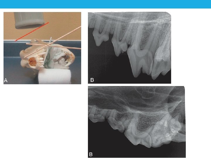
Bisecting-angle Technique • Technique for ALL maxillary teeth & _______ canines and incisors • Placing the beam perpendicular to tooth (or film) results in foreshortening or elongation • Find the bisecting-angle: • Place film parallel to tongue • Draw an imaginary line along the tooth’s axis (use root) • Draw another imaginary line along the axis of the film • Find the angle that is exactly in the middle of these two • Position the primary beam perpendicular to the “bisecting angle”

Bisecting Angle- Mandibular canine

Bisecting Angle- Mandibular incisors

Bisecting Angle- Maxillary incisors

Distortion: Foreshortening

25 Distortion: Elongation FILM
- Slides: 26